. 2021 Feb;95(2):727-747.
doi: 10.1007/s00204-020-02946-5. Epub 2021 Jan 25.

Chronic senescent human mesenchymal stem cells as possible contributor to the wound healing disorder after exposure to the alkylating agent sulfur mustard

Affiliations

Chronic senescent human mesenchymal stem cells as possible contributor to the wound healing disorder after exposure to the alkylating agent sulfur mustard

Simone Rothmiller et al. Arch Toxicol. 2021 Feb.

Abstract

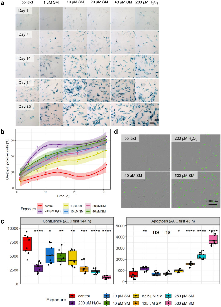
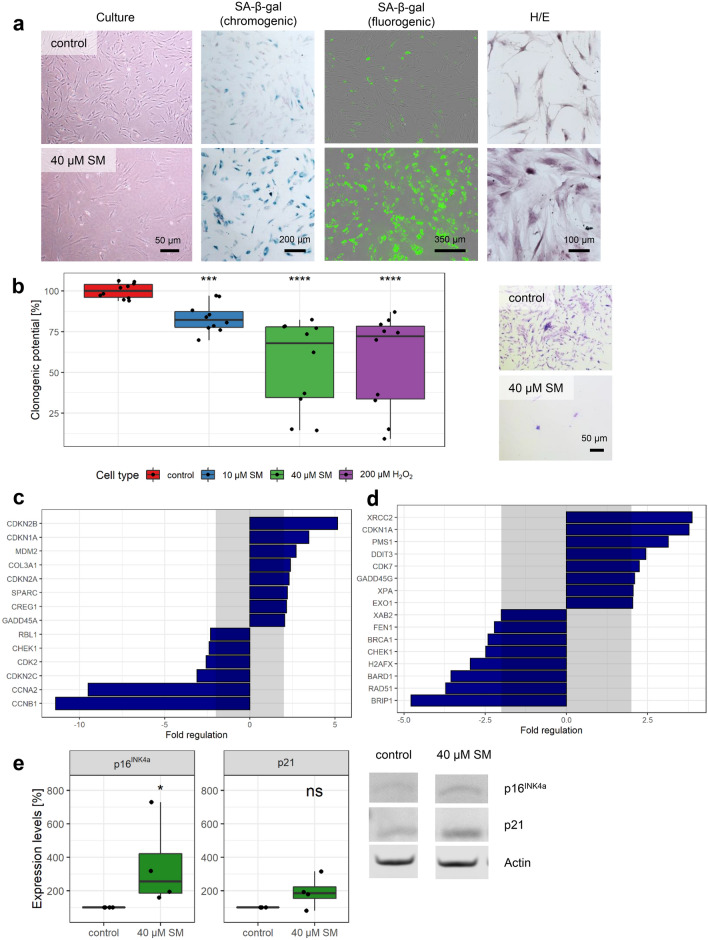
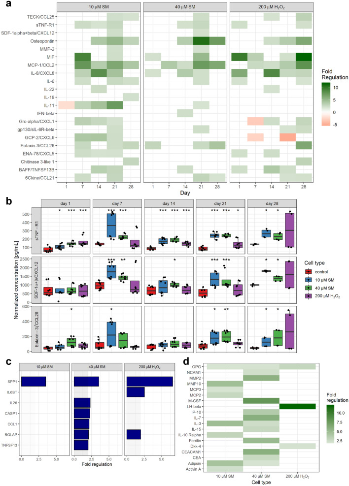
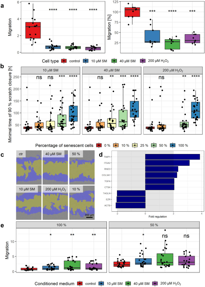
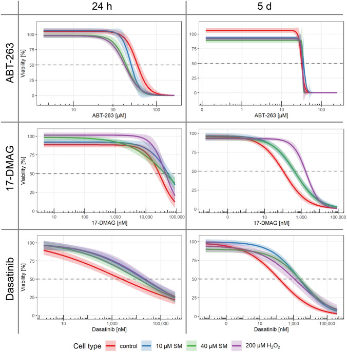
Wound healing is a complex process, and disturbance of even a single mechanism can result in chronic ulcers developing after exposure to the alkylating agent sulfur mustard (SM). A possible contributor may be SM-induced chronic senescent mesenchymal stem cells (MSCs), unable to fulfil their regenerative role, by persisting over long time periods and creating a proinflammatory microenvironment. Here we show that senescence induction in human bone marrow derived MSCs was time- and concentration-dependent, and chronic senescence could be verified 3 weeks after exposure to between 10 and 40 µM SM. Morphological changes, reduced clonogenic and migration potential, longer scratch closure times, differences in senescence, motility and DNA damage response associated genes as well as increased levels of proinflammatory cytokines were revealed. Selective removal of these cells by senolytic drugs, in which ABT-263 showed initial potential in vitro, opens the possibility for an innovative treatment strategy for chronic wounds, but also tumors and age-related diseases.

Keywords: Chemical warfare agents; Mesenchymal stem cells; Senescence; Sulfur mustard; Wound healing disorder.


Conflict of interest statement

The authors declare that they have no conflict of interest.

Figures

Fig. 1
Fig. 1
Senescence and apoptosis induction. a Representative images of concentration- and time-dependent development of SA-β-gal staining (blue) after single dose SM or H2O2 exposure (day 0) in contrast to solvent controls. Counterstaining of all cells with nuclear fast red (red). Scale bar, 200 µm. b The means with a trend including 99% confidence intervals of the percentage of SA-β-gal positive cells counted every 3–4 days over 31 days (n = three randomly selected fields per group from three independent experiments). c Area under the curve (AUC) for confluence (area covered by cells) within the first 144 h and apoptosis (d annexin V positive cells (green), representative images t = 48 h) within the first 48 h. Shown is the significance between solvent controls and 200 µM H2O2 or SM exposed MSCs (n = three randomly selected images per group from three independent experiments). Data are represented as Tukey boxplots; *p < 0.05, **p < 0.01, ***p < 0.001, ****p < 0.0001. See also Supplementary Figs. 1–3
Fig. 2
Fig. 2
Senescence markers. a Representative images from both chromogenic (PromoCell) and fluorogenic (Cell Biolabs) SA-β-gal staining in MSCs 21 days after 40 µM SM exposure show a very high percentage of senescent cells with high staining intensity compared to solvent controls. Flattening and enlargement shown already in culture as well as by hematoxylin/eosin (H/E) staining. b Loss of replicative potential shown by clonogenicity assay. Colonies formed and stained with crystal violet could be observed for controls, but only single cells for 40 µM SM senescent cells. After destaining with methanol, crystal violet absorbance examined at 570 nm by plate reader and mean of controls set to 100% (n = duplicates per group from five independent experiments). Genes associated with c senescence biomarkers or d DNA damage and DNA repair were tested 21 days after 40 µM SM exposure and compared to controls. After RNA extraction from MSCs, a RT-qPCR assay was performed and fold regulation of up- or downregulated genes (≥ 2.0 or ≤ − 2.0 in combination with p < 0.05, outside grey box) are shown as means (n = three independent experiments). e The upregulation of p16INK4a and p21 was also observed using Western Blot. Representative bands and expression levels are shown (n = four independent experiments). Data are represented as Tukey boxplots; *p < 0.05, **p < 0.01, ***p < 0.001, ****p < 0.0001. See also Supplementary Fig. 4
Fig. 3
Fig. 3
Senescence-associated increase in proinflammatory factors. a, b The concentration of over 70 chemokines, cytokines and growth factors was determined by Bio-Plex assays in cell culture supernatants and normalized for the cell number (n = biological duplicates per group from three independent experiments). a Fold regulation of up- or downregulated factors (≥ 2.0 or ≤ − 2.0 in combination with normalized concentrations above 20 pg/mL) of senescent to non-senescent controls are shown. b Three factors showed an upregulation in SM- but not in H2O2-exposed MSCs. c After RNA extraction from MSCs at day 21 after exposure to 10 and 40 µM SM, 200 µM H2O2 or solvent, a RT-qPCR assay of a custom designed cytokine and chemokine gene panel was performed and fold regulation of up- or downregulated genes (≥ 2.0 or ≤ -2.0 in combination with p < 0.05, outside grey box) are shown as means (n = three independent experiments). d Cell culture supernatants were collected 21 day post-exposure and freshly transferred to glass slide based ELISA cytokine arrays. Images were recorded with Odyssey scanner and spot intensities of 279 different factors calculated. After normalization with the positive controls, fold regulation was calculated for each senescent cell type against control and fold regulation of up- or downregulated factors are shown (≥ 2.0 or ≤ -2.0 in combination with normalized concentrations above 5%; n = biological duplicates per group from two independent experiments). Data are represented as Tukey boxplots; *p < 0.05, **p < 0.01, ***p < 0.001. See also Supplementary Fig. 5
Fig. 4
Fig. 4
Senescence-associated decreased migration and scratch closure. a Senescent cells showed a significant reduction in migration using IncuCyte migration (left) as well as modified Boyden chamber assay (right). Left: Different cells in FBS-reduced medium in inserts migrated towards the reservoir plate filled with standard medium. For normalization, the confluence on the bottom was divided by the confluence on the top for each time point and results are shown after 160 h (n = up to 8 biological replicates per group from three independent experiments). Right: Cells were added to the Boyden chamber and incubated for 8 h, fixed, stained with DAPI, and migrated cells were counted. Migration was normalized to the mean of controls (n = 3 biological replicates per group from three independent experiments). b, c Increasing amounts of senescent cells were added to non-senescent controls to observe the influence on scratch closure (0% = only non-senescent solvent controls, 10% = 10% senescent cells + 90% controls, and so on). b Scratch assay was performed using the wound maker and IncuCyte microscope, and the minimal time of 90% scratch closure was determined (n = up to 8 biological replicates per group from four independent experiments). c Representative images at t = 24 h (top right 50% and top bottom 10% 40 µM SM, blue initial scratch and yellow cell-free area). d Genes associated with wound healing and cell motility were tested in 40 µM SM exposed senescent and non-senescent cells. After RNA extraction from MSCs, a RT-qPCR assay was performed and fold regulation of up- or downregulated genes (≥ 2.0 or ≤ -2.0 in combination with p < 0.05, outside grey box) are shown as means (n = three independent experiments). e The migration of healthy MSCs was increased towards conditioned medium from senescent in comparison to that from non-senescent (control) cells. Conditioned medium was added into the reservoir plate (left) or diluted half and half with culture medium (right). For normalization, the confluence on the bottom was divided by the confluence on the top for each time point and results are shown after 188 h (n = up to 8 biological replicates per group from three independent experiments). Data are represented as Tukey boxplots; *p < 0.05, **p < 0.01, ***p < 0.001, ****p < 0.0001. See also Supplementary Fig. 6
Fig. 5
Fig. 5
Elimination with senolytics. The selectivity towards senescent cells of literature reported senolytic drugs was tested. Senescent and non-senescent (control) MSC were treated with increasing concentrations of ABT-263, 17-DMAG or dasatinib for 5 days or 24 h. Viability was assessed by XTT assay and normalized for each cell type to the corresponding solvent controls (n = 4 biological replicates per group from two or three independent experiments). Data are represented as linear regression including 99% confidence intervals. See also Supplementary Fig. 7

References

    1. Abreu-Blanco MT, Watts JJ, Verboon JM, Parkhurst SM. Cytoskeleton responses in wound repair. Cell Mol Life Sci. 2012;69:2469–2483. doi: 10.1007/s00018-012-0928-2. - DOI - PMC - PubMed
    1. Alessio N, Del Gaudio S, Capasso S, et al. Low dose radiation induced senescence of human mesenchymal stromal cells and impaired the autophagy process. Oncotarget. 2015;6:8155–8166. doi: 10.18632/oncotarget.2692. - DOI - PMC - PubMed
    1. Augustin M, Maier K. Psychosomatic aspects of chronic wounds. Dermatol Psychosom/Dermatol Psychosom. 2003;4:5–13. doi: 10.1159/000070529. - DOI
    1. Baar MP, Brandt RMC, Putavet DA, et al. Targeted apoptosis of senescent cells restores tissue homeostasis in response to chemotoxicity and aging. Cell. 2017;169:132–147.e16. doi: 10.1016/j.cell.2017.02.031. - DOI - PMC - PubMed
    1. Baker DJ, Wijshake T, Tchkonia T, et al. Clearance of p16Ink4a-positive senescent cells delays ageing-associated disorders. Nature. 2011;479:232–236. doi: 10.1038/nature10600. - DOI - PMC - PubMed

Publication types

MeSH terms

LinkOut - more resources