Review
. 2021 Mar 1;320(3):F378-F403.
doi: 10.1152/ajprenal.00634.2020. Epub 2021 Jan 25.

WNK4 kinase: from structure to physiology

Affiliations
Review

WNK4 kinase: from structure to physiology

Adrián Rafael Murillo-de-Ozores et al. Am J Physiol Renal Physiol. .

Abstract

With no lysine kinase-4 (WNK4) belongs to a serine-threonine kinase family characterized by the atypical positioning of its catalytic lysine. Despite the fact that WNK4 has been found in many tissues, the majority of its study has revolved around its function in the kidney, specifically as a positive regulator of the thiazide-sensitive NaCl cotransporter (NCC) in the distal convoluted tubule of the nephron. This is explained by the description of gain-of-function mutations in the gene encoding WNK4 that causes familial hyperkalemic hypertension. This disease is mainly driven by increased downstream activation of the Ste20/SPS1-related proline-alanine-rich kinase/oxidative stress responsive kinase-1-NCC pathway, which increases salt reabsorption in the distal convoluted tubule and indirectly impairs renal K+ secretion. Here, we review the large volume of information that has accumulated about different aspects of WNK4 function. We first review the knowledge on WNK4 structure and enumerate the functional domains and motifs that have been characterized. Then, we discuss WNK4 physiological functions based on the information obtained from in vitro studies and from a diverse set of genetically modified mouse models with altered WNK4 function. We then review in vitro and in vivo evidence on the different levels of regulation of WNK4. Finally, we go through the evidence that has suggested how different physiological conditions act through WNK4 to modulate NCC activity.

Keywords: blood pressure; distal convoluted tubule; distal nephron; epithelial transport; familial hyperkalemic hypertension; potassium.


Figures
None
Graphical abstract
Figure 1.
Figure 1.
Conserved regions in with no lysine kinase 4 (WNK4). A: sequence conservation analysis was performed including the sequences of WNK1-4 kinases from the human, mouse, rat, zebrafish, pig, and clawed frog. Multiple sequence analysis was performed in Clustal Omega, and the conservation score was then calculated by https://compbio.cs.princeton.edu/conservation/score.html. The top graph shows the conservation score where three major conserved regions were identified. The first conserved region encompasses the kinase domain, the PF2a domain, and the acidic motif. The second and third conserved regions encompass the PF2b domain and the COOH-terminal coiled-coil domain (CT-CCD), respectively. The middle graph shows the conservation score observed along the WNK4 sequence in an analysis performed with the sequences of 27 WNK4 orthologs (including mammals, fishes, birds, reptiles, and amphibians). The analysis shows that, in addition to the domains and motifs that are conserved among different WNK kinases, a high degree of conservation was observed in the COOH-terminal segment, from the CT-CCD until the end of the protein. The bottom graph shows the results of the analysis of disordered protein regions. Many of these disordered regions overlap with low-complexity regions denoted by a green line. Low-complexity regions were predicted in http://smart.embl.de/. Disordered protein region probability was calculated in https://iupred2a.elte.hu/. B: amino acid sequence alignment of the COOH-terminal region of WNK4 of the human (KEGG entry 65266), rat (KEGG entry 287715), mouse (KEGG entry 69847), cat (KEGG entry 101100264), chicken (KEGG entry 777580), American alligator (KEGG entry 102565768), African clawed frog (KEGG entry 108701024), zebrafish (KEGG entry 100330953), and whale shark (KEGG entry 109912281). Numbers at the top represent the residue numbers of human WNK4. Different sites are indicated by arrows, such as critical residues for WNK-WNK binding located within the CT-CCD (153), familial hyperkalemic hypertension (FHHt) mutations (172, 188), calmodulin (CaM)-binding site (101), PKC/PKA/serum/glucocorticoid regulated kinase-1 (SGK1) phosphorylation sites (16, 124, 129), and a protein phosphatase-1 (PP1)-binding site (100). Alignment was generated in Clustal Omega (EMBL-EBI). KEGG, Kyoto Encyclopedia of Genes and Genomes.
Figure 2.
Figure 2.
The with no lysine kinase (WNK) 4 kinase domain shares predicted structural features with WNK1. A: structural alignment of the WNK1 kinase domain (PDB 4Q2A) with the predicted structure of the kinase domain of WNK4 obtained from the sequence homology-based server http://raptorx.uchicago.edu/. B, inset: residues whose backbones amides and lateral chains are involved in the coordination of the Cl anion. Black labels indicate these amino acid residues in WNK1. Leu322 in blue denotes the experimentally validated residue involved in Cl sensing in WNK4.
Figure 3.
Figure 3.
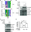
Sequence, structural, and functional analysis of the predicted PF2 domains in with no lysine kinase (WNK)4. A: sequence homology analysis guided by structural information of the PF2a domain of WNK1 (2LRU) reveals high degree of conservation with PF2 domains of all WNK members and oxidative stress responsive kinase-1 (OSR1). Arrows indicate the conservation of the residues involved in the recognition of the RFxV/I motifs. All sequences correspond to human proteins. B: structural alignment of the PF2a domain of WNK1 with the predicted structure of WNK4 PF2a and PF2b domains. Structural prediction was obtained by http://raptorx.uchicago.edu/. The predicted structures for both WNK4 PF2 domains show the groove where the RFxV/I motifs bind. R2 and F3 indicate the positions of the Arg and Phe residues of the GRFQVT peptide (93). C: representative Western blot of coimmunoprecipitation of human WNK4 and STE20/SPS1-related proline-alanine-rich protein kinase (SPAK) coexpressed in human embryonic kidney (HEK)-293 cells. While the second RFxV/I motif found in mWNK4 (site 2, residues 1016-1019) drives the association of WNK4-SPAK, mutation of key residues within the PF2a (F476A,F478A) or PF2b (F703A,F705A) domains impacts the ability of WNK4 to optimally phosphorylate SPAK at Ser373. Similar results were observed in two independent experiments. D: the attributed function of WNK4’s PF2 domains remain to be discovered. To assess whether these domains are important for WNK multimerization, we used a FLAG-tagged full-length mouse WNK4 clone (FLAG-mWNK4 F.L.) and a FLAG-tagged truncated mWNK4 clone at residue 996 (FLAG-mWNK4 996X) that lacks the coiled-coil domain (CCD) as well as RFxV/I site 2. In this last clone, mutations of PF2 domains’ key residues were introduced individually or together (same mutations as those tested in C). E: by means of coimmunoprecipitation, we tested the interaction between FLAG-tagged WNK4 proteins and a HA-tagged mWNK4 full-length protein. We found that HA-mWNK4 F.L. interacted strongly with FLAG-mWNK4 F.L. (lane 2), but its ability to interact with mWNK4-996X decreased considerably (lane 3), probably due to lack of the COOH-terminal (CT)-CCD (153). However, mutation of the PF2 domains in FLAG-mWNK4-996X individually or together did not further decrease the interaction (lanes 4−6). This suggests that PF2 domains are not essential for multimerization of WNK4, and their function remains to be uncovered. Similar results were observed in two independent experiments.
Figure 4.
Figure 4.
Sequence conservation and functional analysis of RFxV/I motifs in with no lysine kinase 4 (WNK4). A: multiple sequence alignment of WNK4 regions encompassing RFxV/I sites 1 and 2. Site 2 shows high degree of conservation, whereas site 1 is conserved in mammals but diverges in the second position (F changes to Y) in other classes. B: coimmunoprecipitation assay of STE20/SPS1-related proline-alanine-rich protein kinase (SPAK) and WNK4 proteins with mutated RFxV/I sites. SPAK and human (h)WNK4 clones were coexpressed in human embryonic kidney (HEK)-293 cells. Wild-type and mutant WNK4 proteins were immunoprecipitated, and their interaction with SPAK was assessed by Western blot. Appreciable interaction of SPAK with wild-type WNK4 and the WNK4 site 1 mutant (F421A) was observed. Nevertheless, only wild-type WNK4 was able to mediate SPAK phosphorylation at Ser373. Conversely, the WNK4 RFxV/I site 2 mutant (RF-1016,1017-AA) and the double mutant were unable to interact with SPAK. Similar results were observed in three independent experiments. C: the first RFXV/I motif in WNK4 is not a SPAK-binding site. Mutations affecting RFxV/I site 1 were introduced in a WNK4 clone that also carries the L321F mutation that affects the Cl-binding site and thus impairs inhibition of kinase activity by Cl. Expression of hWNK4-L321F in HEK-293 cells promoted SPAK Ser373 phosphorylation, whereas this effect was decreased with the F421A but not with F421Y mutation. As Tyr in this position would also affect binding to a PF2 domain (117), this suggests that RFxV/I site 1 is not a SPAK-binding site. Instead, it seems that the aromatic rings of Tyr and Phe establish key interactions that allow maintaining a functional structure. D: densitometric analysis of experiments corresponding to Fig. 1C (n =3). *P < 0.05.
Figure 5.
Figure 5.
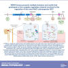
Proposed roles of with no lysine kinase 4 (WNK4) in the nephron. Works by different groups have suggested the participation of WNK4 in the regulation of multiple kidney transport proteins. A: in the thick ascending limb of the loop of Henle, WNK4 has been reported to positively regulate the activity of Na+-K+-2Cl cotransporter (NKCC2) through the phosphorylation of STE20/SPS1-related proline-alanine-rich protein kinase (SPAK)/oxidative stress-responsive 1 (OSR1). Although a role for WNK4 in the regulation of renal outer medullary K+ channels (ROMK) has been postulated, it is unknown whether WNK4 specifically modulates this channel in the thick ascending limb. B: in the distal convoluted tubule (DCT), WNK4 is a positive regulator of NaCl cotransporter (NCC) through the phosphorylation of SPAK/OSR1. It also seems to play a role in Ca2+ handling through the positive regulation of transient receptor potential vanilloid 5 (TRPV5) channels. C: in the principal cells of the aldosterone-sensitive distal nephron (ASDN), WNK4 has been implicated as a negative regulator of electrogenic Na+ reabsorption and K+ secretion through epithelial Na+ channels (ENaC) and ROMK/large-conductance K+ (BK) channels, respectively. D: the Cl/HCO3 antiporter pendrin has been postulated to be upregulated by WNK4, at least in the context of familial hyperkalemic hypertension (FHHt). No mechanisms are known for this phenomenon (figure created with Biorender.com).
Figure 6.
Figure 6.
Regulatory mechanisms of with no lysine kinase (WNK)4 function. In the distal convoluted tubule (DCT), WNK4 protein levels are regulated by the activity of the cullin 3 (CUL3)-Kelch-like family member 3 (KLHL3) E3 complex, which targets WNK4 for degradation by promoting its ubiquitylation at several sites (94, 136). KLHL3 binds to the acidic motif of WNK kinases through its propeller domain. Another level of regulation involves the modulation of WNK4 kinase activity. Several mechanisms have been described that participate in this regulation, which are the following. 1) Binding of Cl to a pocket within the active site of the kinase stabilizes an inactive conformation and prevents kinase autophosphorylation (4, 115). Intracellular Cl concentration levels are thus determinant on WNK4 activity. 2) WNK4 contains five phosphorylation sites within a RRxS motif, two located in the NH2-terminal domain and three located in a COOH-terminal region. All five sites can be phosphorylated in vitro by PKC and PKA (16), and at least the three COOH-terminal sites can be phosphorylated by serum/glucocorticoid regulated kinase-1 (SGK1) (101, 124, 129). Phosphorylation levels of two of these sites (Ser64 and Ser1196) correlate with levels of kinase activity [measured by its ability to autophosphorylate and to phosphorylate STE20/SPS1-related proline-alanine-rich protein kinase (SPAK)]. Phosphoablative mutations at Ser64 and Ser1196 prevent WNK4-mediated phosphorylation of SPAK in response to cretain stimuli, like ANG II (16). 3) A protein phosphatase 1 (PP1)-binding site has been described close to the COOH-terminal end of the protein. Absence of this motif promotes WNK4 hyperphosphorylation and constitutive activation in cultured cells (100). 4) Kidney-specific (KS-)WNK1 interacts with WNK4 through the COOH-terminal coiled-coil domain (CT-CCD). Coexpression of KS-WNK1 in Xenopus laevis oocytes promotes WNK4 autophosphorylation and activation by a mechanism that is currently unknown (3). WNK1 mutations that produce amino acid substitutions within the acidic domain produce a mild familial hyperkalemic hypertension (FHHt) phenotype in humans and mice that has been speculated to be due to increased KS-WNK1-mediated activation of WNK4 (83) (figure created with Biorender.com).
Figure 7.
Figure 7.
Role of with no lysine kinase 4 (WNK4) in the regulation of NaCl cotransporter (NCC) by extracellular K+. In the face of hypokalemia, the extracellular K+ concentration ([K+]e) gradient favors K+ exit from the distal convoluted tubule (DCT) cell through basolateral K+ channels formed by Kir4.1 and Kir5.1 subunits. This leads to membrane hyperpolarization, which, in turn, promotes exit of Cl. The resulting decrease in intracellular Cl concentration ([Cl-]i) promotes WNK4 activation due to release of Cl from the Cl-binding site, leading to increased STE20/SPS1-related proline-alanine-rich protein kinase (SPAK)/oxidative stress-responsive 1 (OSR1) and NCC phosphorylation and activation (152). An opposite mechanism may operate in hyperkalemia, although additional mechanisms are thought to participate in this setting. The decrease in [Cl]i induced by hypokalemia appears also to be responsible for the increase in WNK4 phosphorylation levels at RRxS sites that is observed in response to decreases in [K+]e (99). The detailed mechanism is currently under investigation, but it may involve activation of PKC and/or PKA, given that these kinases can phosphorylate RRxS sites in vitro. In addition, phosphorylation of Kelch-like family member 3 (KLHL3) in the RRxS site located within the substate-binding propeller domain is also induced by decreases in [K+]e (58). Given the similarity of this last mechanism to the regulation of WNK4 by phosphorylation at RRxS sites, it is possible that KLHL3-RRxS phosphorylation may also be secondary to [Cl]i depletion in the setting of hypokalemia (figure created with Biorender.com).
Figure 8.
Figure 8.
Role of with no lysine kinase 4 (WNK4) in the regulation of NaCl cotransporter (NCC) by ANG II and by extracellular Ca2+. Stimulation of ANG II type 1 receptors (AT1) or calcium-sensing receptor (CaSR) in human embryonic kidney (HEK)-293 cells promotes PKC activation that, in turn, phosphorylates RRxS sites in WNK4 and Kelch-like family member 3 (KLHL3) (5, 16, 135). KLHL3 phosphorylation prevents WNK4 degradation (135), and WNK4 phosphorylation promotes kinase activation (16). In mice, high circulating ANG II levels correlate with increased KLHL3 and WNK4 phosphorylation levels at RRXS sites as well as increased levels of WNK4 protein expression (16, 135). Higher levels of WNK4 expression and phosphorylation at an RRxS site are also observed in mice administered the calcimimetic R-568, which acts as a positive allosteric modulator of CaSR (5). On the basis of this evidence, our group has proposed that activation of ANG II receptors and CaSR receptors in distal convoluted tubule cells promotes NCC activation via the depicted pathway (figure created with Biorender.com).

References

    1. Cao-Pham AH, Urano D, Ross-Elliott TJ, Jones AM. Nudge-nudge, WNK-WNK (kinases), say no more? New Phytol 220: 35–48, 2018. doi:10.1111/nph.15276. - DOI - PubMed
    1. Xu B, English JM, Wilsbacher JL, Stippec S, Goldsmith EJ, Cobb MH. WNK1, a novel mammalian serine/threonine protein kinase lacking the catalytic lysine in subdomain II. J Biol Chem 275: 16795–16801, 2000. doi:10.1074/jbc.275.22.16795. - DOI - PubMed
    1. Veríssimo F, Jordan P. WNK kinases, a novel protein kinase subfamily in multi-cellular organisms. Oncogene 20: 5562–5569, 2001. doi:10.1038/sj.onc.1204726. - DOI - PubMed
    1. Wilson FH, Disse-Nicodème S, Choate KA, Ishikawa K, Nelson-Williams C, Desitter I, Gunel M, Milford DV, Lipkin GW, Achard JM, Feely MP, Dussol B, Berland Y, Unwin RJ, Mayan H, Simon DB, Farfel Z, Jeunemaitre X, Lifton RP. Human hypertension caused by mutations in WNK kinases. Science 293: 1107–1112, 2001. doi:10.1126/science.1062844. - DOI - PubMed
    1. Piala AT, Moon TM, Akella R, He H, Cobb MH, Goldsmith EJ. Chloride sensing by WNK1 involves inhibition of autophosphorylation. Sci Signal 7: ra41, 2014. doi:10.1126/scisignal.2005050. - DOI - PMC - PubMed

Publication types

MeSH terms

LinkOut - more resources