. 2021 Mar 1;42(3):e332-e340.
doi: 10.1097/MAO.0000000000002985.

The Pathology of the Vestibular System in CANVAS

Affiliations

The Pathology of the Vestibular System in CANVAS

Reuven Ishai et al. Otol Neurotol. .

Abstract

Objective: To describe the site of lesion responsible for the severe, bilateral, symmetrical, selective loss of vestibular function in Cerebellar Ataxia with Neuronopathy and Vestibular Areflexia Syndrome (CANVAS), an adult-onset recessively-inherited ataxia, characterized by progressive imbalance due to a combination of cerebellar, somatosensory, and selective vestibular impairment with normal hearing.

Methods: Histologic examination of five temporal bones and the brainstems from four CANVAS patients and the brainstem only from one more, each diagnosed and followed from diagnosis to death by one of the clinician authors.

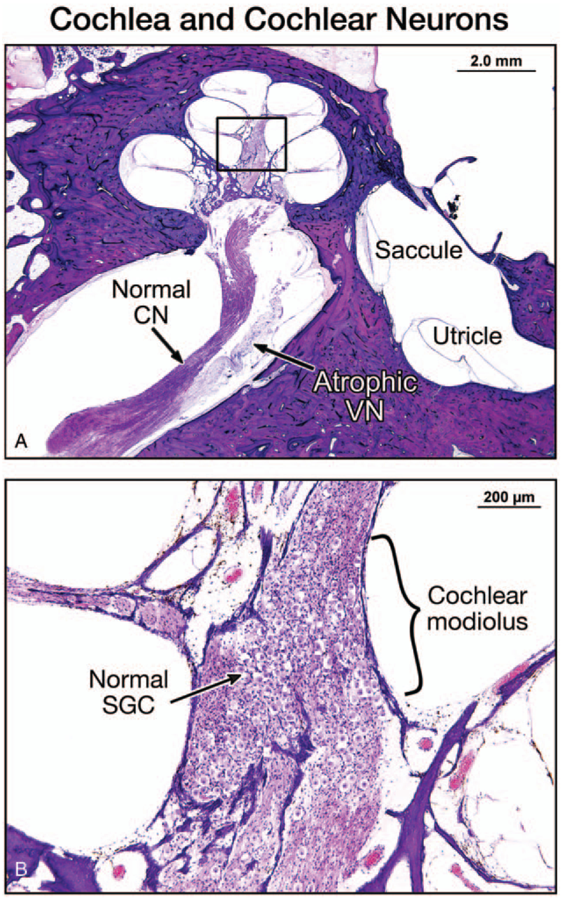
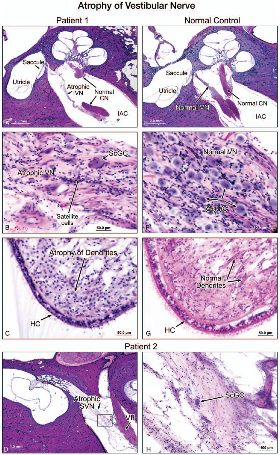
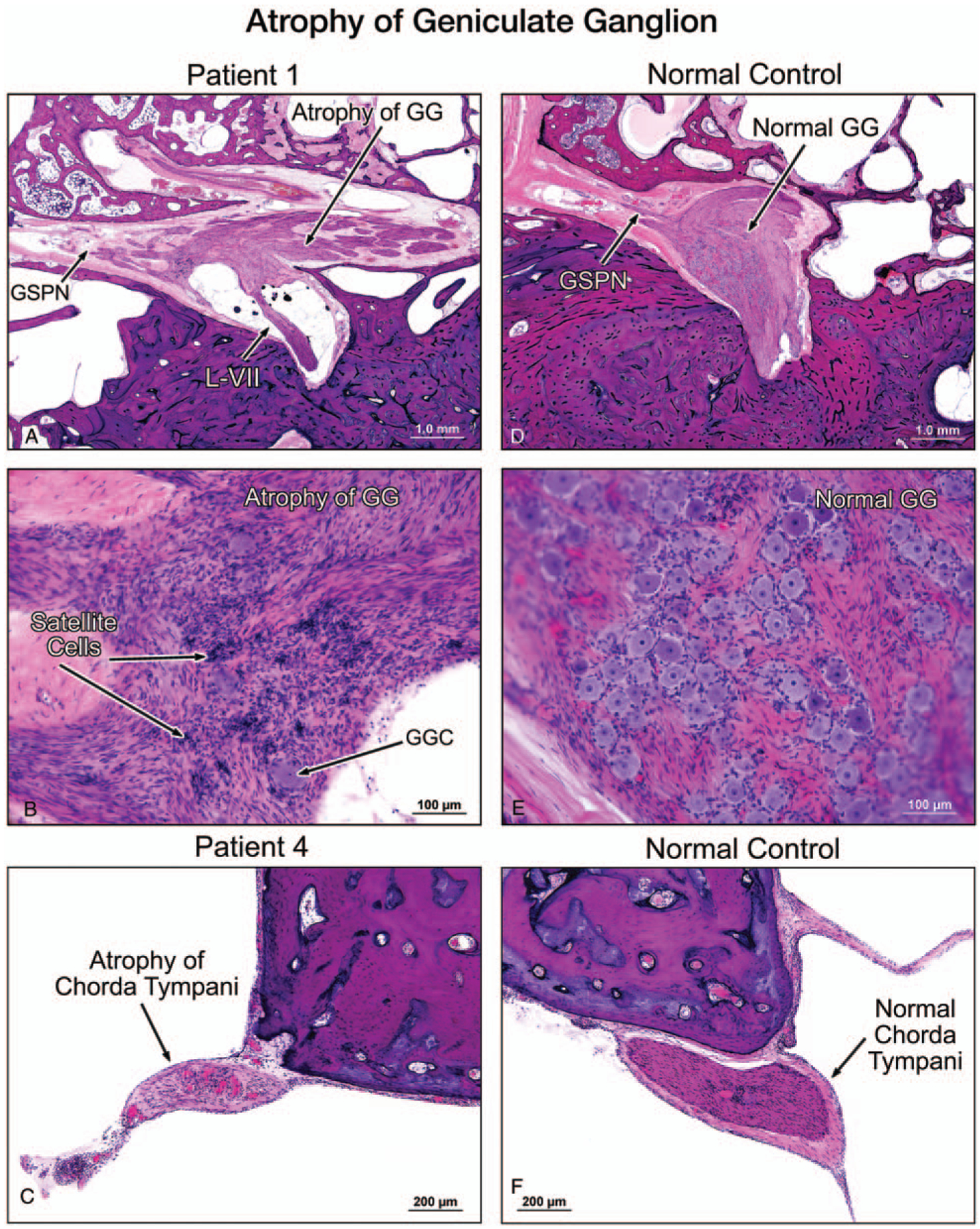
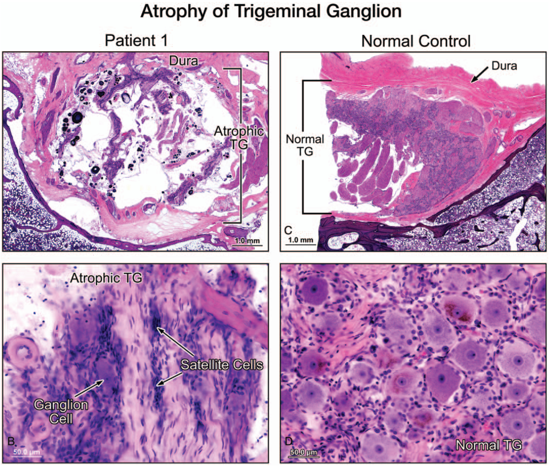
Results: All five temporal bones showed severe loss of vestibular ganglion cells (cell counts 3-16% of normal), and atrophy of the vestibular nerves, whereas vestibular receptor hair cells and the vestibular nuclei were preserved. In contrast, auditory receptor hair cells, the auditory ganglia (cell counts 51-100% of normal), and the auditory nerves were relatively preserved. In addition, the cranial sensory ganglia (geniculate and trigeminal), present in two temporal bones, also showed severe degeneration.

Conclusions: In CANVAS there is a severe cranial sensory ganglionopathy neuronopathy (ganglionopathy) involving the vestibular, facial, and trigeminal ganglia but sparing the auditory ganglia. These observations, when coupled with the known spinal dorsal root ganglionopathy in CANVAS, indicate a shared pathogenesis of its somatosensory and cranial nerve manifestations. This is the first published account of both the otopathology and neuropathology of CANVAS, a disease that involves the central as well as the peripheral nervous system.


Conflict of interest statement

The authors disclose no conflicts of interest.

Figures

FIG. 1.
FIG. 1.
A, A low-power view of the right temporal bone of Patient 1 showed a normal cochlea and a normal cochlear nerve (CN) in a mid-modiolar section, and atrophy of the vestibular nerve (VN). The saccule and utricle were normal. B, A high-power view of the cochlear modiolus showed a normal population of spiral ganglion cells (SGC).
FIG. 2.
FIG. 2.
A, A low-power view of a midmodiolar section of the left temporal bone in Patient 1. In the internal auditory canal (IAC), the inferior vestibular nerve (VN) was atrophied and the cochlear nerve appeared normal. For comparison, the normal appearance of the vestibular nerve is demonstrated in a section from a normal temporal bone selected from our collection (E–G). B, A high-power view of the inferior vestibular nerve (VN) within the internal auditory canal. The nerve was severely degenerated, and there was severe loss of Scarpa’s ganglion cells (ScGC), compared with the inferior vestibular nerve in the normal control (F). No evidence of inflammation or vasculitis could be seen. C, The crista of the lateral semicircular canal in patient 1 showed a normal population of hair cells (HC) but a significantly reduced number of dendrites. Dendrites in a normal TB is shown for comparison (G). HC indicates vestibular hair cells. D, A low-power view (D) and a higher power view (H) of the superior vestibular nerve (SVN), of Patient 2 (left ear) showed severe atrophy and significant reduction of the number of Scarpa’s ganglion cells (ScGC). There was no evidence of inflammation or vasculitis.
FIG. 3.
FIG. 3.
A low-power view (A) and a higher power view (B) of the facial nerve of the left temporal bone of Patient 1 showed severe atrophy of both the labyrinthine segment of the nerve (L-VII) and the geniculate ganglion (GG), as compared with a normal control which showed a normal population of GG cells, (D, E). There was no evidence of inflammation or vasculitis. GSPN indicates greater superficial petrosal nerve. A high-power view showed an atrophy of the chorda tympani nerve (C) in Patient 4, as compared with the chorda tympani nerve in a normal control (F).
FIG. 4.
FIG. 4.
A low-power view (A) and a higher power view (B) of the trigeminal ganglion (TG) (left ear, Patient 1) showed severe atrophy and significant reduction of the number of TG cells, as compared with a normal temporal bone (C, D). There was no evidence of inflammation or vasculiti.

References

    1. Cortese A, Simone R, Sullivan R, et al. Biallelic expansion of an intronic repeat in RFC1 is a common cause of late-onset ataxia. Nat Genet 2019;51:649–58. - PMC - PubMed
    1. Rafehi H, Szmulewicz DJ, Bennett MF, et al. Bioinformatics-based identification of expanded repeats: A non-reference intronic pentamer expansion in RFC1 causes CANVAS. Am J Hum Genet 2019;105:151–65. - PMC - PubMed
    1. Szmulewicz DJ, Waterston JA, Halmagyi GM, et al. Sensory neuropathy as part of the cerebellar ataxia neuropathy vestibular areflexia syndrome. Neurology 2011;76:1903–10. - PMC - PubMed
    1. Szmulewicz DJ, Merchant SN, Halmagyi GM. Cerebellar ataxia with neuropathy and bilateral vestibular areflexia syndrome: A histopathologic case report. Otol Neurotol 2011;32:63–5. - PMC - PubMed
    1. Szmulewicz DJ, McLean CA, Rodriguez ML, et al. Dorsal root ganglionopathy is responsible for the sensory impairment in CANVAS. Neurology 2014;82:. 1410–1415. - PMC - PubMed