Review
. 2021 Jan 25;12(1):e00308.
doi: 10.14309/ctg.0000000000000308.

Human Intestinal Barrier: Effects of Stressors, Diet, Prebiotics, and Probiotics

Affiliations
Review

Human Intestinal Barrier: Effects of Stressors, Diet, Prebiotics, and Probiotics

Michael Camilleri. Clin Transl Gastroenterol. .

Abstract

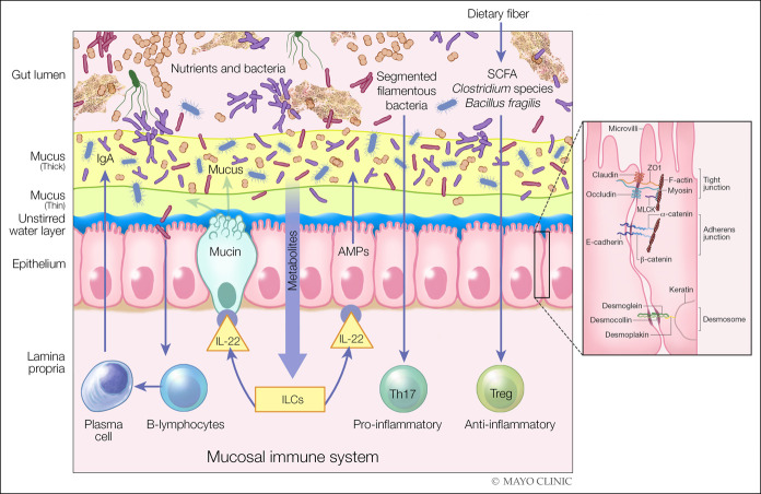
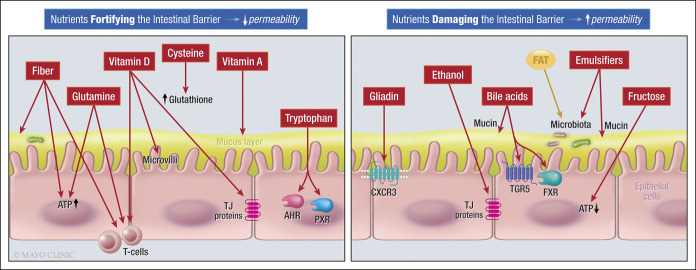
The objectives of this article are to understand the effects of stressors (nonsteroidal antiinflammatory drug, exercise, and pregnancy) and components in the diet, specifically prebiotics and probiotics, on intestinal barrier function. Stressors generally reduce barrier function, and these effects can be reversed by supplements such as zinc or glutamine that are among the substances that enhance the barrier. Other dietary factors in the diet that improve the barrier are vitamins A and D, tryptophan, cysteine, and fiber; by contrast, ethanol, fructose, and dietary emulsifiers increase permeability. Effects of prebiotics on barrier function are modest; on the other hand, probiotics exert direct and indirect antagonism of pathogens, and there are documented effects of diverse probiotic species, especially combination agents, on barrier function in vitro, in vivo in animal studies, and in human randomized controlled trials conducted in response to stress or disease. Clinical observations of benefits with combination probiotics in inflammatory diseases have simultaneously not appraised effects on intestinal permeability. In summary, probiotics and synbiotics enhance intestinal barrier function in response to stressor or disease states. Future studies should address the changes in barrier function and microbiota concomitant with assessment of clinical outcomes.


Conflict of interest statement

Guarantor of the article: Michael Camilleri, MD.

Specific author contributions: M. Camilleri conceived the idea for this article, collected the data, and drafted and finalized the manuscript.

Financial support: M. Camilleri is supported by grant R01-DK115950 from National Institutes of Health for permeability studies in bile acid diarrhea and by a research grant from ILSI North America to develop optimal methods to study human intestinal permeability.

Potential competing interests: M. Camilleri has submitted a patent application entitled “Methods and Materials for Assessing Intestinal Permeability,” Publication number: 20190145953.

Figures

Figure 1.
Figure 1.
Components of intestinal barrier. Components in the diet and specific immune mechanisms are involved in maintaining the integrity of the barrier, for example, through the production of short-chain fatty acids (SCFAs) by the gut microbiota. The SCFA are used by the colonic epithelium as a source of energy and can, independently, induce immune tolerance through T regulatory (Treg) cells. Other metabolites in diet can activate innate lymphoid cells to produce IL-22, which, in turn, can enhance the production of mucin and antimicrobial peptides (AMPs) by the intestinal epithelium to fortify gut barrier function. Plasma cells can also produce and secrete sIgA into the intestinal mucus layer to protect the host from the luminal contents of the intestinal tract. The intercellular space is sealed by the tight junction, which is a component of the apical junctional complex, the key elements being the zona occludens (ZO) and zona adherens, each made up of different components. Myosin light chain kinase (MLCK) is associated with the perijunctional actomyosin ring, and desmosomes reinforce the barrier. In general, diffusion through claudins and occludin is energy independent, whereas ZO-1 facilitates exchange energy-dependent mechanisms.(2,3) Left panel is reproduced from Camilleri M, Lyle BJ, Madsen KL, Sonnenburg J, Verbeke K, Wu GD. Role for diet in normal gut barrier function: Developing guidance within the framework of food labeling regulations. Am J Physiol 2019;317:G17-G39, which is published under a CC-BY license. Right panel is reprinted by permission from Springer Nature, Nature Reviews Immunology. Intestinal mucosal barrier function in health and disease. Turner JN. 2009;9:799–809.
Figure 2.
Figure 2.
Dietary components that impact the intestinal barrier function (37).

References

    1. Keita AV, Soderholm JD. The intestinal barrier and its regulation by neuroimmune factors. Neurogastroenterol Motil 2010;22:718–33. - PubMed
    1. Camilleri M, Lyle BJ, Madsen KL, et al. . Role for diet in normal gut barrier function: Developing guidance within the framework of food labeling regulations. Am J Physiol 2019;317:G17–G39. - PMC - PubMed
    1. Turner JR. Intestinal mucosal barrier function in health and disease. Nat Rev Immunol 2009;9:799–809. - PubMed
    1. Paone P, Cani PD. Mucus barrier, mucins and gut microbiota: The expected slimy partners? Gut 2020;0:1–12. - PMC - PubMed
    1. Strocchi A, Levitt MD. A reappraisal of the magnitude and implications of the intestinal unstirred layer. Gastroenterology 1991;101:843–7. - PubMed

Publication types

MeSH terms

Substances