

Incorporating radiomics into clinical trials: expert consensus endorsed by the European Society of Radiology on considerations for data-driven compared to biologically driven quantitative biomarkers
- PMID: 33492473
- PMCID: PMC8270834
- DOI: 10.1007/s00330-020-07598-8
Incorporating radiomics into clinical trials: expert consensus endorsed by the European Society of Radiology on considerations for data-driven compared to biologically driven quantitative biomarkers
Erratum in
-
Correction to: Incorporating radiomics into clinical trials: expert consensus endorsed by the European Society of Radiology on considerations for data-driven compared to biologically driven quantitative biomarkers.Eur Radiol. 2021 Aug;31(8):6408-6409. doi: 10.1007/s00330-021-07721-3. Eur Radiol. 2021. PMID: 33693997 Free PMC article. No abstract available.
Abstract
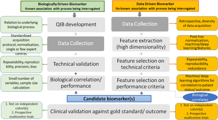
Existing quantitative imaging biomarkers (QIBs) are associated with known biological tissue characteristics and follow a well-understood path of technical, biological and clinical validation before incorporation into clinical trials. In radiomics, novel data-driven processes extract numerous visually imperceptible statistical features from the imaging data with no a priori assumptions on their correlation with biological processes. The selection of relevant features (radiomic signature) and incorporation into clinical trials therefore requires additional considerations to ensure meaningful imaging endpoints. Also, the number of radiomic features tested means that power calculations would result in sample sizes impossible to achieve within clinical trials. This article examines how the process of standardising and validating data-driven imaging biomarkers differs from those based on biological associations. Radiomic signatures are best developed initially on datasets that represent diversity of acquisition protocols as well as diversity of disease and of normal findings, rather than within clinical trials with standardised and optimised protocols as this would risk the selection of radiomic features being linked to the imaging process rather than the pathology. Normalisation through discretisation and feature harmonisation are essential pre-processing steps. Biological correlation may be performed after the technical and clinical validity of a radiomic signature is established, but is not mandatory. Feature selection may be part of discovery within a radiomics-specific trial or represent exploratory endpoints within an established trial; a previously validated radiomic signature may even be used as a primary/secondary endpoint, particularly if associations are demonstrated with specific biological processes and pathways being targeted within clinical trials. KEY POINTS: • Data-driven processes like radiomics risk false discoveries due to high-dimensionality of the dataset compared to sample size, making adequate diversity of the data, cross-validation and external validation essential to mitigate the risks of spurious associations and overfitting. • Use of radiomic signatures within clinical trials requires multistep standardisation of image acquisition, image analysis and data mining processes. • Biological correlation may be established after clinical validation but is not mandatory.
Keywords: Clinical trial; Radiology; Standardization; Statistics and numerical data; Validation studies.
© 2021. The Author(s).
Conflict of interest statement
Congress sponsorship from Guerbet. Industrial grant on radiomics from Invectys, Novartis. Co-investigator in grant with Philips, Ariana Pharma, Evolucare.
Figures
References
-
- Santamaria G, Velasco M, Bargallo X, Caparros X, Farrus B, Luis Fernandez P. Radiologic and pathologic findings in breast tumors with high signal intensity on T2-weighted MR images. Radiographics. 2010;30:533–548. - PubMed
-
- Parghane RV, Basu S. PET/computed tomography in treatment response assessment in cancer: an overview with emphasis on the evolving role in response evaluation to immunotherapy and radiation therapy. PET Clin. 2020;15:101–123. - PubMed
-
- Lee SH, Moon WK, Cho N, et al. Shear-wave elastographic features of breast cancers: comparison with mechanical elasticity and histopathologic characteristics. Invest Radiol. 2014;49:147–155. - PubMed
-
- de Bazelaire C, Calmon R, Chapellier M, Pluvinage A, Frija J, de Kerviler E. CT and MRI imaging in tumoral angiogenesis. Bull Cancer. 2010;97:79–90. - PubMed
-
- Ammari S, Thiam R, Cuenod CA, et al. Radiological evaluation of response to treatment: application to metastatic renal cancers receiving anti-angiogenic treatment. Diagn Interv Imaging. 2014;95:527–539. - PubMed
Publication types
MeSH terms
Substances
Grants and funding
LinkOut - more resources
Full Text Sources
Other Literature Sources
Medical
