. 2021 Jan 25;16(1):e0245171.
doi: 10.1371/journal.pone.0245171. eCollection 2021.

Manganese exposure in juvenile C57BL/6 mice increases glial inflammatory responses in the substantia nigra following infection with H1N1 influenza virus

Affiliations

Manganese exposure in juvenile C57BL/6 mice increases glial inflammatory responses in the substantia nigra following infection with H1N1 influenza virus

Collin M Bantle et al. PLoS One. .

Abstract

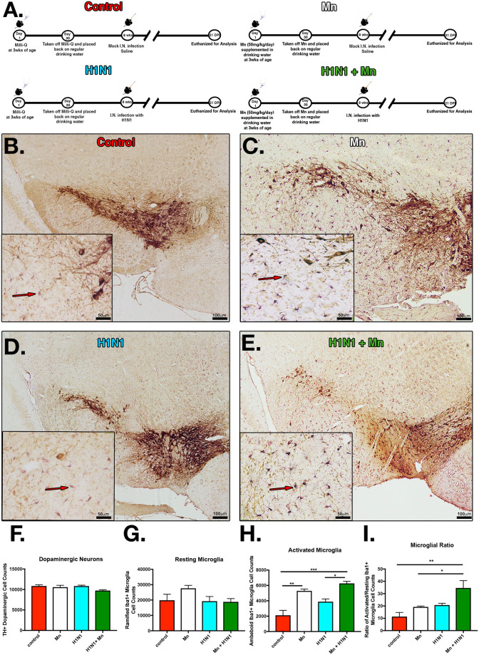
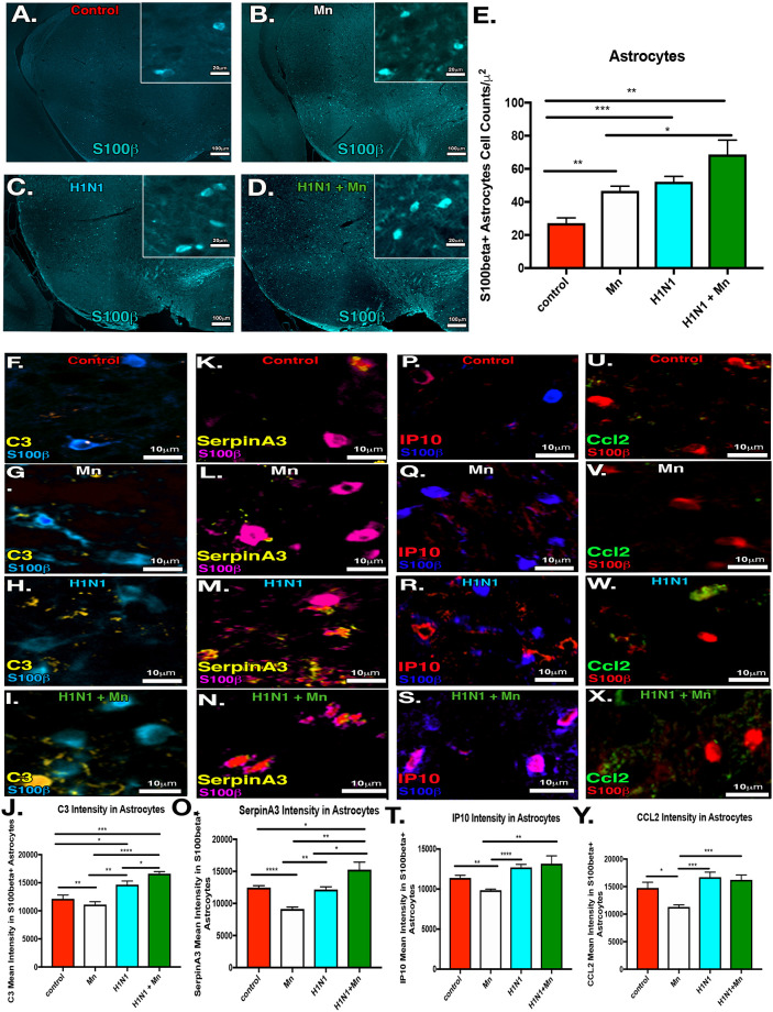
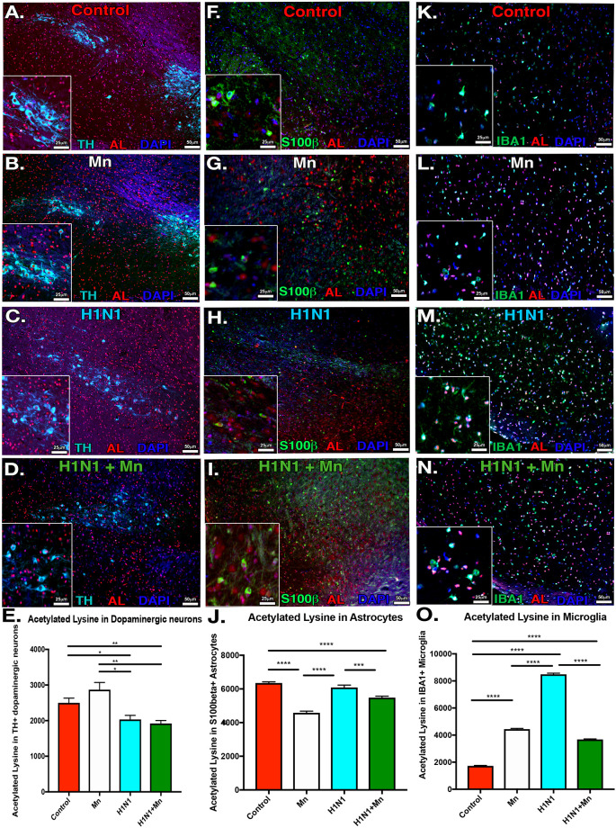
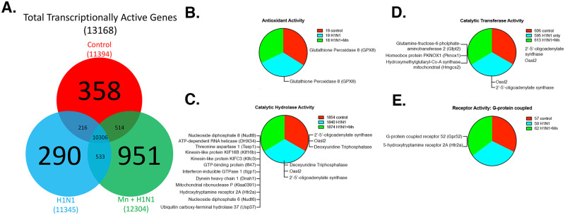
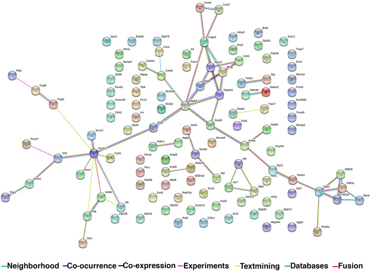
Infection with Influenza A virus can lead to the development of encephalitis and subsequent neurological deficits ranging from headaches to neurodegeneration. Post-encephalitic parkinsonism has been reported in surviving patients of H1N1 infections, but not all cases of encephalitic H1N1 infection present with these neurological symptoms, suggesting that interactions with an environmental neurotoxin could promote more severe neurological damage. The heavy metal, manganese (Mn), is a potential interacting factor with H1N1 because excessive exposure early in life can induce long-lasting effects on neurological function through inflammatory activation of glial cells. In the current study, we used a two-hit model of neurotoxin-pathogen exposure to examine whether exposure to Mn during juvenile development would induce a more severe neuropathological response following infection with H1N1 in adulthood. To test this hypothesis, C57BL/6 mice were exposed to MnCl2 in drinking water (50 mg/kg/day) for 30 days from days 21-51 postnatal, then infected intranasally with H1N1 three weeks later. Analyses of dopaminergic neurons, microglia and astrocytes in basal ganglia indicated that although there was no significant loss of dopaminergic neurons within the substantia nigra pars compacta, there was more pronounced activation of microglia and astrocytes in animals sequentially exposed to Mn and H1N1, as well as altered patterns of histone acetylation. Whole transcriptome Next Generation Sequencing (RNASeq) analysis was performed on the substantia nigra and revealed unique patterns of gene expression in the dual-exposed group, including genes involved in antioxidant activation, mitophagy and neurodegeneration. Taken together, these results suggest that exposure to elevated levels of Mn during juvenile development could sensitize glial cells to more severe neuro-immune responses to influenza infection later in life through persistent epigenetic changes.


Conflict of interest statement

The authors have declared that no competing interests exist.

Figures

Fig 1
Fig 1. Pre-treatment with manganese increases microglial activation in the substantia nigra following infection with H1N1.
(A) C57BL/6 mice were divided into groups with and without Mn in drinking water (50 mg/Kg) from day 21–51 PN and then exposed to H1N1 influenza virus three weeks later. Groups: Control, Mn, H1N1, H1N1+MnCl2. (B-E) IHC labeling of dopamine neurons (TH) and microglia (IBA1). (F) Stereological determination of the number of TH+ neurons in the substantia nigra pars compacta (SNpc). (G) Resting microglia in the SNpc. (H) Activated microglia in the SNpc. (I) Ration of activated/resting microglia in the SNpc. *P<0.05 **P<0.01 ***P<0.001****P<0.0001. n = 6 mice/group.
Fig 2
Fig 2. Pre-treatment with manganese induces proliferation and inflammatory activation in astrocytes in the substantia nigra following infection with H1N1.
(A-D) IF labeling of astrocytes (s100β). Groups: Control, Mn, H1N1, H1N1+ MnCl2. (E) Cell counts of the s100β+ astrocytes in the substantia nigra pars compacta (SNpc). (F-I) IF colocalization of astrocytes (s100β) with complement C3 (C3). (J) C3 mean intensity measurements in s100β+ astrocytes in the SNpc. (K-N) IF colocalization of astrocytes (s100β) with SerpinA3. (O) SerpinA3 mean intensity measurements in S100β+ astrocytes in the SNpc. (P-S) IF colocalization of astrocytes (s100β) with complement IP-10/Cxcl10 (IP-10). (T) IP-10 mean intensity measurements in s100β+ astrocytes in the SNpc. (U-X) IF colocalization of astrocytes (s100β) with complement Ccl2. (J) Ccl2 mean intensity measurements in s100β+ astrocytes in the SNpc. *P<0.05 **P<0.01 ***P<0.001****P<0.0001. n = 6 mice/group.
Fig 3
Fig 3. Dual treatment with MnCl2 and H1N1 alters histone acetylation in dopaminergic neurons, microglia, and astrocytes in the SNpc.
(A-D) Immunofluorescence co-localization of dopaminergic neurons (TH) and total acetylated lysine residues (AL). Groups: Control, Mn, H1N1, H1N1+ MnCl2. (E) AL mean intensity measurements in TH+ dopaminergic neurons in the substantia nigra pars compacta (SNpc). (F-G) IF colocalization of astrocytes (s100β) with AL. (J) AL mean intensity measurements in s100β+ astrocytes in SNpc. (K-N) IF colocalization of microglia (Iba1) with AL. (O) AL mean intensity measurements in Iba1+ microglia in the SNpc. *P<0.05 **P<0.01 ***P<0.001****P<0.0001. n = 6 mice/group.
Fig 4
Fig 4. RNA sequencing of the H1N1+MnCl2 treatment group highlights uniquely transcribed genes annotated to functionally relevant biological processes.
(A) Venn diagram depicting unique and overlapping transcripts. (B-E) GO Consortium & PANTHER Classification System pathway analysis: Pie charts describe the relative proportions of RNAseq transcripts annotated to each GO Term within the molecular function Gene Ontology. The exact gene lists unique or uniquely absent from the H1N1+ Mn treatment group are listed for each category. Genes were classified according to molecular function and biological process with Panther overrepresentation and Functional Enrichment analyses. Groups: Control, H1N1, H1N1+ Mn.
Fig 5
Fig 5. Transcriptional connectome of dual treatment group with associated gene products.
(A) Search Tool for the Retrieval of Interacting Genes database (STRING) diagram of functional associations between protein products of unique genes within the H1N1+Mn treatment group. Nodes represent proteins and edges correspond to functional interactions. Edge colors differ according to a scored confidence scale based on the extent and type of evidence supporting that particular association. Evidence channels: Green Line: Neighborhood, Navy Line: Co-occurrence, Black line: Co-expression, Pink line: Experiments, Yellow line: Textmining, Light blue line: Databases, Red line: Fusion.

References

    1. Beal MF. Parkinson’s disease: a model dilemma. Nature. 2010;466(7310). - PubMed
    1. Amberger JS, Bocchini CA, Schiettecatte F, Scott AF, Hamosh A. OMIM.org: Online Mendelian Inheritance in Man (OMIM(R)), an online catalog of human genes and genetic disorders. Nucleic Acids Res. 2015;43:D789–98. 10.1093/nar/gku1205 - DOI - PMC - PubMed
    1. Reid AH, McCall S, Henry JM, Taubenberger JK. Experimenting on the past: the enigma of von Economo’s encephalitis lethargica. J Neuropathol Exp Neurol. 2001;60(7):663–70. 10.1093/jnen/60.7.663 - DOI - PubMed
    1. Dourmashkin RR. What caused the 1918–30 epidemic of encephalitis lethargica? J R Soc Med. 1997;90(9):515–20. 10.1177/014107689709000916 - DOI - PMC - PubMed
    1. Elizan TS, Schwartz J, Yahr MD, Casals J. Antibodies against arboviruses in postencephalitic and idiopathic Parkinson’s disease. Arch Neurol. 1978;35(5):257–60 10.1001/archneur.1978.00500290003001 - DOI - PubMed

Publication types

LinkOut - more resources