

Review
doi: 10.1016/j.kint.2021.01.004.
Epub 2021 Jan 22.
Best practices for correctly identifying coronavirus by transmission electron microscopy
Affiliations
- PMID: 33493525
- PMCID: PMC7825881
- DOI: 10.1016/j.kint.2021.01.004
Item in Clipboard
Review
Best practices for correctly identifying coronavirus by transmission electron microscopy
Kidney Int.
2021 Apr.
Abstract
This guidance provides clear, concise strategies for identifying coronaviruses by transmission electron microscopy of ultrathin sections of tissues or infected tissue cultures. These include a description of virus morphology as well as cell organelles that can resemble viruses. Biochemical testing and caveats are discussed. Numerous references provide information for documentation and further study.
Keywords: coronavirus; coronavirus electron microscopy; coronavirus misidentification; coronavirus morphology; coronavirus ultrastructure; microbiology.
Copyright © 2021 International Society of Nephrology. All rights reserved.
Figures
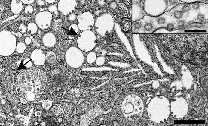
Severe acute respiratory syndrome coronavirus 2 (SARS-CoV-2)–infected Vero cell containing abundant viral particles held within intracellular vesicular/vacuolar structures (arrows). Bar = 1 μm. Inset: Higher magnification of intracellular SARS-CoV-2 particles with cross sections through the helical nucleocapsid visible as internal black dots. Bar = 200 nm. To optimize viewing of this image, please see the online version of this article at www.kidney-international.org .
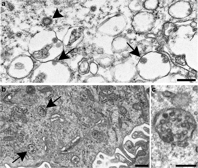
Normal subcellular organelles mimicking coronavirus. (a) Clathrin-coated vesicle free in the cytoplasm of a cell (arrowhead) and nearby severe acute respiratory syndrome coronavirus 2 (SARS-CoV-2) particles inside intracellular vacuoles (arrows). Cross sections through the viral nucleocapsid are visible in the SARS-CoV-2 particles (small dots inside the virus). Bar = 200 nm. (b) Membrane bound collections of vesicles (arrows) making up multivesicular bodies (MVBs) within the cytoplasm. Bar = 500 nm. (c) Higher magnification of an MVB; note the absence of dots inside the vesicles corresponding to cross sections through the viral nucleocapsid. Bar = 200 nm. Figure 2b and c are courtesy of Dr. Ricardo Vancini. To optimize viewing of this image, please see the online version of this article at www.kidney-international.org .
References
Publication types
MeSH terms
Grants and funding
LinkOut - more resources
Full Text Sources
Other Literature Sources
Medical
