. 2021 Jan 20;13(3):4335-4356.
doi: 10.18632/aging.202394. Epub 2021 Jan 20.

CUEDC2 ablation enhances the efficacy of mesenchymal stem cells in ameliorating cerebral ischemia/reperfusion insult

Affiliations

CUEDC2 ablation enhances the efficacy of mesenchymal stem cells in ameliorating cerebral ischemia/reperfusion insult

Yan Huang et al. Aging (Albany NY). .

Abstract

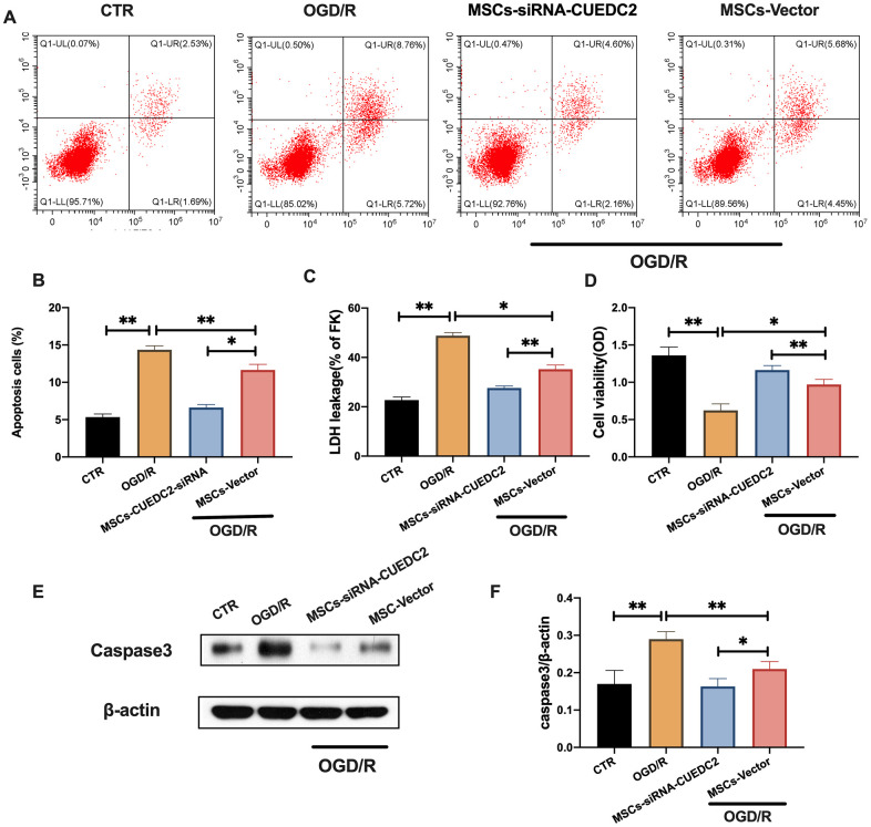
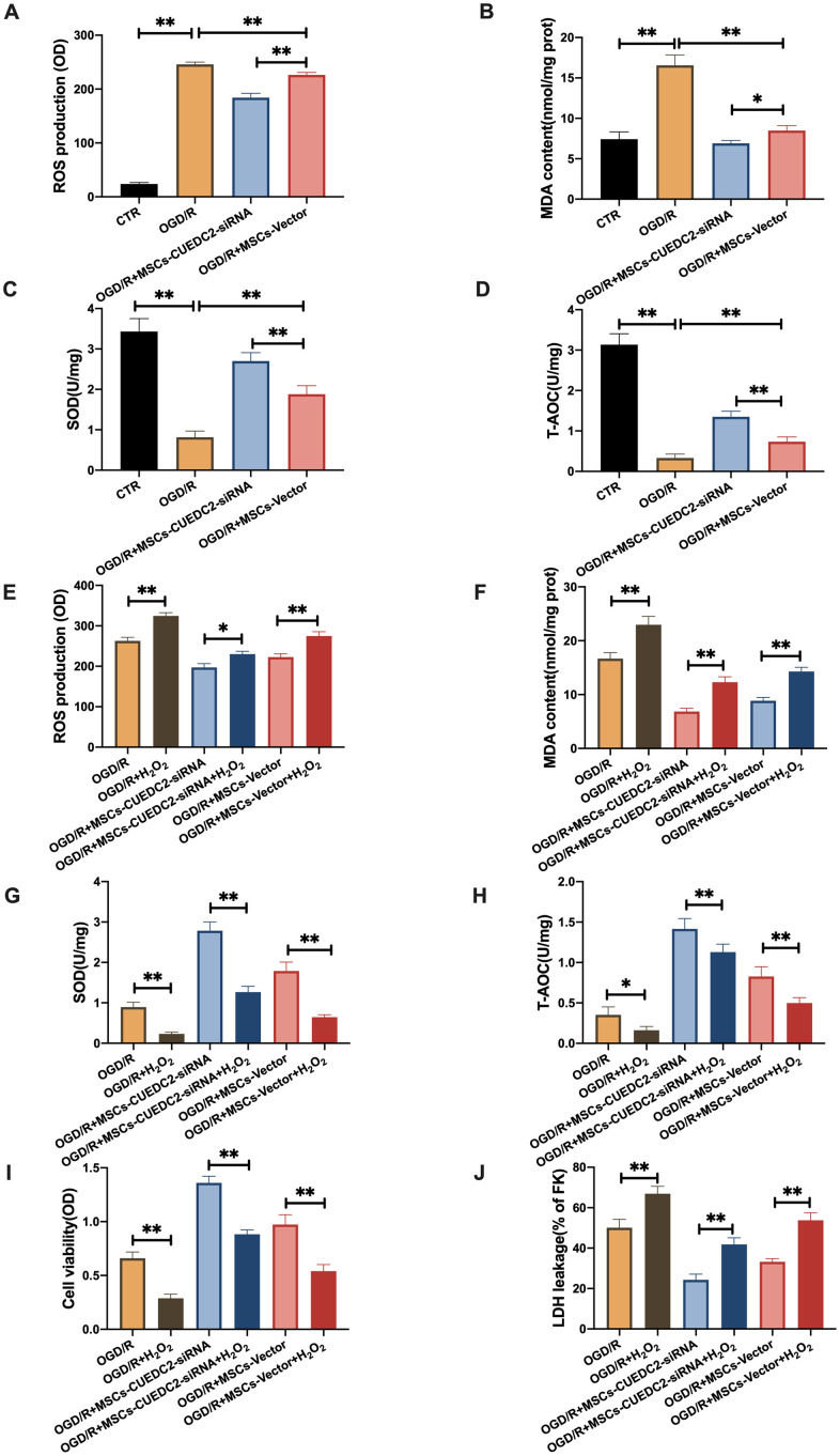
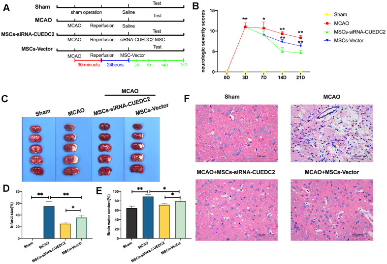
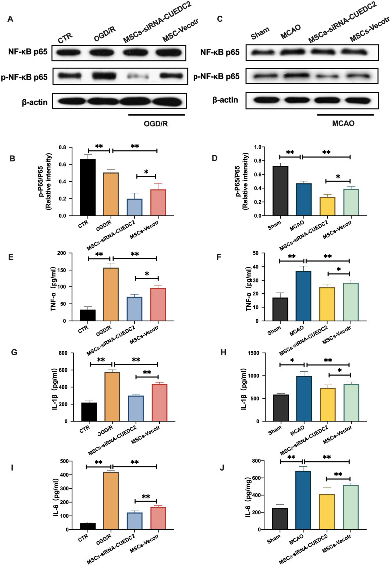
Mesenchymal stem cell (MSC) therapy has been reported to be a promising therapeutic option for cerebral ischemia/reperfusion (I/R) insult. However, the poor survival rate of engrafted MSCs under unfavorable cerebral I/R-induced microenvironment inhibits their efficiency during clinical application. CUE domain-containing 2(CUECD2) exhibits its protective role on cardiomyocytes by mediating the antioxidant capacity. Our study explored the functional role of CUEDC2 in cerebral I/R challenge and determined whether CUECD2-modified MSCs could improve the efficacy of treatment of the insulted neurons. We also evaluated the possible mechanisms involved in cerebral I/R condition. Cerebral I/R stimulation suppressed CUEDC2 levels in brain tissues and neurons. siRNA-CUEDC2 in neurons significantly inhibited cerebral I/R-induced apoptosis and oxidative stress levels invitro. Moreover, siRNA-CUEDC2 in the MSCs group remarkably enhanced the therapeutic efficacies in cerebral I/R-induced neuron injury and brain tissue impairment when compared to the non-genetic MSCs treatment group. At the molecular level, siRNA-CUEDC2 in MSCs markedly enhanced its antioxidant and anti-inflammatory effect in co-cultured neurons by upregulating glutathione peroxidase 1 (GPX1) expression levels while suppressing NF-kB activation. These findings provide a novel strategy for the utilization of MSCs to promote cerebral ischemic stroke outcomes.

Keywords: CUEDC2; MCAO; MSCs; OGD/R.


Conflict of interest statement

CONFLICTS OF INTEREST: The authors declare no conflicts of interest.

Figures

Figure 1
Figure 1
Cerebral I/R induced the downregulation of CUEDC2 expression in brain tissues. (AC) Protein and mRNA expression of CUEDC2 in neurons as detected by western blotting and PCR assays. (DF) Protein and mRNA expression of CUEDC2 in brain tissues as assayed by western blotting and PCR. (G, H) Protein expression of CUEDC2 in brain tissues as assayed by immunohistochemical staining. Scale bar = 50 μm. CTR, control; CUEDC2: CUE domain-containing 2; OGD/R: oxygen-glucose deprivation (4 hours) and reperfusion (0,4,8,12, and 24 hours); MCAO, middle cerebral artery occlusion. All data are presented as the mean value ± SD. *p<0.05, **p<0.01; compared to the control group, sham group.
Figure 2
Figure 2
Effects of CUEDC2 on cerebral I/R-induced neuron insult. (A, B) Apoptotic cell death in neurons as detected by flow cytometry with Annexin V/PI staining. (C) Apoptotic cell death in neurons as detected by LDH-leakage assay. (D) Neuron viability as assessed by the MTT assay. (E, F) Protein expression of caspase-3 in neurons as assayed by western blotting. (G) ROS production in neurons as detected by DCFH-DA assay. (H) MDA production in neurons as evaluated by lipid peroxidation MDA assay. (I) SOD production in neurons as determined by WST-8 assay. (J) T-AOC level in neurons as detected by ABTS assay. (K) Viability of H2O2-treated neurons as analyzed by MTT assay. (L) Apoptotic cell death in H2O2-treated neurons as detected by LDH leakage assay. CTR, control; CUEDC2: CUE domain-containing 2; OGD/R: oxygen-glucose deprivation (4 hours) and reperfusion (24 hours); neurons-siRNA-CUEDC2: small interfering RNA silencing CUEDC2 in neurons; neurons-vector: the vector of neurons. All data are presented as the mean value ± SD. *p<0.05, **p<0.01; compared to the vector group, the H2O2 treatment group.
Figure 3
Figure 3
MSCs attenuate cerebral I/R-induced injury in co-cultured neurons. (A) Viability of co-cultured neurons as assessed by MTT assay. (B) Apoptotic cell death in co-cultured neurons as detected by LDH-leakage assay. (C, D) Apoptotic cell death in co-cultured neurons as detected by Hoechst staining. (E) ROS production in co-cultured neurons as detected by DCFH-DA assay. (F) MDA production in co-cultured neurons as evaluated by lipid peroxidation MDA assay. (G) SOD production in co-cultured neurons as determined by WST-8 assay. (H) T-AOC level in co-cultured neurons as detected by ABTS assay. (I, J) CUEDC2 protein expression in co-cultured neurons as assayed by western blotting. (K, L) CUEDC2 protein expression in MSCs-treated brain tissues as assayed by western blotting. CTR, control; CUEDC2: CUE domain-containing 2; OGD/R: oxygen-glucose deprivation (4 hours) and reperfusion (24 hours); MSCs: mesenchymal stem cells; MCAO, middle cerebral artery occlusion. All data are presented as the mean value ± SD. *p<0.05, **p<0.01; compared to OGD/R + MSCs group, MCAO + MSCs treatment group.
Figure 4
Figure 4
CUEDC2 knockdown in MSCs enhances the inhibitory effect on OGD/R-induced apoptotic cell death in co-cultured neurons. (A, B) Apoptotic cell death in co-cultured neurons after treatment with different MSCs as detected by flow cytometry with Annexin V/PI staining. (C) Apoptotic cell death in co-cultured neurons after treatment with different MSCs as analyzed by LDH-leakage assay. (D) Viability of co-cultured neurons after treatment with different MSCs as evaluated by MTT assay. (E, F) Caspase-3 protein expression in co-cultured neurons after treatment with different MSCs as assayed by western blotting. CTR: control; MSCs: mesenchymal stem cells; CUEDC2: CUE domain-containing 2; OGD/R: oxygen-glucose deprivation (4 hours) and reperfusion (24 hours); MSCs- siRNA-CUEDC2: small interfering RNA silencing CUEDC2 in MSCs; MSCs-vector: the vector of MSCs. All data are presented as the mean value ± SD. *p<0.05, **p<0.01; compared to the control group, MSCs-vector group.
Figure 5
Figure 5
CUEDC2 degradation in MSCs improves protection against OGD/R-induced apoptotic cell death in co-cultured neurons by suppressing oxidative toxicity. (A) ROS production in co-cultured neurons after treatment with different MSCs as detected by DCFH-DA assay. (B) MDA production in co-cultured neurons after treatment with different MSCs as evaluated by lipid peroxidation MDA assay. (C) SOD production in co-cultured neurons after treatment with different MSCs as determined by WST-8 assay. (D) T-AOC level in co-cultured neurons after treatment with different MSCs as detected by ABTS assay. (E) ROS production in co-cultured neurons after treatment with different MSCs exposed to H2O2 as analyzed by DCFH-DA assay. (F) MDA production in co-cultured neurons after treatment with different MSCs exposed to H2O2 as evaluated by lipid peroxidation MDA assay. (G) SOD production in co-cultured neurons after treatment with different MSCs exposed to H2O2 as determined by WST-8 assay. (H) T-AOC level in co-cultured neurons after treatment with different MSCs exposed to H2O2 as detected by ABTS assay. (I) Co-cultured neuron viability after treatment with different MSCs subjected to H2O2 as evaluated by MTT analysis. (J) Apoptosis in co-cultured neurons after treatment with different MSCs subjected to H2O2 as evaluated by LDH leakage assay. CTR: control; MSCs: mesenchymal stem cells; CUEDC2: CUE domain-containing 2; OGD/R: oxygen-glucose deprivation (4 hours) and reperfusion (24 hours). MSCs- siRNA-CUEDC2: small interfering RNA silencing CUEDC2 in MSCs; MSCs-vector: the vector of MSCs. All data are presented as the mean value ± SD. *P<0.05, **P<0.01; compared with the control, MSCs-vector, and H2O2 treatment groups.
Figure 6
Figure 6
CUEDC2 ablation promotes the efficacy of MSCs in I/R-induced brain damages. (A) Illustration of MCAO-induced model of cerebral I/R in rats and different MSCs treatment. SD rats were exposed to MCAO for 90 minutes and subsequent reperfusion. After 24-hours of reperfusion, the CUEDC2-modified MSCs and the MSCs vector were infused on MCAO model rats. (B) Evaluation of neurologic severity scores. (C, D) Assessment of infarct size with TTC staining in whole-brain tissues. (E) Determination of brain water content. (F) H&E staining in brain tissues. Scale bar = 100 μm. MSCs: mesenchymal stem cells; MSCs- siRNA-CUEDC2: small interfering RNA silencing CUEDC2 in MSCs; MSCs-vector: the vector of MSCs; MCAO, middle cerebral artery occlusion. All data are presented as the mean value ± SD. *p<0.05, **p<0.01; compared to sham group, the vector of MSC group.
Figure 7
Figure 7
CUEDC2 silencing in MSCs upregulates the antioxidant effect on co-cultured neurons by increasing GPX1 expression. (A, B) GPX1 protein expression in co-cultured neurons under non-OGD/R condition as detected by western blotting. (C, D) GPX1 protein expression in co-cultured neurons under OGD/R condition as detected by western blotting. (E) ROS production in co-cultured neurons after treatment with various MSCs as analyzed by DCFH-DA assay. (F) MDA production in co-cultured neurons after treatment with various MSCs as evaluated by lipid peroxidation MDA assay. (G) SOD production in co-cultured neurons after treatment with various MSCs as determined by WST-8 assay. (H) T-AOC level in co-cultured neurons after treatment with various MSCs as detected by ABTS assay. (I) Viability of co-cultured neurons after treatment with various MSCs as evaluated by MTT analysis. (J) Apoptosis in co-cultured neurons after treatment with various MSCs as evaluated by LDH leakage assay. (K, L) Apoptosis in co-cultured neurons after treatment with various MSCs as detected by Hoechst staining. CUEDC2: CUE domain-containing 2; OGD/R: oxygen-glucose deprivation (4 hours) and reperfusion (12- or 24-hours). MSCs-siRNA-CUEDC2: small interfering RNA silencing CUEDC2 in MSCs; MSCs-vector: the vector of MSCs; siRNA-GPX1: small interfering RNA knockdown of GPX1 in MSC; All data are presented as the mean value ± SD. *p<0.05, **p<0.01; comparison to MSC-vector group.
Figure 8
Figure 8
CUEDC2 silencing in MSCs suppresses NF-kB activity following cerebral I/R injury. (A, B) NF-kB and p-NF-kB protein expression in co-cultured neurons as detected by western blotting. (C, D) NF-kB and p-NF-kB protein expression in brain tissues as detected by western blotting. (E) TNF-α level in co-cultured neurons as detected by ELISA assay. (F) TNF-α level in brain tissues as analyzed by ELISA assay. (G) IL-1β level in co-cultured neurons as determined by ELISA assay. (H) IL-1β level in brain tissues as analyzed by ELISA assay. (I) IL-6 level in co-cultured neurons as evaluated by ELISA assay. (J) IL-6 level in brain tissues as determined by ELISA assay. CTR: control; CUEDC2: CUE domain-containing 2; OGD/R: oxygen-glucose deprivation (4 hours) and reperfusion (24 hours). MSCs: mesenchymal stem cells; MSCs-siRNA-CUEDC2: small interfering RNA silencing CUEDC2 in MSCs; MSCs-vector: the vector of MSCs; MCAO, middle cerebral artery occlusion. All data are presented as the mean value ± SD. *p<0.05, **p<0.01; compared to the control, sham and the vector of MSC-group.

Similar articles

Cited by

References

    1. Randolph SA. Ischemic stroke. Workplace Health Saf. 2016; 64:444. 10.1177/2165079916665400 - DOI - PubMed
    1. Amani H, Habibey R, Shokri F, Hajmiresmail SJ, Akhavan O, Mashaghi A, Pazoki-Toroudi H. Selenium nanoparticles for targeted stroke therapy through modulation of inflammatory and metabolic signaling. Sci Rep. 2019; 9:6044. 10.1038/s41598-019-42633-9 - DOI - PMC - PubMed
    1. Chen S, Chen H, Du Q, Shen J. Targeting myeloperoxidase (MPO) mediated oxidative stress and inflammation for reducing brain ischemia injury: potential application of natural compounds. Front Physiol. 2020; 11:433. 10.3389/fphys.2020.00433 - DOI - PMC - PubMed
    1. Amani H, Habibey R, Hajmiresmail SJ, Latifi S, Pazoki-Toroudi H, Akhavan O. Antioxidant nanomaterials in advanced diagnoses and treatments of ischemia reperfusion injuries. J Mater Chem B. 2017; 5:9452–76. 10.1039/c7tb01689a - DOI - PubMed
    1. Krause M, Phan TG, Ma H, Sobey CG, Lim R. Cell-based therapies for stroke: are we there yet? Front Neurol. 2019; 10:656. 10.3389/fneur.2019.00656 - DOI - PMC - PubMed

Publication types

MeSH terms

LinkOut - more resources