. 2021 Jan 21;10(2):101.
doi: 10.3390/antibiotics10020101.

Effects of Antimicrobial Photodynamic Therapy on Organic Solution and Root Surface In Vitro

Affiliations

Effects of Antimicrobial Photodynamic Therapy on Organic Solution and Root Surface In Vitro

Yuji Matsushima et al. Antibiotics (Basel). .

Abstract

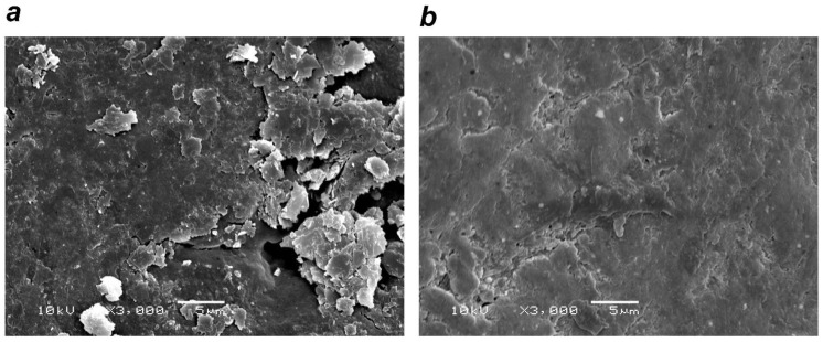
Antimicrobial photodynamic therapy (a-PDT) is attracting attention as a new form of dental treatment. While it is primarily applied to produce an antibacterial effect, it decreases lipopolysaccharide (LPS) and protease activity. Here, we evaluated differences in the antibacterial activity of a-PDT on three types of bacteria and the effects on the organic substances (i.e., albumin and LPS). Furthermore, we investigated the effects of a-PDT on root surfaces. A FotoSan630® and toluidine blue were used to perform a-PDT in this study. We measured its antimicrobial activity against Porphyromonas gingivalis, Streptococcus mutans, and Enterococcus faecalis. Antimicrobial testing revealed strong antimicrobial action and P. gingivalis, E. faecalis, and S. mutans were almost undetectable after 50, 120, and 100 s, respectively. In organic resolution tests, albumin was significantly decreased from 1 min after a-PDT application onward, while LPS significantly decreased at 5 min after the application. The root surfaces after a-PDT were confirmed to be cleaner than the controls without suffering any damage. Depending on the bacterial species, a-PDT exhibited antimicrobial activity against various types of bacteria and sensitivity differed. Moreover, we reported that a-PDT resolves protein and LPS, enabling the formation of a healthy root surface without any damage.

Keywords: antibacterial therapy; bacterial flora; dental treatment; lipopolysaccharide; organic resolution; photodynamic therapy.


Conflict of interest statement

The authors declare no conflict of interest.

Figures

Figure 1
Figure 1
Effects of (antimicrobial photodynamic therapy) a-PDT on the root surface. (a) Control group (rinsing after scaling and root planing (SRP)): smeared layer and sediment noted on the root surface. (b) a-PDT group: decreased sediment on the root surface. (Scanning Electron Microscope image (SEM) × 3000)
Figure 2
Figure 2
Effects of a-PDT on albumin (electrophoresis image). Light was applied with FotoSan630® to the albumin suspension for 0, 1, 2, and 5 min. The albumin bandwidth decreased in an irradiation time-dependent manner. MW: molecular weight marker.
Figure 3
Figure 3
Effects of a-PDT on albumin (BCA (bicinchoninic acid) protein assay). A spin gel filtration column was used to remove the toluidine blue pigment from the specimens. Although gel filtration reduced the amount of albumin by ~30%, application of a-PDT for 1 min or longer significantly reduced the amount of albumin (p < 0.05). (Albumin concentration was 2 mg/mL).
Figure 4
Figure 4
Effects of a-PDT on lipopolysaccharide (LPS) (chromogenic LAL (limulus amebocyte lysate) endotoxin assay). LPS measurement did not reveal any effects from a spin gel filtration column for pigment removal. Few changes in LPS were noted with a-PDT application of 1 or 2 min. However, LPS was decreased significantly (p < 0.05) with an application of 5 min. (LPS concentration was 1 EU/mL).
Figure 5
Figure 5
Antibacterial activity of a-PDT (bacterial culture testing). Bacteria were adjusted to 1 × 10⁶ CFU (colony forming unit)/mL; 0.01% concentration toluidine blue was added, and light was applied. Consequently, no additional colony formation was noted for P. gingivalis at 50 s, E. faecalis at 120 s, and S. mutans at 100 s.
Figure 6
Figure 6
Bactericidal effects of bactericidal on each bacterial species (bacteria counts). After light application, specimens were cultured for seven days, after which colonies were counted. The results revealed that (a) P. gingivalis significantly decreased with 30 s of application and became undetectable with 50 s of application. Similarly, (b) E. faecalis was significantly decreased with 90 s of application and became undetectable with 120 s of application, while (c) S. mutans was significantly decreased with 80 s of application and became undetectable with 100 s of application.

References

    1. Yilmaz S., Kuru B., Kuru L., Noyan U., Argun D., Kadir T. Effect of gallium arsenide diode laser on human periodontal disease: A microbiological and clinical study. Lasers Surg. Med. 2002;30:60–66. doi: 10.1002/lsm.10010. - DOI - PubMed
    1. Andersen R., Loebel N., Hammond D., Wilson M. Treatment of periodontal disease by photodisinfection compared to scaling and root planing. J. Clin. Dent. 2007;18:34–38. - PubMed
    1. Chambrone L., Wang H.-L., Romanos G.E. Antimicrobial photodynamic therapy for the treatment of periodontitis and peri-implantitis: An American Academy of Periodontology best evidence review. J. Perodontol. 2018;89:783–803. - PubMed
    1. Vohra F., Akram Z., Safii S.H., Vaithilingam R.D., Ghanem A., Sergis K., Javed F. Role of antimicrobial photodynamic therapy in the treatment of aggressive periodontitis: A systematic review. Photodiagnosis Photodyn. Ther. 2016;13:139–147. doi: 10.1016/j.pdpdt.2015.06.010. - DOI - PubMed
    1. Souza E.G.M., da Rocha T.E., Toro L.F., Guiati I.Z., Ervolino E., Garcia V.G., Wainwright M., Theodoro L.M. Antimicrobial photodynamic therapy compared to systemic antibiotic therapy in non-surgical treatment of periodontitis: Systematic review and meta-analysis. Photodiagnosis Photodyn. Ther. 2020;31:101808. doi: 10.1016/j.pdpdt.2020.101808. - DOI - PubMed

LinkOut - more resources