Review
. 2021 Jan 21;13(3):388.
doi: 10.3390/cancers13030388.

Surgical Advances in Osteosarcoma

Affiliations
Review

Surgical Advances in Osteosarcoma

Marcus J Brookes et al. Cancers (Basel). .

Abstract

Osteosarcoma (OS) is the most common primary bone cancer in children and, unfortunately, is associated with poor survival rates. OS most commonly arises around the knee joint, and was traditionally treated with amputation until surgeons began to favour limb-preserving surgery in the 1990s. Whilst improving functional outcomes, this was not without problems, such as implant failure and limb length discrepancies. OS can also arise in areas such as the pelvis, spine, head, and neck, which creates additional technical difficulty given the anatomical complexity of the areas. We reviewed the literature and summarised the recent advances in OS surgery. Improvements have been made in many areas; developments in pre-operative imaging technology have allowed improved planning, whilst the ongoing development of intraoperative imaging techniques, such as fluorescent dyes, offer the possibility of improved surgical margins. Technological developments, such as computer navigation, patient specific instruments, and improved implant design similarly provide the opportunity to improve patient outcomes. Going forward, there are a number of promising avenues currently being pursued, such as targeted fluorescent dyes, robotics, and augmented reality, which bring the prospect of improving these outcomes further.

Keywords: osteosarcoma; sarcoma; surgery.


Conflict of interest statement

The authors declare no conflict of interest.

Figures

Figure 1
Figure 1
MRI and CT scans of a patient with left sided pelvic OS.
Figure 2
Figure 2
(a) Shows the resected femur and OS contained within vastus medialis. (b) Shows the specimen through the infrared camera (Stryker), with the OS glowing bright. (c) Shows the resected specimen next to the three-dimensional (3D) printed model produced prior to the procedure (Axial3D). (d) A sample of the tumour was dissected out of the specimen to demonstrate higher fluorescence compared to a piece of fat.
Figure 3
Figure 3
Huygen’s construction of a conical Cherenkov wavefront-a charged particle traveling in a given direction transmits its kinetic energy to the surrounding media, depicted by the larger circles trailing behind the particle. Cherenkov radiation is generated at an angle to the direction of the travelling particle, defined as θ, which is related to the energy of particle [35]. Therefore, the higher the kinetic energy of the particle, the wider the generated wavefront, and hence the more easily the radiation can be detected.
Figure 4
Figure 4
CANS flowchart: adapted from Wong et al. [56].
Figure 5
Figure 5
CANS in use for a pelvic tumour resection. The screen on the left shows the bone-tumour model with the position of the navigation probe superimposed.
Figure 6
Figure 6
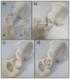
Images demonstrate the 3D printed components used for the accurate resection of a pelvic tumour. (a) shows 3D printed components involved—(i) is a 3D printed model of the patient’s pelvis post-osteotomies, (ii) is the guide for the posterior and inferior cuts, (iii) is a template of the 3D printed implant, (iv) is the drill bit guide jig. The superior pubic ramus was cut under computer assisted navigation surgery (CANS) guidance, before the posterior and inferior cut saw guide was positioned as shown in (b) and held in place with pins. After the cuts were made, the template was positioned in the patient as shown in (c). The drill bit guide was the positioned as shown in (d) which, in conjunction with the implant template, ensured the screw holes are drilled correctly for the custom implant. Implants from Implantcast GmbH.
Figure 7
Figure 7
Images show a 3D printed model of a pelvic tumour with a large soft tissue component, demonstrating both the extent of the tumour, and its relationship to the blood vessels (model from Axial3D).
Figure 8
Figure 8
Images show the planning stages of a 3D implant. (a) Shows the identification of the margins required on a 3D image of the patient’s pelvis, whilst (b) Shows an image of the patient’s pelvis post resection. This identifies the deficit that needs to be replaced, which can then be designed, as shown in (c).
Figure 9
Figure 9
Images show the reconstruction of a mandibular OS. (a) Shows the reconstruction plan including the pre-determined screw lengths and locations. (b) Shows the 3D cutting guides, 3D model and the custom reconstruction plate. (c) Shows custom cutting guide on free fibula osseocutaneous flap. (d) custom reconstruction plate attached to free fibular osseocutaneous flap. (e) Shows the follow up orthopantomogram with free fibula osseocutaneous flap fixed in defect.

References

    1. Takeuchi A., Yamamoto N., Hayashi K., Matsubara H., Miwa S., Igarashi K., Tsuchiya H. Joint-preservation surgery for pediatric osteosarcoma of the knee joint. Cancer Metastasis Rev. 2019;38:709–722. doi: 10.1007/s10555-019-09835-z. - DOI - PubMed
    1. NCIN . Bone Sarcoma Incidence and Survival. Tumours Diagnosed between 1985 and 2009. NCIN; London, UK: 2012.
    1. Whelan J.S., Jinks R.C., McTiernan A., Sydes M.R., Hook J.M., Trani L., Uscinska B., Bramwell V., Lewis I.J., Nooij M.A., et al. Survival from high-grade localised extremity osteosarcoma: Combined results and prognostic factors from three European Osteosarcoma Intergroup randomised controlled trials. Ann. Oncol. 2012;23:1607–1616. doi: 10.1093/annonc/mdr491. - DOI - PMC - PubMed
    1. Gerrand C., Athanasou N., Brennan B., Grimer R., Judson I., Morland B., Peake D., Seddon B., Whelan J., British Sarcoma G. UK guidelines for the management of bone sarcomas. Clin. Sarcoma Res. 2016;6:7. doi: 10.1186/s13569-016-0047-1. - DOI - PMC - PubMed
    1. Casali P.G., Bielack S., Abecassis N., Aro H.T., Bauer S., Biagini R., Bonvalot S., Boukovinas I., Bovee J.V.M.G., Brennan B., et al. Bone sarcomas: ESMO-PaedCan-EURACAN Clinical Practice Guidelines for diagnosis, treatment and follow-up. Ann. Oncol. 2018;29:iv79–iv95. doi: 10.1093/annonc/mdy310. - DOI - PubMed

LinkOut - more resources