Review
. 2021 Jan 21;9(2):103.
doi: 10.3390/biomedicines9020103.

Insights into the Roles of the Sideroflexins/SLC56 Family in Iron Homeostasis and Iron-Sulfur Biogenesis

Affiliations
Review

Insights into the Roles of the Sideroflexins/SLC56 Family in Iron Homeostasis and Iron-Sulfur Biogenesis

Nesrine Tifoun et al. Biomedicines. .

Abstract

Sideroflexins (SLC56 family) are highly conserved multi-spanning transmembrane proteins inserted in the inner mitochondrial membrane in eukaryotes. Few data are available on their molecular function, but since their first description, they were thought to be metabolite transporters probably required for iron utilization inside the mitochondrion. Such as numerous mitochondrial transporters, sideroflexins remain poorly characterized. The prototypic member SFXN1 has been recently identified as the previously unknown mitochondrial transporter of serine. Nevertheless, pending questions on the molecular function of sideroflexins remain unsolved, especially their link with iron metabolism. Here, we review the current knowledge on sideroflexins, their presumed mitochondrial functions and the sparse-but growing-evidence linking sideroflexins to iron homeostasis and iron-sulfur cluster biogenesis. Since an imbalance in iron homeostasis can be detrimental at the cellular and organismal levels, we also investigate the relationship between sideroflexins, iron and physiological disorders. Investigating Sideroflexins' functions constitutes an emerging research field of great interest and will certainly lead to the main discoveries of mitochondrial physio-pathology.

Keywords: ferritinophagy; ferroptosis; heme biosynthesis; iron homeostasis; iron-sulfur cluster; mitochondria; mitochondrial transporters; one-carbon metabolism; sideroflexin.


Conflict of interest statement

The authors declare no conflict of interest.

Figures

Figure A1
Figure A1
Predicted HBMs in human SFXN1. Left panel: The putative HBMs on SFXN1 predicted structure are highlighted (in green). Right panel: Sequences of the 9mer motifs in SFXN1 corresponding to predicted HBMs. The position of the potential heme-coordination site (Cys, His or Tyr) is shown (bold).
Figure 1
Figure 1
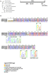
Sideroflexins (SFXNs) form a family of conserved proteins in Eukarya. (A). Left panel: Phylogenetic tree obtained using the MultiAlin software (http://multalin.toulouse.inra.fr/multalin/) [10]. Right panel: scheme of the SFXN1 protein and its conserved motifs. (B). Alignment of human SFXNs protein sequences. Red amino acids are for high consensus levels (90%), the blue ones are for low consensus levels (50%). Meaning of symbols found in the consensus line: “!” is for Ile or Val, “$” is for Leu or Met, “%” is for Phe or Tyr,” #” is anyone of Asn, Asp, Glu, Gln. Conserved motifs are shown and highlighted using an HMM logo created using Skyline (http://skylign.org/) with consensus colors for amino acids according to the ClustalX coloring scheme.
Figure 2
Figure 2
Predicted structure of human SFXN1. Structure prediction was obtained using trRosetta. The confidence of the predicted model shown here is very high (with estimated TM-score = 0.806). The model was built by trRosetta based on de novo folding, guided by deep learning restraints. iCn3D was used for the visualization of 3D structure [13]. (A). SFXN1 predicted structure reveals several alpha helices and beta strands. N and C termini are labelled. The inlet shows the position of the HPDT motif (aa 80–83), located just after the fourth helix. (B). Two views highlighting secondary structures (helices in red, beta sheets in green). (C). Models for SFXN1 insertion in the inner mitochondrial membrane.
Figure 3
Figure 3
Scheme of the mitochondrial respiratory chain. For each complex, Iron Sulfur Cluster (ISC) and heme numbers are given.
Figure 4
Figure 4
Iron homeostasis and utilization at the cell level. Iron cellular uptake is controlled by transferrin and its receptor (TF and TFR1, respectively). Afterwards, in the endosome, iron is reduced thanks to the action of STEAP3 (which converts the insoluble Fe3+ to soluble Fe2+) and released from the endosome into the cytoplasm by the DMT1 channel. Free iron can be stored by ferritin in the cytoplasm or can be transported into the mitochondria, thanks to Mitoferrin 1 and 2 transporters (Mfrn1/2). Excess of iron is released out of the cell by Ferroportin (FPN). Inside the mitochondrion, iron can be stored in FTMT (mitochondrial ferritin) or incorporated in heme or Fe-S clusters. IRP1 and 2 (Iron Related Protein 1 and 2) are the major regulators of iron metabolism. In iron-depleted cells, IRP1 can bind IRE (Iron Response Elements) motifs to promote or repress mRNA translation. If IREs are located in the 5′UTR, IRP1 binding represses mRNA translation under low iron levels. On the contrary, transcripts with IREs at the 3′UTR are stabilized and translated upon IRP binding. Hence, low iron levels lead to decreased Ferritin and FPN levels, but promote TFR1 and DMT1 synthesis. High levels of iron prevent IRP1 binding to IREs (see the main text for details).
Figure 5
Figure 5
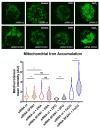
Depleting SFXN1 in HT1080 human cells leads to an intramitochondrial iron accumulation. Top panel: mitochondrial labile Fe(II) staining using the Mito-FerroGreen probe [70] after transient transfection with a control siRNA (siRNA ct) or a pool of SFXN1-targeting siRNA (siRNA SFXN1). Cells were further treated with DMSO (vehicle), erastin, DFO or FeCl3. Erastin is a drug that is widely used to trigger ferroptosis, DFO (deferoxamine) is an iron chelator that lowers mitochondrial iron levels and is used as a negative control. FeCl3 increases intracellular iron levels and served as a positive control. SFXN1 depleted cells show higher mitochondrial iron levels than control cells (siRNA scramble transfected cells). Same magnification is used for all images and insets from full images taken at 630× are shown here. Bottom panel: quantification of three independent assays (n > 50 cells per condition) in which the fluorescent signal is measured and values are normalized to siRNA ct mean levels (mean = 1). After Mann-Whittney tests, significant differences are shown (** p < 0.01, *** p > 0.001, NS Not Significant). See Appendix A.2. for experimental details.
Figure 6
Figure 6
Regulation of heme biosynthesis by SFXN1. Gly and succinyl CoA are the substrates to generate ALA, the first heme precursor, thanks to ALAS enzyme. Gly can enter directly into the mitochondria by SLC25A38, or can be the result of Ser transformation (previously imported by SFXN1) by SHMT2. ALA is further exported to the cytosol where the next steps of heme biosynthesis are catalysed by ALAD, PBGB, UROS and UROD. CPOX, PPOX and FECH are the three mitochondrial enzymes that catalyze the three last steps of heme synthesis (see main text). The last step corresponds to the incorporation of iron into the protoporphyrin PPIX to complete the heme synthesis. Cells lacking SFXN1 show decreased CPOX and FECH mRNA and protein levels (orange box), but a higher amount of ALAS protein (green box), according to Acoba et al. [16].
Figure 7
Figure 7
IRE sequences from known proteins involved in iron metabolism. IRE sequences can be localized at 5′ or 3′. In the absence of iron, IRP1 binds the sequences located at 5′ of blocking the translation of the RNA. Ferritin, ALAS and Ferroportin are proteins involved in iron storage, heme synthesis and iron export, respectively. In the same situation, IRP binding to 3′ sequences, stabilizes the RNA promoting the translation of, for example, Transferrin receptor, involved in iron import. In the opposite situation, with high iron levels, IRP binds to iron, which unbinds the IREs, thus promoting translation of Ferritin, ALAS and Ferroportin and leading to Transferrin receptor RNA decay, which is no more protected by IRP1. Green nucleotides form the six-nucleotide apical loop of IRE.
Figure 8
Figure 8
Predicted IRE in SFXN transcripts. (A). Two IREs were found in the 3′UTR of SFXN1 transcripts using the SIREs Web Server 2.0. The first one is located at the end of the coding sequence. (B). Alignment showing the position of the two IREs in SFXN1 transcripts. All except one shorter SFXN1 transcript variant possess putative IREs. (C). Alignment of the IREs of DMT-1, transferrin receptor (TFRC) and SFXN1 transcripts using MultiAlin. The consensus highlights the position of the six-nucleotide apical loop (5′-CAGWGH-3′) as shown in the yellow box. D, E. Schemes (D) and RNA fold prediction (E) for the IREs from TFRC, DMT-1, SFXN1 and SFXN5 transcripts generated by the SIREs Web Server 2.0.

References

    1. Fleming M.D. A Mutation in a Mitochondrial Transmembrane Protein Is Responsible for the Pleiotropic Hematological and Skeletal Phenotype of Flexed-Tail (f/f) Mice. Genes Dev. 2001;15:652–657. doi: 10.1101/gad.873001. - DOI - PMC - PubMed
    1. Lenox L.E., Perry J.M., Paulson R.F. BMP4 and Madh5 Regulate the Erythroid Response to Acute Anemia. Blood. 2005;105:2741–2748. doi: 10.1182/blood-2004-02-0703. - DOI - PubMed
    1. Hegde S., Lenox L.E., Lariviere A., Porayette P., Perry J.M., Yon M., Paulson R.F. An Intronic Sequence Mutated in Flexed-Tail Mice Regulates Splicing of Smad5. Mamm. Genome. 2007;18:852–860. doi: 10.1007/s00335-007-9074-9. - DOI - PubMed
    1. SLC56 Sideroflexins IUPHAR/BPS Guide to PHARMACOLOGY. [(accessed on 20 January 2021)];2020 Available online: http://www.guidetopharmacology.org/GRAC/FamilyDisplayForward?familyId=1008.
    1. Li X., Han D., Kin Ting Kam R., Guo X., Chen M., Yang Y., Zhao H., Chen Y. Developmental Expression of Sideroflexin Family Genes in Xenopus Embryos. Dev. Dyn. 2010;239:2742–2747. doi: 10.1002/dvdy.22401. - DOI - PubMed

LinkOut - more resources