. 2021 Jan 25;19(1):14.
doi: 10.1186/s12958-021-00699-z.

The role of Chito-oligosaccharide in regulating ovarian germ stem cells function and restoring ovarian function in chemotherapy mice

Affiliations

The role of Chito-oligosaccharide in regulating ovarian germ stem cells function and restoring ovarian function in chemotherapy mice

Yaoqi Huang et al. Reprod Biol Endocrinol. .

Abstract

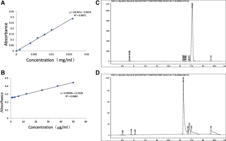
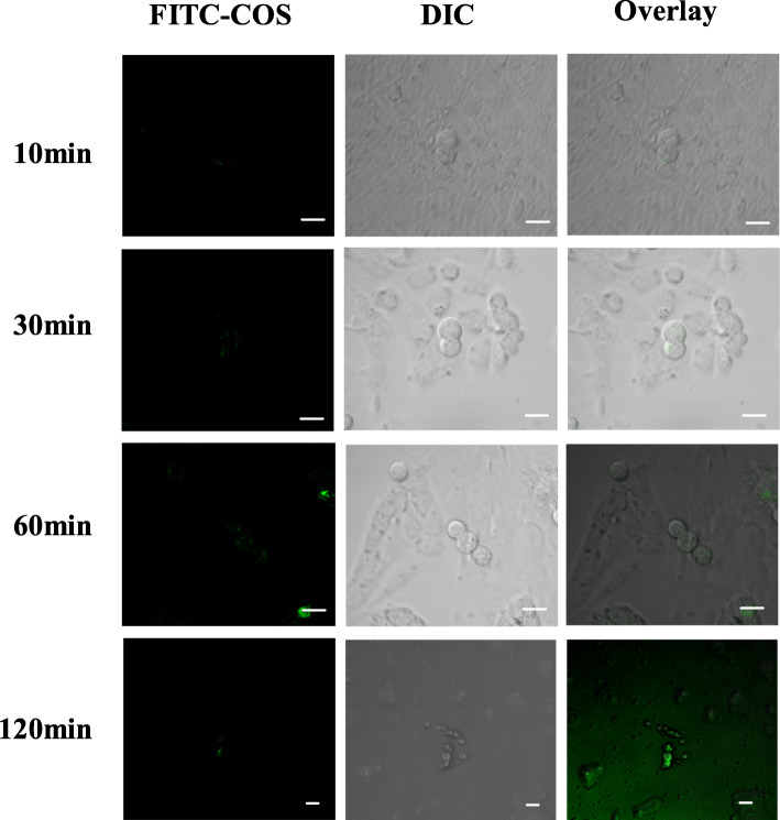
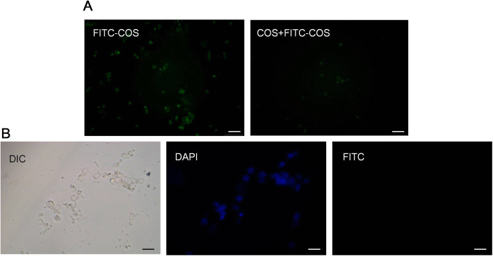
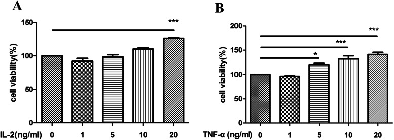
In recent years, the discovery of ovarian germ stem cells (OGSCs) has provided a new research direction for the treatment of female infertility. The ovarian microenvironment affects the proliferation and differentiation of OGSCs, and immune cells and related cytokines are important components of the microenvironment. However, whether improving the ovarian microenvironment can regulate the proliferation of OGSCs and remodel ovarian function has not been reported. In this study, we chelated chito-oligosaccharide (COS) with fluorescein isothiocyanate (FITC) to track the distribution of COS in the body. COS was given to mice through the best route of administration, and the changes in ovarian and immune function were detected using assays of organ index, follicle counting, serum estrogen (E2) and anti-Mullerian hormone (AMH) levels, and the expression of IL-2 and TNF-α in the ovaries. We found that COS significantly increased the organ index of the ovary and immune organs, reduced the rate of follicular atresia, increased the levels of E2 and AMH hormones, and increased the protein expression of IL-2 and TNF-α in the ovary. Then, COS and OGSCs were co-cultured to observe the combination of COS and OGSCs, and measure the survival rate of OGSCs. With increasing time, the fluorescence intensity of cells gradually increased, and the cytokines IL-2 and TNF-α significantly promoted the proliferation of OGSCs. In conclusion, COS could significantly improve the ovarian and immune function of chemotherapy model mice, and improve the survival rate of OGSCs, which provided a preliminary blueprint for further exploring the mechanism of COS in protecting ovarian function.

Keywords: COS; Chemotherapy; Inflammation; OGSCs.


Conflict of interest statement

The authors declare that there is no conflict of interest.

Figures

Fig. 1
Fig. 1
Preparation and chromatographic analysis of FITC- chitosan oligosaccharides a The standard curve of FITC solution; b Typical standard calibration curve for the determination of FITC-COS in the ovary. c HPLC chromatograms of FITC; d HPLC chromatograms of FITC-COS
Fig. 2
Fig. 2
The distribution of chitooligosaccharides in the ovary. a-c The distribution of chitosan oligosaccharide in ovary at different time after intraperitoneal injection, gavage administration and tail vein injection; d Distribution of ovaries and kidneys after intraperitoneal injection. Scale bar = 100 μm
Fig. 3
Fig. 3
Protective effects of chitooligosaccharides on reproductive and immune function. a-c The index of different organs in control, CY/BUS and CY/BUS+COS group after 28 days of continuous administration; d, e Effects of COS on AMH and E2 hormone levels in mice; f-h Expression of IL-2 and TNF-α protein in ovaries. ** P < 0.01, and ***P < 0.001, vs. the control group, #P < 0.05, ##P < 0.01 and ###P < 0.001 vs. the CY/BUS group
Fig. 4
Fig. 4
Chitooligosaccharides (COS) administration prevents the loss of different follicle types and decreases atresia caused by cyclophosphamide (CY)/ busulfan (BUS) treatment. a Representative images of HE stained sections from control, CY/BUS and CY/BUS+COS mice, the scale bar is 200 μm. b The number of follicles at different levels was counted by tissue section. c Quantity changes of the percentage of primordial and atretic follicles in total follicles. Data are presented as mean ± SEM for three independent experiments. ** P < 0.01, and ***P < 0.001, vs. the control group, #P < 0.05, ##P < 0.01 and ###P < 0.001 vs. the CY/BUS group
Fig. 5
Fig. 5
Identification of ovarian reproductive stem cells. a, b Morphology of ovarian germline stem cells in early stage of culture; c Alkaline phosphatase staining; d Identification of ovarian germline stem cells and whole ovary by RT-PCR; (E,F):Immunofluorescence of Ovarian Germline Stem Cells
Fig. 6
Fig. 6
The distribution of FITC-COS into OGSCs changed with time (Bar:10 μm)
Fig. 7
Fig. 7
Distribution of FITC and COS in OGSCs. a After 1 h, the fluorescence distribution of FITC-COS group and COS + FITC-COS group in OGSCs was observed. b Fluorescence distribution of FITC in OGSCs (Bar:20 μm)
Fig. 8
Fig. 8
The proliferation effect of different concentrations of cytokines on OGSCs. a The cytokine IL-2; b The cytokine TNF-α. *p < 0.05, ***p < 0.001, vs. the control group

Similar articles

Cited by

References

    1. Raymond J Rodgers, Joop S E Laven. Genetic relationships between early menopause and the behaviour of theca interna during follicular atresia [J]. Human Reproduction. 2020;35(10):2185–7. - PubMed
    1. Wang XY, Zhang XY, Dang YJ. Long noncoding RNA HCP5 participates in premature ovarian insufficiency by transcriptionally regulating MSH5 and DNA damage repair via YB1. Nucleic Acids Res. 2020;48(8):4480–4491. doi: 10.1093/nar/gkaa127. - DOI - PMC - PubMed
    1. Reiner A. Veitia. Primary ovarian insufficiency, meiosis and DNA repair. Biom J. 2020;43(2):115–123. - PMC - PubMed
    1. Johnson J, Canning J, Kaneko T, et al. Germline stem cells and follicular renewal in the postnatal mammalian ovary. Nature. 2004;428(6797):145–150. doi: 10.1038/nature02316. - DOI - PubMed
    1. Niikura Y, Niikura T, Tilly JL. Aged mouse ovaries possess rare premeiotic germ cells that can generate oocytes following transplantation into a young host environment. Aging (Albany NY) 2009;1(12):971–978. doi: 10.18632/aging.100105. - DOI - PMC - PubMed

MeSH terms