. 2021 Jan 25;12(1):83.
doi: 10.1186/s13287-021-02136-9.

Glucocorticoid agonists enhance retinal stem cell self-renewal and proliferation

Affiliations

Glucocorticoid agonists enhance retinal stem cell self-renewal and proliferation

Kenneth N Grisé et al. Stem Cell Res Ther. .

Abstract

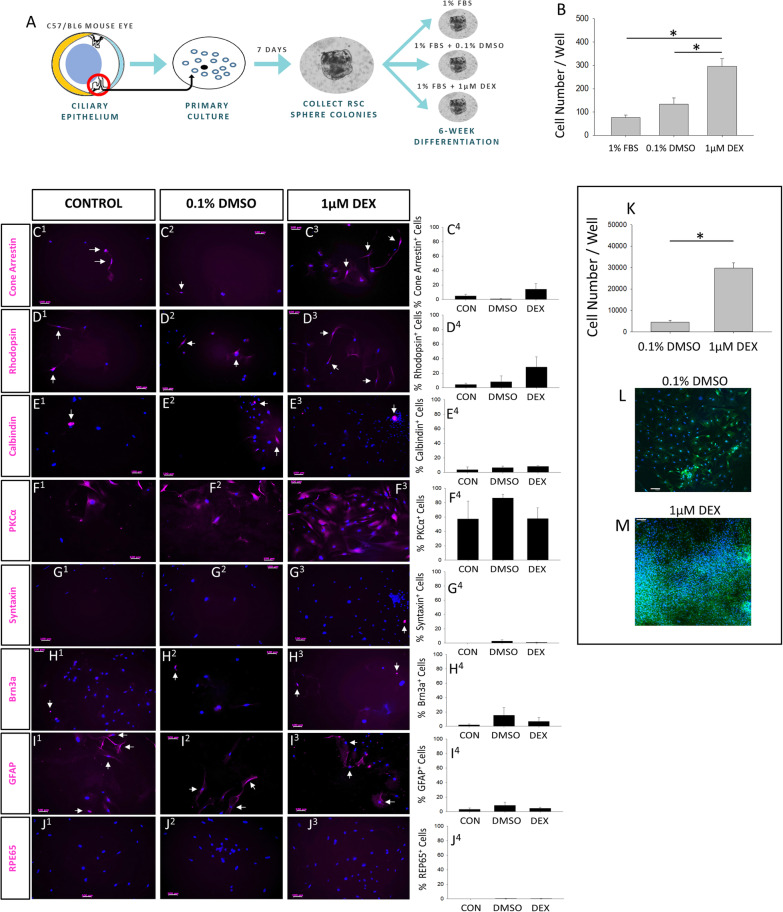
Background: Adult mammalian retinal stem cells (RSCs) readily proliferate, self-renew, and generate progeny that differentiate into all retinal cell types in vitro. RSC-derived progeny can be induced to differentiate into photoreceptors, making them a potential source for retinal cell transplant therapies. Despite their proliferative propensity in vitro, RSCs in the adult mammalian eye do not proliferate and do not have a regenerative response to injury. Thus, identifying and modulating the mechanisms that regulate RSC proliferation may enhance the capacity to produce RSC-derived progeny in vitro and enable RSC activation in vivo.

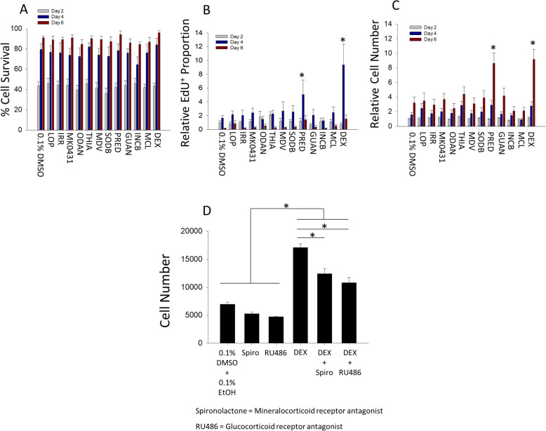
Methods: Here, we used medium-throughput screening to identify small molecules that can expand the number of RSCs and their progeny in culture. In vitro differentiation assays were used to assess the effects of synthetic glucocorticoid agonist dexamethasone on RSC-derived progenitor cell fate. Intravitreal injections of dexamethasone into adult mouse eyes were used to investigate the effects on endogenous RSCs.

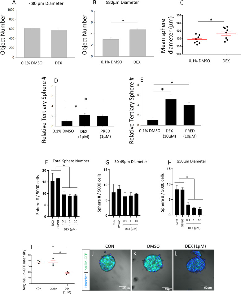
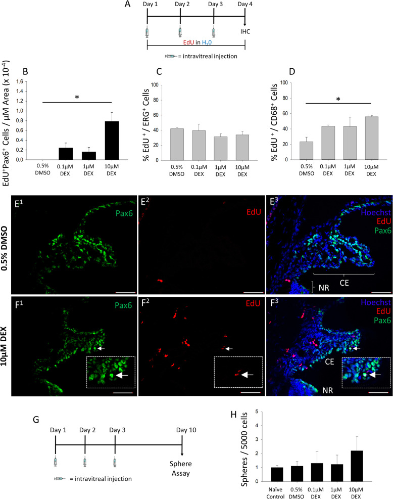
Results: We discovered that high-affinity synthetic glucocorticoid agonists increase RSC self-renewal and increase retinal progenitor proliferation up to 6-fold without influencing their differentiation in vitro. Intravitreal injection of synthetic glucocorticoid agonist dexamethasone induced in vivo proliferation in the ciliary epithelium-the niche in which adult RSCs reside.

Conclusions: Together, our results identify glucocorticoids as novel regulators of retinal stem and progenitor cell proliferation in culture and provide evidence that GCs may activate endogenous RSCs.

Keywords: Cell differentiation; Dexamethasone; Drug discovery; Glucocorticoid; Neurogenesis; Photoreceptor; Retina; Self-renewal; Stem cell; progenitor; proliferation.


Conflict of interest statement

The authors declare that they have no competing interests.

Figures

Fig. 1
Fig. 1
Medium-throughput screening identifies several unique compound classes that increase retinal stem and progenitor cell number. a Schematic overview of the MTS pipeline to detect RSPC expanding compounds. be Quantification as a percent of control (POC) of all compounds that met the hit criteria of having a relative number of nuclei or total area of actin-GFP expression over 3xSD above the 1× control mean. Hits that were identified in multiple criteria and/or screens are color-coded. b, c Seven compounds in screen 1 were found to be hits in both the number of nuclei and actin-GFP area and were selected as lead compounds. 1× control: N = 80 technical replicates. 2× control: N = 12 technical replicates. Compounds: N = 1 technical replicate. d, e Eight compounds in screen 2 were found to be hits in both the number of nuclei and actin-GFP area and were selected as lead compounds. 1× control: N = 80 technical replicates. 2× control: N = 15 technical replicates. Compounds: N = 1 technical replicate. f A dose-response assay of the hit compounds identified via MTS. N = 3 for all compounds at each concentration. Data are mean ± SEM
Fig. 2
Fig. 2
Glucocorticoid agonists increase retinal stem and progenitor cell proliferation through glucocorticoid receptor and mineralocorticoid receptor signaling in mice. ac Quantification of growth parameters for RSPCs at day 2, day 4, and day 6 of a monolayer growth assay. *Significantly different from control within that time point. a The proportion of live cells was not significantly different across drug treatments (p = 0.55), and there was no interaction between drug and time (p = 0.99). There was a significant effect of time on the proportion of live cells, and all times were significantly different (two-way ANOVA F (2, 195) = 261.9, p < 0.001; Holm-Sidak post hoc test, *p < 0.05). N = 6 for all conditions at each time point. b The proportion of proliferating EdU-labeled cells relative to control. There was a significant interaction between drug and time (two-way ANOVA F (24, 195) = 2.59, p < 0.001; Holm-Sidak post hoc test, *p < 0.05). N = 6 for all conditions at each time point. c The total number of cells relative to control. There was a significant interaction between drug and time (two-way ANOVA F (24, 429) = 3.79, p < 0.001; Holm-Sidak post hoc test, *p < 0.05). N = 12 for all conditions at each time point. d Total RSPC number at the end of a 7-day monolayer growth assay with 0.1% DMSO and 0.1% EtOH in all conditions. Control was significantly different from all Dex treatment conditions but not spironolactone (Spiro) or RU486 alone (one-way ANOVA F (5, 12) = 67.88, p < 0.001; Holm-Sidak post hoc test, *p < 0.05). N = 3 for RU486 alone. N = 4 for all other groups. Data are mean ± SEM
Fig. 3
Fig. 3
Dexamethasone increases retinal stem cell sphere size, number, and self-renewal but inhibits growth and insulin expression of pancreatic multipotent progenitor spheres. ac Quantification of sphere diameter after a 7-day free-floating clonal sphere assay with actin-GFP mouse-derived secondary RSPCs in 0.1% DMSO vehicle or 1 μM Dex. a The number of cell colonies less than 80 μm in diameter was not significantly different between 0.1% DMSO vehicle control and 1 μM Dex treatment. N = 8 per condition. b Cell colonies greater than 80 μm in diameter increased in number by 1.63-fold with Dex treatment. (t test t (14) = − 5, p < .001; N = 8 per condition). c RSC sphere colonies 80 μm or above in diameter demonstrated an overall increase in size with Dex treatment (t test t (14) = 2.56, p < .05; N = 8 per condition). *Significantly different from 0.1% DMSO control. d, e Quantification of the number of tertiary RSC sphere colonies grown in drug-free media after prior exposure of secondary cells to the indicated compounds at 1 μM or 10 μM during a 7-day free-floating clonal sphere assay. Exposure to Dex and Pred increased the number of RSC spheres after passaging by 2.2-fold and 2-fold, respectively, at 1 μM (d; one-way ANOVA F (2, 15) = 4.02, p = 0.04; Fisher LSD post hoc test, *p < 0.05; N = 6 per group), and increased RSC sphere number by 5.2-fold and 4-fold, respectively, at 10 μM (e; one-way ANOVA F (2, 6) = 9.6, p = 0.014; Fisher LSD post hoc test, *p < 0.05; N = 3 per group). *Significantly different from 0.1% DMSO control. fh Quantification of adult PMP spheres after a 7-day free-floating clonal sphere assay. f The total number of spheres ≥ 30 μm was significantly reduced by all concentrations of Dex tested (one-way ANOVA F (4, 10) = 4.76, p = 0.02; post hoc test, Holm-Sidak post hoc test, *p < 0.05; N = 3 experiments). g The number of spheres 30–49 μm in diameter was not influenced by Dex treatment at any concentration tested. N = 3 experiments. h The number of spheres ≥ 50 μm was significantly reduced by all concentrations of Dex tested (one-way ANOVA F (4, 10) = 16.12, p < 0.001; Holm-Sidak post hoc test, *p < 0.05; N = 3 experiments). *Significantly different from indicated conditions. il The intensity of MIP-GFP expression in PMP spheres. i Quantification of the intensity of MIP-GFP expression in PMP spheres (one-way ANOVA F (2, 6) = 18.35, p = 0.0028; Holm-Sidak post hoc test, *p < 0.05; N = 3 replicates per condition). jl Representative confocal projection images of MIP-GFP expression in PMP spheres. Data are mean ± SEM
Fig. 4
Fig. 4
Dexamethasone does not affect the differentiation profile of retinal progenitor cells. a Schematic overview of the RSPC differentiation assay. b Quantification of cell number per well after 6 weeks in the indicated differentiation conditions (one-way ANOVA F (2, 8) = 16.46, p = 0.001; Holm-Sidak post hoc test, *p < 0.05; N = 3–4 per group). *Significantly different between indicated conditions. cj IHC images and quantification of mature retinal cell type markers following 6 weeks of differentiation across the indicated conditions. N = 3–4 per group. The nuclei labeled with Hoechst stain (blue). White arrows indicate cells positive for cell type markers. km Images and quantification of secondary RSPCs from actin-GFP mice grown as monolayers for 6 weeks in 1% FBS differentiation media (t test t (14) = − 9.54, p < .001; N = 8 wells per condition). *Significantly different between indicated conditions. Data are mean ± SEM
Fig. 5
Fig. 5
Intravitreal dexamethasone injection induces ciliary epithelium proliferation but does not expand the retinal stem cell population in vivo. a Schematic of the intravitreal injection paradigm followed by endpoint IHC. b Quantification of Pax6+EdU co-labeled cells relative to the total CE area in the eyes treated with 0.5% DMSO vehicle or indicated Dex concentrations (one-way ANOVA F (3, 15) = 6.21, p = 0.006; Holm-Sidak post hoc test, *p < 0.05; N = 3–6 eyes per condition). *Significantly different between indicated conditions. c The proportion of ERG + EdU-co-labeled endothelial cells relative to the total number of EdU-labeled cells in the ciliary body for each indicated condition. N = 3–6 eyes per condition. d The proportion of CD68 + EdU-co-labeled microglia/macrophages relative to the total number of EdU-labeled cells in the ciliary body for each indicated condition (one-way ANOVA F (3, 14) = 6.89, p = 0.004; Fisher Holm-Sidak post hoc test, *p < 0.05; N = 3–6 eyes per condition). *Significantly different between indicated conditions. e, f Representative images of Pax6 IHC and EdU labeling in the ciliary body and peripheral retina of mouse eyes exposed to 0.5% DMSO vehicle or indicated Dex concentrations. The nuclei labeled with Hoechst stain. White arrows indicate Pax6 + EdU co-labeled cells; 10-μm-thick sections; 50-μm scale bars. g Schematic of intravitreal injection paradigm followed by endpoint primary sphere-forming assay. h Quantification of RSC sphere frequency relative to naive un-injected control after a 7-day clonal sphere growth assay following intravitreal injection of indicated conditions. N = 6 eyes per group. Data are mean ± SEM

References

    1. Berry M, Ahmed Z, Lorber B, Douglas M, Logan A. Regeneration of axons in the visual system. Restor Neurol Neurosci. 2008;26:147–174. - PubMed
    1. Kubota R, Hokoc JN, Moshiri A, McGuire C, Reh TA. A comparative study of neurogenesis in the retinal ciliary marginal zone of homeothermic vertebrates. Brain Res Dev Brain Res. 2002;134:31–41. doi: 10.1016/S0165-3806(01)00287-5. - DOI - PubMed
    1. Gasparini SJ, Llonch S, Borsch O, Ader M. Transplantation of photoreceptors into the degenerative retina: current state and future perspectives. Prog Retin Eye Res. 2019;69:1–37. doi: 10.1016/j.preteyeres.2018.11.001. - DOI - PubMed
    1. Miller FD, Kaplan DR. Mobilizing endogenous stem cells for repair and regeneration: are we there yet? Cell Stem Cell. 2012;10:650–652. doi: 10.1016/j.stem.2012.05.004. - DOI - PubMed
    1. Aladdad AM, Kador KE. Adult stem cells, tools for repairing the retina. Curr Ophthalmol Rep. 2019;7:21–29. doi: 10.1007/s40135-019-00195-z. - DOI - PMC - PubMed

Publication types

Substances

Grants and funding