. 2021 Jan 25;25(1):36.
doi: 10.1186/s13054-020-03424-1.

Sepsis is associated with mitochondrial DNA damage and a reduced mitochondrial mass in the kidney of patients with sepsis-AKI

Affiliations

Sepsis is associated with mitochondrial DNA damage and a reduced mitochondrial mass in the kidney of patients with sepsis-AKI

Elisabeth C van der Slikke et al. Crit Care. .

Abstract

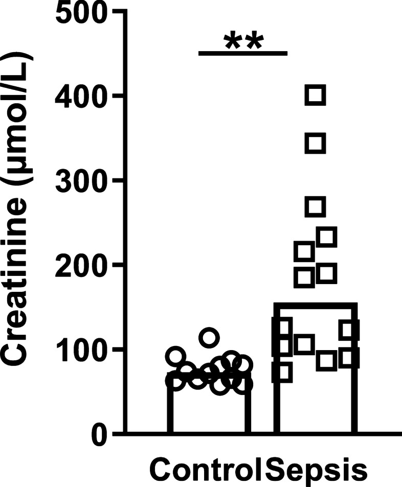
Background: Sepsis is a life-threatening condition accompanied by organ dysfunction subsequent to a dysregulated host response to infection. Up to 60% of patients with sepsis develop acute kidney injury (AKI), which is associated with a poor clinical outcome. The pathophysiology of sepsis-associated AKI (sepsis-AKI) remains incompletely understood, but mitochondria have emerged as key players in the pathogenesis. Therefore, our aim was to identify mitochondrial damage in patients with sepsis-AKI.

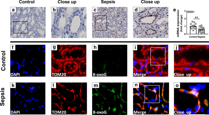
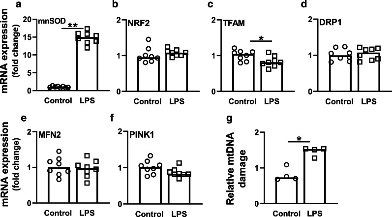
Methods: We conducted a clinical laboratory study using "warm" postmortem biopsies from sepsis-associated AKI patients from a university teaching hospital. Biopsies were taken from adult patients (n = 14) who died of sepsis with AKI at the intensive care unit (ICU) and control patients (n = 12) undergoing tumor nephrectomy. To define the mechanisms of the mitochondrial contribution to the pathogenesis of sepsis-AKI, we explored mRNA and DNA expression of mitochondrial quality mechanism pathways, DNA oxidation and mitochondrial DNA (mtDNA) integrity in renal biopsies from sepsis-AKI patients and control subjects. Next, we induced human umbilical vein endothelial cells (HUVECs) with lipopolysaccharide (LPS) for 48 h to mimic sepsis and validate our results in vitro.

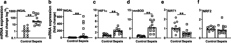
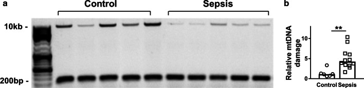
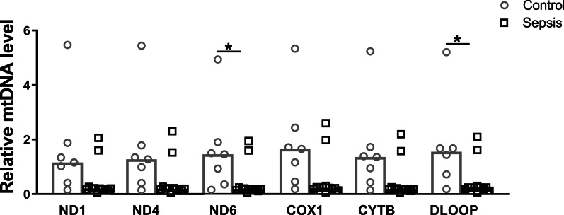
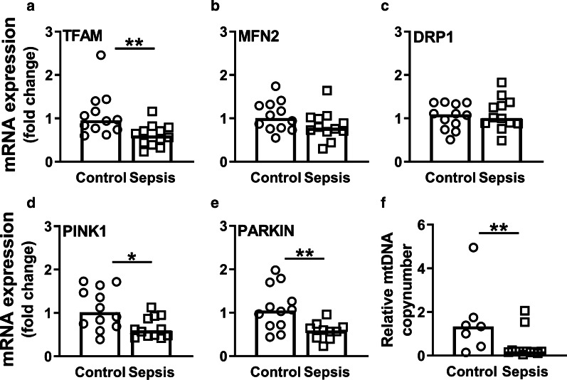
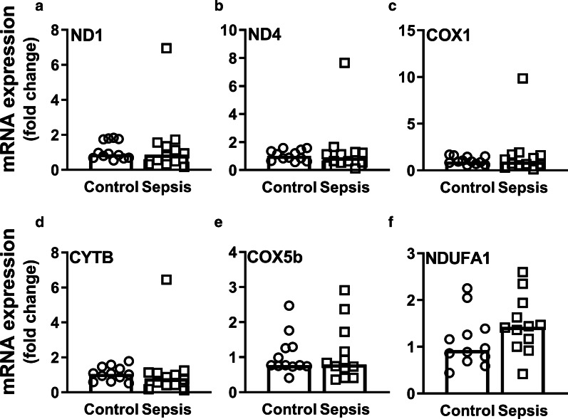
Results: Compared to control subjects, sepsis-AKI patients had upregulated mRNA expression of oxidative damage markers, excess mitochondrial DNA damage and lower mitochondrial mass. Sepsis-AKI patients had lower mRNA expression of mitochondrial quality markers TFAM, PINK1 and PARKIN, but not of MFN2 and DRP1. Oxidative DNA damage was present in the cytosol of tubular epithelial cells in the kidney of sepsis-AKI patients, whereas it was almost absent in biopsies from control subjects. Oxidative DNA damage co-localized with both the nuclei and mitochondria. Accordingly, HUVECs induced with LPS for 48 h showed an increased mnSOD expression, a decreased TFAM expression and higher mtDNA damage levels.

Conclusion: Sepsis-AKI induces mitochondrial DNA damage in the human kidney, without upregulation of mitochondrial quality control mechanisms, which likely resulted in a reduction in mitochondrial mass.

Keywords: Acute kidney injury; Mitochondria; Reactive oxygen species; Sepsis.


Conflict of interest statement

The authors declare that they have no competing interests.

Figures

Fig. 1
Fig. 1
Serum creatinine concentrations. Serum concentrations of creatinine (µmol/L) in control subjects and sepsis-AKI patients before nephrectomy or prior to extirpation, respectively
Fig. 2
Fig. 2
Oxidative DNA damage in controls and sepsis-AKI subjects. ad Immunohistochemistry of 8-oxoG (DNA oxidation marker) e mRNA expression of OGG1 as quantified by RT-qPCR fo immunohistochemistry of TOM20 (mitochondrial marker) and 8-oxoG (DNA oxidation marker). Bars represent median, dots represent individual levels; ** means p < 0.005
Fig. 3
Fig. 3
Gene expression of oxidative damage- and antioxidant markers. mRNA expression of a NGAL (log10), b NGAL, c HIF1α, d mnSOD, e SIRT1, and f NRF2 as quantified by RT-qPCR. Bars represent median, dots represent individual levels; ** means p < 0.005
Fig. 4
Fig. 4
Changes in mitochondrial biogenesis, fusion, fission and mitophagy after 48-h LPS induction in HUVECs. Cells were induced with 10 µg/mL LPS for 48 h. mRNA expression of a mnSOD, b NRF2, c TFAM, d DRP1, e MFN2, and f PINK1; g Relative mtDNA damage levels of control and LPS-induced HUVECs. Bars represent median, dots represent individual levels; * means p < 0.05 and ** p <0.005
Fig. 5
Fig. 5
Mitochondrial DNA damage in control and sepsis-AKI subjects. A 10-kb-long mtDNA sequence was amplified by long-range PCR, while a 222-bp short mtDNA sequence was amplified with qPCR. a Amplified mtDNA sequences were loaded on agarose gel from representative samples of control subjects and sepsis-AKI subjects; b relative mtDNA damage of the control subjects compared to the sepsis-AKI patients. Bars represent median, dots represent individual levels; ** means p < 0.005
Fig. 6
Fig. 6
Relative mitochondrial DNA levels. Relative mtDNA levels of ND1, ND4, ND6, COX1, CYTB, and D-loop as quantified by qPCR. Bars represent the median, dots represent individual levels; * means p < 0.05
Fig. 7
Fig. 7
Changes in mitochondrial biogenesis, fusion, fission, mitophagy and copy number in response to sepsis-AKI. mRNA expression of a TFAM, b MFN2, c DRP1, d PINK1, and e PARKIN as quantified by RT-qPCR; f mtDNA levels were quantified by RT-qPCR to calculate the mitochondrial copy number. Bars represent median, dots represent individual levels; */** means p < 0.05/0.005; mtDNA mitochondrial DNA
Fig. 8
Fig. 8
mRNA expression of mitochondrial electron transport chain complexes. mRNA expression of a ND1, b ND4, c COX1, d CYTB, e COX5b, and f NDUFA1 as quantified by RT-qPCR. Bars represent median, dots represent individual levels

References

    1. Angus DC, van der Poll T. Severe sepsis and septic shock. N Engl J Med. 2013;369(9):840–851. doi: 10.1056/NEJMra1208623. - DOI - PubMed
    1. Cecconi M, Evans L, Levy M, Rhodes A. Sepsis and septic shock. Lancet. 2018;392(10141):75–87. doi: 10.1016/S0140-6736(18)30696-2. - DOI - PubMed
    1. Reinhart K, Daniels R, Kissoon N, Machado FR, Schachter RD, Finfer S. Recognizing sepsis as a global health priority—a WHO resolution. N Engl J Med. 2017;377(5):414–417. doi: 10.1056/NEJMp1707170. - DOI - PubMed
    1. Gotts JE, Matthay MA. Sepsis: pathophysiology and clinical management. BMJ. 2016;353:i1585. doi: 10.1136/bmj.i1585. - DOI - PubMed
    1. Schrier RW, Wang W. Acute renal failure and sepsis. N Engl J Med. 2004;351(2):159–169. doi: 10.1056/NEJMra032401. - DOI - PubMed

Publication types

Substances