. 2021 Feb 19;371(6531):850-854.
doi: 10.1126/science.abf9302. Epub 2021 Jan 25.

Prospective mapping of viral mutations that escape antibodies used to treat COVID-19

Affiliations

Prospective mapping of viral mutations that escape antibodies used to treat COVID-19

Tyler N Starr et al. Science. .

Abstract

Antibodies are a potential therapy for severe acute respiratory syndrome coronavirus 2 (SARS-CoV-2), but the risk of the virus evolving to escape them remains unclear. Here we map how all mutations to the receptor binding domain (RBD) of SARS-CoV-2 affect binding by the antibodies in the REGN-COV2 cocktail and the antibody LY-CoV016. These complete maps uncover a single amino acid mutation that fully escapes the REGN-COV2 cocktail, which consists of two antibodies, REGN10933 and REGN10987, targeting distinct structural epitopes. The maps also identify viral mutations that are selected in a persistently infected patient treated with REGN-COV2 and during in vitro viral escape selections. Finally, the maps reveal that mutations escaping the individual antibodies are already present in circulating SARS-CoV-2 strains. These complete escape maps enable interpretation of the consequences of mutations observed during viral surveillance.


Figures

Fig. 1
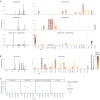
Fig. 1. Complete maps of mutations that escape binding by the REGN-COV2 antibodies and Ly-CoV016.
(A) Maps for antibodies in REGN-COV2. Line plots at left show escape at each site in the RBD (summed effects of all mutations at each site). Sites of strong escape (purple underlines) are shown in logo plots at right. The height of each letter is proportional to how strongly that amino acid mutation mediates escape, with a per-mutation “escape fraction” of 1 corresponding to complete escape. The y-axis scale is different for each row, so, for instance, E406W escapes all REGN antibodies but is most visible for the cocktail as it is swamped out by other sites of escape for the individual antibodies. See https://jbloomlab.github.io/SARS-CoV-2-RBD_MAP_clinical_Abs/ for zoomable versions. Letters are colored according to how mutations affect the RBD’s affinity for ACE2 as measured via yeast display (7), with yellow indicating poor affinity and brown indicating good affinity; see fig. S2, A and B, for maps colored by how mutations affect expression of folded RBD and fig. S2, C and D, for distribution of effects on ACE2 affinity and RBD expression across all mutations observed among circulating viral isolates. (B) Map, as in (A), for LY-CoV016. (C) Validation of key mutations in neutralization assays using spike-pseudotyped lentiviral particles. We chose to validate mutations predicted to have large effects or that are present at high frequency among circulating SARS-CoV-2 isolates (e.g., N439K). Each point indicates the fold increase in the median inhibitory concentration (IC50) for a mutation relative to the unmutated wild-type (WT) spike, which contains D614G. The dotted blue line at 1 indicates WT-like neutralization, and values >1 indicate increased neutralization resistance. Point colors indicate whether escape was expected at that site from the maps. Point shapes indicate that the fold change is censored (an upper or lower limit) owing to the IC50 being outside the dilution series used. Most mutants were tested in duplicate and thus have two points. Full neutralization curves are shown in fig. S3. Single-letter abbreviations for the amino acid residues are as follows: A, Ala; C, Cys; D, Asp; E, Glu; F, Phe; G, Gly; H, His; I, Ile; K, Lys; L, Leu; M, Met; N, Asn; P, Pro; Q, Gln; R, Arg; S, Ser; T, Thr; V, Val; W, Trp; and Y, Tyr.
Fig. 2
Fig. 2. Escape maps are consistent with viral mutations selected in cell culture and a persistently infected patient.
(A) Viral escape mutations selected by Regeneron with spike-pseudotyped VSV in cell culture in the presence of antibody (12). (B) Escape maps like those in Fig. 1A, but showing only mutations accessible by single-nucleotide changes to the Wuhan-Hu-1 sequence, with nongray colors indicating mutations in cell culture (red), the infected patient (blue), or both (purple). Figure S5 shows these maps colored by how mutations affect ACE2 affinity or RBD expression. (C) Dynamics of RBD mutations in a patient treated with REGN-COV2 at day 145 of infection (black dot-dash vertical line). E484A rose in frequency in linkage with F486I, but because E484A is not an escape mutation in our maps, it is not shown in other panels. See also fig. S4. (D) The escape mutations that arise in cell culture and the infected patient are single-nucleotide–accessible and escape antibody binding without imposing a large cost on ACE2 affinity [as measured using yeast display (7)]. Each point is a mutation, with shape and color indicating whether it is accessible and selected during viral growth. Points farther to the right on the x axis indicate stronger escape from antibody binding; points higher up on the y axis indicate higher ACE2 affinity.
Fig. 3
Fig. 3. Antibody escape mutations in circulating SARS-CoV-2.
For each antibody or antibody combination, the escape score for each mutation is plotted versus its frequency among the 317,866 high-quality human-derived SARS-CoV-2 sequences on GISAID (26) as of 11 January 2021. Escape mutations with notable GISAID frequencies are labeled. The REGN-COV2 cocktail escape mutation E406W requires multiple nucleotide changes from the Wuhan-Hu-1 RBD sequence and is not observed among sequences in GISAID. Other mutations to residue E406 (E406Q and E406D) are observed with low frequency counts, but neither of these mutant amino acids is a single-nucleotide mutation away from W either.
Fig. 4
Fig. 4. Structural context of escape mutations.
(A) Escape maps projected on antibody-bound RBD structures. [REGN10933 and REGN10987: Protein Data Bank (PDB) ID 6XDG (11); LY-CoV016: PDB ID 7C01 (13)]. Antibody heavy- and light-chain variable domains are shown as blue cartoons, and the RBD surface is colored to indicate how strongly mutations at that site mediate escape (white indicates no escape, red indicates strongest escape site for that antibody or cocktail). Sites where no mutations are functionally tolerated are colored gray. (B) For each antibody, sites were classified as direct antibody contacts (non-hydrogen atoms within 4 Å of antibody), antibody-proximal (4 to 8 Å), or antibody-distal (>8 Å). Each point indicates a site, classified as escape (red) or non-escape (black). The dashed gray line indicates the cutoff used to classify sites as escape or non-escape (see materials and methods for details). Red and black numbers indicate how many sites in each category are escape or non-escape sites, respectively. Interactive visualizations are at https://jbloomlab.github.io/SARS-CoV-2-RBD_MAP_clinical_Abs/, and hypothesized mechanisms of escape and additional structural details for labeled points are shown in fig. S6.

Update of

References

    1. Renn A., Fu Y., Hu X., Hall M. D., Simeonov A., Fruitful neutralizing antibody pipeline brings hope to defeat SARS-Cov-2. Trends Pharmacol. Sci. 41, 815–829 (2020). 10.1016/j.tips.2020.07.004 - DOI - PMC - PubMed
    1. Caskey M., Klein F., Lorenzi J. C. C., Seaman M. S., West A. P. Jr.., Buckley N., Kremer G., Nogueira L., Braunschweig M., Scheid J. F., Horwitz J. A., Shimeliovich I., Ben-Avraham S., Witmer-Pack M., Platten M., Lehmann C., Burke L. A., Hawthorne T., Gorelick R. J., Walker B. D., Keler T., Gulick R. M., Fätkenheuer G., Schlesinger S. J., Nussenzweig M. C., Viraemia suppressed in HIV-1-infected humans by broadly neutralizing antibody 3BNC117. Nature 522, 487–491 (2015). 10.1038/nature14411 - DOI - PMC - PubMed
    1. Kugelman J. R., Kugelman-Tonos J., Ladner J. T., Pettit J., Keeton C. M., Nagle E. R., Garcia K. Y., Froude J. W., Kuehne A. I., Kuhn J. H., Bavari S., Zeitlin L., Dye J. M., Olinger G. G., Sanchez-Lockhart M., Palacios G. F., Emergence of Ebola virus escape variants in infected nonhuman primates treated with the MB-003 antibody cocktail. Cell Rep. 12, 2111–2120 (2015). 10.1016/j.celrep.2015.08.038 - DOI - PubMed
    1. Simões E. A. F., Forleo-Neto E., Geba G. P., Kamal M., Yang F., Cicirello H., Houghton M. R., Rideman R., Zhao Q., Benvin S. L., Hawes A., Fuller E. D., Wloga E., Pizarro J. M. N., Munoz F. M., Rush S. A., McLellan J. S., Lipsich L., Stahl N., Yancopoulos G. D., Weinreich D. M., Kyratsous C. A., Sivapalasingam S., Suptavumab for the prevention of medically attended respiratory syncytial virus infection in preterm infants. Clin. Infect. Dis. ciaa951 (2020). 10.1093/cid/ciaa951 - DOI - PMC - PubMed
    1. Wrapp D., Wang N., Corbett K. S., Goldsmith J. A., Hsieh C.-L., Abiona O., Graham B. S., McLellan J. S., Cryo-EM structure of the 2019-nCoV spike in the prefusion conformation. Science 367, 1260–1263 (2020). 10.1126/science.abb2507 - DOI - PMC - PubMed

Publication types

MeSH terms