. 2021 Feb 2;118(5):e2019622118.
doi: 10.1073/pnas.2019622118.

Drosophila Sex Peptide controls the assembly of lipid microcarriers in seminal fluid

Affiliations

Drosophila Sex Peptide controls the assembly of lipid microcarriers in seminal fluid

S Mark Wainwright et al. Proc Natl Acad Sci U S A. .

Abstract

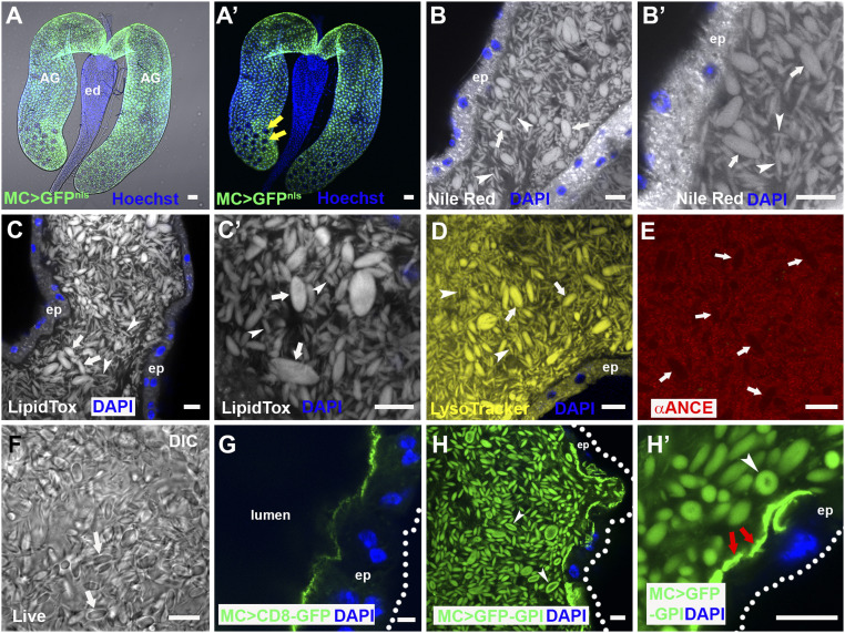
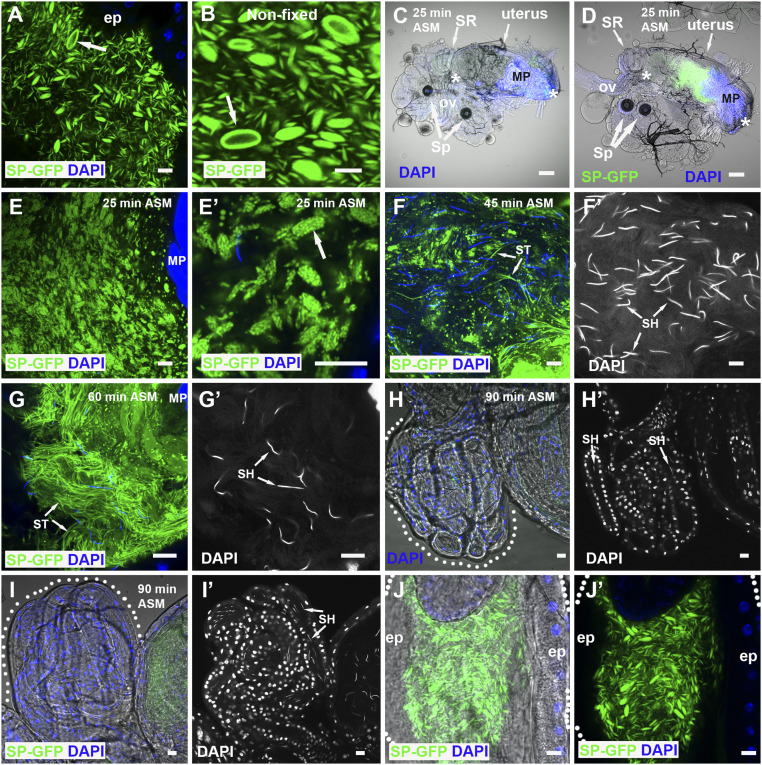
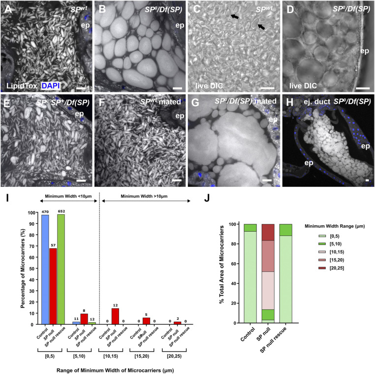
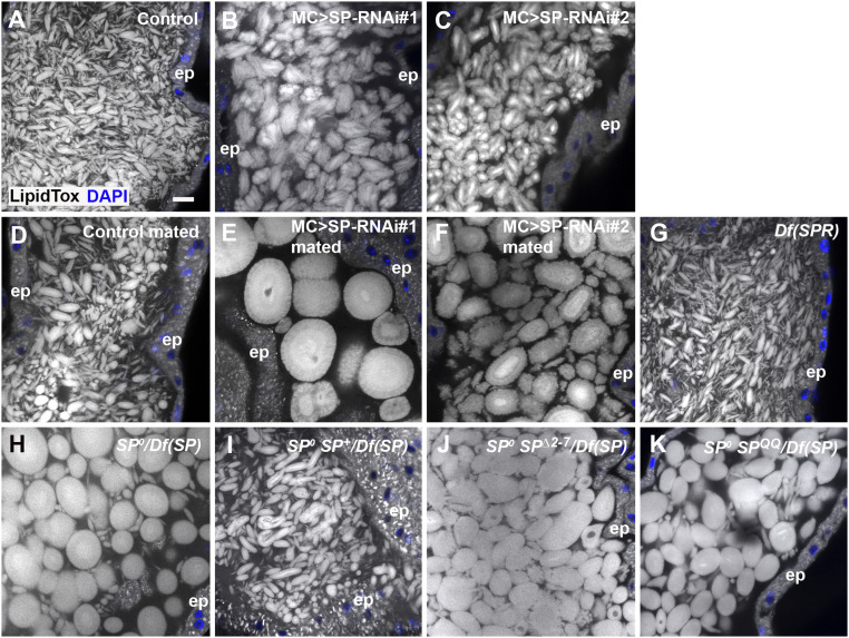
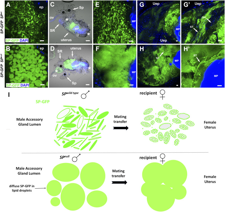
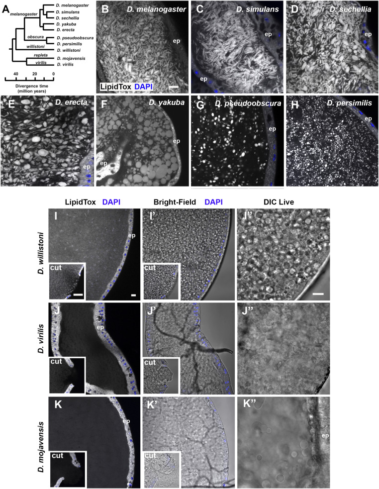
Seminal fluid plays an essential role in promoting male reproductive success and modulating female physiology and behavior. In the fruit fly, Drosophila melanogaster, Sex Peptide (SP) is the best-characterized protein mediator of these effects. It is secreted from the paired male accessory glands (AGs), which, like the mammalian prostate and seminal vesicles, generate most of the seminal fluid contents. After mating, SP binds to spermatozoa and is retained in the female sperm storage organs. It is gradually released by proteolytic cleavage and induces several long-term postmating responses, including increased ovulation, elevated feeding, and reduced receptivity to remating, primarily signaling through the SP receptor (SPR). Here, we demonstrate a previously unsuspected SPR-independent function for SP. We show that, in the AG lumen, SP and secreted proteins with membrane-binding anchors are carried on abundant, large neutral lipid-containing microcarriers, also found in other SP-expressing Drosophila species. These microcarriers are transferred to females during mating where they rapidly disassemble. Remarkably, SP is a key microcarrier assembly and disassembly factor. Its absence leads to major changes in the seminal proteome transferred to females upon mating. Males expressing nonfunctional SP mutant proteins that affect SP's binding to and release from sperm in females also do not produce normal microcarriers, suggesting that this male-specific defect contributes to the resulting widespread abnormalities in ejaculate function. Our data therefore reveal a role for SP in formation of seminal macromolecular assemblies, which may explain the presence of SP in Drosophila species that lack the signaling functions seen in Dmelanogaster.

Keywords: Sex Peptide; reproduction; secretion; seminal proteins; triacylglycerides.


Conflict of interest statement

The authors declare no competing interest.

Figures

Fig. 1.
Fig. 1.
The AG lumen contains abundant lipophilic microcarriers. (A and A′) Fluorescence image with (A) and without (A′) bright-field illumination of paired D. melanogaster male AGs connecting to the ejaculatory duct (ed). Main cells express nuclear GFP under Acp26Aa-GAL4 main cell-specific control (green), but secondary cells in distal tip (two of which are marked by yellow arrows in A′) do not. (BE) Confocal sections through AG lumen stained with Nile Red (B, B′; latter is high magnification view), LipidTox (C and C′), LysoTracker Deep Red (D; yellow) and anti-ANCE (red), a soluble secreted protein (E). White arrows mark representative large microcarriers, and arrowheads mark small microcarriers. (F) DIC image of lumen from living AG also reveals microcarriers (white arrows). (G) Transmembrane CD8-GFP expressed in main cells marks the apical plasma membrane, but not luminal microcarriers. (H and H′) Main cell-expressed GFP-GPI labels microcarriers at their surface (H and H′, white arrowheads) together with the apical surface of the epithelial monolayer (H′, red arrows). Nuclei marked with Hoechst (A and A′, blue) or DAPI (B, E, G, and H, blue). AG epithelium (ep) (dotted white line in G and H marks approximate basal surface). Main cell-specific Acp26Aa-GAL4 driver (MC>). (Scale bars: 10 µm.)
Fig. 2.
Fig. 2.
SP-GFP is loaded on microcarriers, which disassemble when transferred to females. (A and B) SP-GFP (green) marks microcarriers in fixed (A) and nonfixed (B) AG lumen, coating the surface of the largest structures (arrows). (C and D) Combined fluorescence and bright-field images of reproductive tract of female mated to a control (C) or SP-GFP (D) male dissected 25 min ASM. Anterior (Left) and posterior (Right) limits of uterus are demarcated by white asterisks, and seminal receptacle (SR), paired spermathecae (Sp), common oviduct (ov), and mating plug (MP), which autofluoresces in the DAPI channel, are marked. (E and F) Higher magnification views of posterior uterus at this time reveal microcarrier structures have changed (E) with SP-GFP concentrated in microdomains (E′, arrow). (F and G) Later (45 min ASM), many microcarriers have disassembled, and some SP-GFP has associated with sperm tails (ST, F and F′) while, later still (60 min ASM), few recognizable microcarriers remain, and many more strongly labeled sperm tails are observed in the anterior uterus (ST, G and G′). Sperm heads (SH) are marked by DAPI. (H and I) Labeled sperm tails (ST) are not present in the seminal receptacles (60 to 90 min ASM), which contain sperm heads (SH), both in females mated with control (H and H′) and SP-GFP males (I and I′). (J and J′) Microcarriers remaining in the ejaculatory duct after mating maintain their structure. Outlines of seminal receptacles (H and I) and ejaculatory duct (J and J′) are marked by dotted lines. Nuclei marked with DAPI (blue). AG and ejaculatory duct epithelia (ep). (Scale bars: 10 µm, except C and D, 30 µm.)
Fig. 3.
Fig. 3.
SP is essential for proper assembly of microcarriers. (A and B) Confocal images of LipidTox-labeled microcarriers in lumen of AG from control (A) and SP0/Df(SP) null (B) males. Mutant male has grossly enlarged microcarriers. (C and D) DIC images of living AGs dissected from control (C, black arrows) and SP0/Df(SP) (D) males. (E) Microcarrier structural defects in SP0/Df(SP) null males are rescued by a genomic SP construct SP0 SP+/Df(SP). (F and G) Microcarriers enlarge further after multiple matings in SP0/Df(SP) null (G), but not in wild-type (F) males. (H) In SP0/Df(SP) null males, these enlarged microcarriers are observed when seminal fluid remains in the lumen of the ejaculatory duct after mating. (I and J) Microcarrier size and area profiles for glands shown in A, B, and E. Microcarrier outlines were detected in images of the AG lumen using CellProfiler Software version 2.2.0 (Materials and Methods) and then grouped according to minimum width range (I) or percentage of luminal area occupied by microcarriers in each width range (J). Numbers of microcarriers within each size range are shown above bars (I). SP0/Df(SP) null glands have considerably fewer small microcarriers (<10 µm) and more large microcarriers (>10 µm) than the other genotypes. The enlarged microcarriers in SP0/Df(SP) null glands contain most of the lipid in the AG lumen, as estimated by LipidTox staining area. Nuclei marked with DAPI (blue). AG (AG) or ejaculatory duct (H) epithelium (ep). (Scale bars: 10 µm.)
Fig. 4.
Fig. 4.
Knockdown of SP in main cells also produces highly enlarged microcarriers. All specimens are stained with LipidTox. (A) Confocal image of microcarriers in lumen of AG from control male. (B and C) Knockdown of SP in main cells at 29 °C with two RNAis, UAS-SP-RNAi#1 (B; IR2 from ref. 11) and UAS-SP-RNAi#2 (C; TRiP.JF02022) produces enlarged microcarriers. (DF) Multiple mating of SP knockdown males leads to further increases in microcarrier size (E and F), presumably via fusion, which is not observed in controls (D). (G) SPR mutant males [homozygous Df(1)Exel6234] have normal microcarriers. (HK) The SP0/Df(SP) null phenotype (H) is rescued by a wild-type SP genomic construct in SP0 SP+/Df(SP) males (I), but not by genomic constructs expressing mutant SP∆2-7 (J) or SPQQ (K). Nuclei marked with DAPI (blue). AG epithelium (ep). (Scale bar: A, 10 µm, applies to all panels.)
Fig. 5.
Fig. 5.
Microcarriers from SP null males do not dissipate normally when transferred to females during mating. (A and B) A genomic SP-GFP fusion construct labels SP wild-type microcarriers (A), and enlarged defective microcarriers in the AG of SP0/Df(SP) null males although it does not rescue the associated microcarrier phenotype (B). (CF) Combined fluorescence and bright-field images at 25 to 30 min ASM of whole reproductive tracts (anterior on left; C and D) and posterior uterus at higher magnification (E and F) from females mated either with control males expressing SP-GFP (C and E) or with SP0/Df(SP) null males expressing SP-GFP (D and F). Microcarrier-like structures from the SP0/Df(SP) null male are fused together in a globular mass whereas microcarriers from control males do not fuse but carry localized SP-GFP puncta. (G and H) At 45 to 50 min ASM, SP-GFP–positive material remains in a globular mass in females mated with SP0/Df(SP) null males, which extends into the anterior uterus, unlike controls (H and H′). This mass contains a few intensely labeled sperm tails (arrows). By contrast, SP-GFP from wild-type males has dispersed although some intense fluorescent puncta remain (G and G′), and often many sperm tails in the anterior uterus are labeled (arrows in G′). (I) Schematic representing microcarrier structure in AGs of wild-type and SP0/Df(SP) null males, as visualized using the SP-GFP fusion protein, and the changes that take place 25 to 30 min ASM in the female reproductive tract. In C and D, anterior (left) and posterior (right) boundaries of uterus are demarcated by asterisks, and seminal receptacle (SR), one of the two spermathecae (Sp), oviduct (ov), and mating plug (MP) are labeled. Nuclei marked with DAPI (blue). AG epithelium (ep), uterine epithelium (Uep). (Scale bars: 10 µm, except for C and D, 30 µm.)
Fig. 6.
Fig. 6.
SP loss differentially modifies the transfer of specific subclasses of seminal protein. Heat map shows mean log2 abundance taken across three replicates for each SFP. Each protein is plotted for the rescue control virgin (far left) and mated (middle right column), and SP null virgin (middle left) and mated (far right) glands. SP network proteins (SPNPs) are marked (black on the left), in addition to the five clusters. Mean-centered abundance patterns for each protein in clusters 1 to 5 are shown on Right. Red, mated glands; blue, virgin glands. Black dashed lines give the average response for a mating treatment.
Fig. 7.
Fig. 7.
Coevolution of microcarrier morphology and SP in Drosophila species. (A) Phylogenetic tree of Drosophila species used in this study. All species except D. mojavensis have a putative SP homolog. Adapted from flybase.org/blast/. (BK) LipidTox staining of AGs from 6-d-old virgin males from selected Drosophila species, namely D. melanogaster (B), D. simulans (C), D. sechellia (D), D. erecta (E), D. yakuba (F), D. pseudoobscura (G), D. persimilis (H), D. willistoni (I), D. virilis (J), and D. mojavensis (K). For (IK), where LipidTox microcarriers are not readily detectable, bright-field images of the same glands are shown (I′–K′), as well as DIC images (I′′–K′′) of different glands. Insets in I–K) are images of AG with epithelial layer punctured to fully expose luminal contents to LipidTox stain, revealing stained structures only in D. willistoni. Note that different subgroups have noticeably different microcarrier size, shape, and density. Nuclei marked with DAPI (blue). AG epithelium (ep). (All scale bars: 10 µm; scale bar in B applies to BH; scale bar in I, applies to IK and I′–K′; and scale bar in I′′ applies to I′′–K′′.)

References

    1. Montagnon D., Valtat B., Vignon F., Koll-Back M. H., Secretory proteins of human seminal vesicles and their relationship to lipids and sugars. Andrologia 22 (suppl. 1), 193–205 (1990). - PubMed
    1. Veveris-Lowe T. L., Kruger S. J., Walsh T., Gardiner R. A., Clements J. A., Seminal fluid characterization for male fertility and prostate cancer: Kallikrein-related serine proteases and whole proteome approaches. Semin. Thromb. Hemost. 33, 87–99 (2007). - PubMed
    1. Vitku J., Kolatorova L., Hampl R., Occurrence and reproductive roles of hormones in seminal plasma. Basic Clin. Androl. 27, 19 (2017). - PMC - PubMed
    1. Pampalakis G., Sotiropoulou G., Tissue kallikrein proteolytic cascade pathways in normal physiology and cancer. Biochim. Biophys. Acta 1776, 22–31 (2007). - PubMed
    1. Wilson C., Leiblich A., Goberdhan D. C., Hamdy F., The Drosophila accessory gland as a model for prostate cancer and other pathologies. Curr. Top. Dev. Biol. 121, 339–375 (2017). - PMC - PubMed

Publication types

LinkOut - more resources