. 2021 Feb 2;118(5):e2020168118.
doi: 10.1073/pnas.2020168118.

Targeting OCT3 attenuates doxorubicin-induced cardiac injury

Affiliations

Targeting OCT3 attenuates doxorubicin-induced cardiac injury

Kevin M Huang et al. Proc Natl Acad Sci U S A. .

Abstract

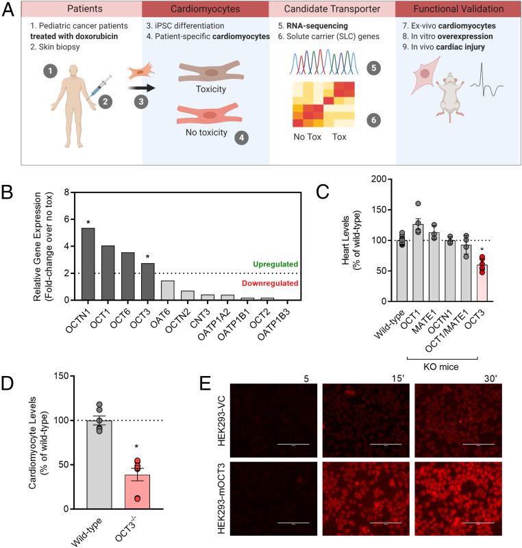
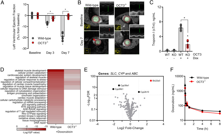
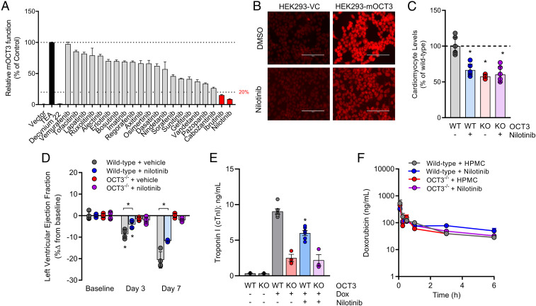
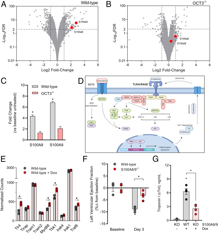
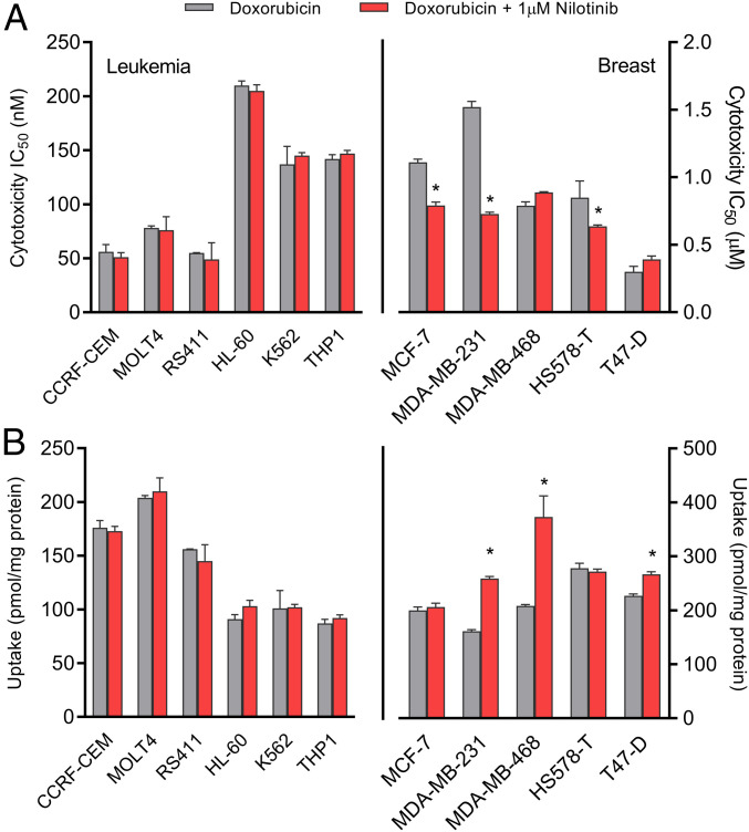
Doxorubicin is a commonly used anticancer agent that can cause debilitating and irreversible cardiac injury. The initiating mechanisms contributing to this side effect remain unknown, and current preventative strategies offer only modest protection. Using stem-cell-derived cardiomyocytes from patients receiving doxorubicin, we probed the transcriptomic landscape of solute carriers and identified organic cation transporter 3 (OCT3) (SLC22A3) as a critical transporter regulating the cardiac accumulation of doxorubicin. Functional validation studies in heterologous overexpression models confirmed that doxorubicin is transported into cardiomyocytes by OCT3 and that deficiency of OCT3 protected mice from acute and chronic doxorubicin-related changes in cardiovascular function and genetic pathways associated with cardiac damage. To provide proof-of-principle and demonstrate translational relevance of this transport mechanism, we identified several pharmacological inhibitors of OCT3, including nilotinib, and found that pharmacological targeting of OCT3 can also preserve cardiovascular function following treatment with doxorubicin without affecting its plasma levels or antitumor effects in multiple models of leukemia and breast cancer. Finally, we identified a previously unrecognized, OCT3-dependent pathway of doxorubicin-induced cardiotoxicity that results in a downstream signaling cascade involving the calcium-binding proteins S100A8 and S100A9. These collective findings not only shed light on the etiology of doxorubicin-induced cardiotoxicity, but also are of potential translational relevance and provide a rationale for the implementation of a targeted intervention strategy to prevent this debilitating side effect.

Keywords: cardiotoxicity; doxorubicin; s100 proteins; slc22a3; solute carriers.


Conflict of interest statement

The authors declare no competing interest.

Figures

Fig. 1.
Fig. 1.
Identification of a doxorubicin uptake transporter. (A) Initial workflow in identifying a doxorubicin uptake transporter from the SLC transportome of pediatric cancer patients receiving doxorubicin-based therapy. (B) Differential gene expression of various SLCs after doxorubicin treatment in hiPSCs-CMs derived from patients who experienced cardiotoxicity and patients who did not experience cardiotoxicity (n = 3 per group) and expressed as a fold change. Statistical analysis was performed using the gene-level differential expression comparison: *P < 0.05; 0.0003, 0.07, 0.43, and 0.04 for OCTN1, OCT1, OCT6, and OCT3, respectively. (C) Relative levels of doxorubicin in heart tissues isolated from wild-type or transporter-deficient mice following a single intravenous injection of doxorubicin at a dose of 5 mg/kg (n = 4 to 6 per group). (D) Ex vivo levels of doxorubicin in cardiomyocytes isolated from wild-type or OCT3-deficient mice (n = 4 to 6 per group). Data presented represent the mean ± SEM and are expressed as a percentage over wild-type (males and females combined). Statistical analysis was performed using an unpaired two-sided Student’s t test with Welch’s correction: *P < 0.05, compared to wild-type values. (E) Fluorescent images illustrating time-dependent accumulation of doxorubicin in HEK293 cells engineered to overexpress a vector control (VC) or mouse OCT3 (mOCT3). (Scale bar: 100 µm.)
Fig. 2.
Fig. 2.
OCT3 deficiency attenuates doxorubicin-induced cardiac injury. (A) Ultrasound evaluation of acute cardiac dysfunction (left ventricular ejection fraction) at baseline, day 3, or day 7 (n = 6 per group). (B) Representative images from cardiac magnetic resonance imaging or ultrasound (Insets) illustrating the ventricular dimensions of the heart. (C) Serum concentrations of cardiac troponin I at day 7 (n = 6 per group). Male wild-type or OCT3-deficient mice received multiple intraperitoneal injections of doxorubicin at a dose of 3 mg/kg consecutively for 7 d (21 mg/kg cumulatively). Data presented represent the mean ± SEM and are expressed as a percentage change compared to baseline values. (D) Categorizing differentially expressed genes using the criteria ≥2 Log10FDR and Log2 fold change of greater and less than positive and negative 2, respectively. Signal transduction pathways were identified using Gene Ontology and KEGG: Kyoto Encyclopedia of Genes and Genomes annotation and analysis with Metascape. (E) Volcano plot of differentially expressed SLC and ABC transporter genes, and CYP metabolizing genes in untreated wild-type or OCT3-deficient mice. Positive fold change indicates higher expression in wild-type mice. Dotted lines indicate a Log10FDR threshold of >2 or Log2FC (fold change) of ±2. RNA-sequencing analysis was performed on cardiomyocytes of naive or doxorubicin-treated wild-type or OCT3-deficient mice (n = 3 per group). (F) Plasma concentration-time profile of doxorubicin in male wild-type or OCT3-defcient mice following a single intraperitoneal dose of 5 mg/kg (n = 6 per group). Statistical analysis was performed using an unpaired two-sided Student’s t test with Welch’s correction: *P < 0.05, compared to baseline or wild-type values or between treatment groups.
Fig. 3.
Fig. 3.
Inhibition of OCT3 with nilotinib preserves cardiovascular function. (A) In vitro identification of nilotinib as a potent inhibitor of mouse OCT3. Tetraethylammonium (TEA) and decynium-22 were used as positive control substrate or inhibitor, respectively (n = 12 per group). (B) Representative fluorescent images of nilotinib-mediated inhibition of doxorubicin uptake in HEK293 cells overexpressing vector control (VC) or mouse OCT3 (mOCT3). (C) Ex vivo levels of doxorubicin in cardiomyocytes isolated from male and female wild-type or OCT3-deficient mice (n = 6 per group) in the presence or absence of nilotinib pretreatment. (D) Ultrasound evaluation of cardiac dysfunction (left ventricular ejection fraction) at baseline, day 3, or day 7 (n = 6 per group). (E) Serum concentrations of cardiac troponin I at day 7 (n = 3 to 6 per group). Male wild-type or OCT3-deficient mice received multiple intraperitoneal injections of doxorubicin at a dose of 3 mg/kg consecutively for 7 d (21 mg/kg cumulatively). Mice were pretreated with vehicle (hydroxypropyl methylcellulose) or nilotinib (15 mg/kg) 30 min before every doxorubicin injection. (F) Plasma concentration-time profile of doxorubicin (5 mg/kg) in male wild-type or OCT3-deficient mice pretreated for 30 min with vehicle or nilotinib (15 mg/kg) (n = 6 per group). Data presented represent the mean ± SEM and are expressed as a percentage change to control, wild-type, or baseline values. Statistical analysis was performed using an unpaired two-sided Student’s t test with Welch’s correction: *P < 0.05, compared to baseline or wild-type values or between treatment groups.
Fig. 4.
Fig. 4.
RNA sequencing reveals OCT3-dependent transcriptional changes. RNA sequencing of heart apexes isolated from male (A) wild-type or (B) OCT3-deficient mice treated with a single intraperitoneal dose (15 mg/kg) of doxorubicin (n = 3 per group). Positive fold change indicates higher expression post doxorubicin treatment in genotype status. Dotted lines indicate a Log10FDR threshold of >2 or Log2FC (fold change) of ±2. (C) Interpolated fold changes in doxorubicin-treated wild-type or OCT3-deficient mice compared to treatment naive mice. (D) Schematic illustrating the proposed mechanism of an OCT3-dependent S100A8/A9 signaling cascade in doxorubicin-induced cardiac injury. (E) Relative expression of genes involved in the TLR4/RAGE-signaling pathway using normalized counts from sequencing data. Values underneath asterisk indicate Log10FDR. (F) Left ejection fraction and (G) serum cardiac troponin I as markers of doxorubicin-induced cardiac toxicity on day 3 following three consecutive doxorubicin injections at a dose of 3 mg/kg (cumulative 9 mg/kg) in dual S100A8/A9 knockout mice (n = 4 per group). All data presented represent the mean ± SEM. Statistical analysis was performed using an unpaired two-sided Student’s t test with Welch’s correction: *P < 0.05, compared to baseline or wild-type values.
Fig. 5.
Fig. 5.
Nilotinib does not influence doxorubicin activity in leukemia or breast cancer. (A) Cytotoxicity of doxorubicin in leukemia (Left) or breast cancer cells (Right) in the presence of absence of nilotinib (1 µM). Cytotoxicity was measured by an MTT assay following continuous 72-h exposure of doxorubicin (n = 12 per group). A nonlinear regression model was used to generate a sigmoidal curve and IC50 values. (B) Uptake of doxorubicin in leukemia (Left) or breast cancer cells (Right) in the presence or absence of nilotinib (1 µM) (n = 6). Uptake data are normalized to protein concentrations. All data presented represent the mean ± SEM. Statistical analysis was performed using a sample Student’s t test: *P < 0.05, compared to doxorubicin alone.

References

    1. Swain S. M., Whaley F. S., Ewer M. S., Congestive heart failure in patients treated with doxorubicin: A retrospective analysis of three trials. Cancer 97, 2869–2879 (2003). - PubMed
    1. Carvalho C., et al. , Doxorubicin: The good, the bad and the ugly effect. Curr. Med. Chem. 16, 3267–3285 (2009). - PubMed
    1. Von Hoff D. D., et al. , Risk factors for doxorubicin-induced congestive heart failure. Ann. Intern. Med. 91, 710–717 (1979). - PubMed
    1. Lionetti V., Ventura C., Regenerative medicine approach to repair the failing heart. Vascul. Pharmacol. 58, 159–163 (2013). - PubMed
    1. Kremer L. C., van Dalen E. C., Offringa M., Ottenkamp J., Voûte P. A., Anthracycline-induced clinical heart failure in a cohort of 607 children: Long-term follow-up study. J. Clin. Oncol. 19, 191–196 (2001). - PubMed

Publication types

MeSH terms

LinkOut - more resources