. 2021 Feb 2;118(5):e2015024118.
doi: 10.1073/pnas.2015024118.

Genome-wide CRISPR screens reveal a specific ligand for the glycan-binding immune checkpoint receptor Siglec-7

Affiliations

Genome-wide CRISPR screens reveal a specific ligand for the glycan-binding immune checkpoint receptor Siglec-7

Simon Wisnovsky et al. Proc Natl Acad Sci U S A. .

Abstract

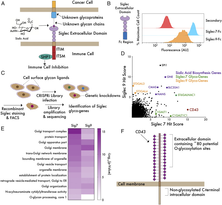
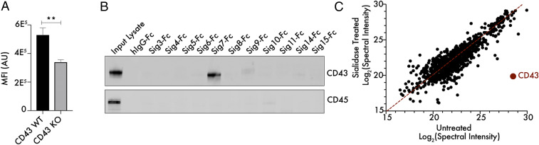
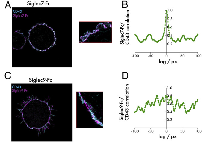
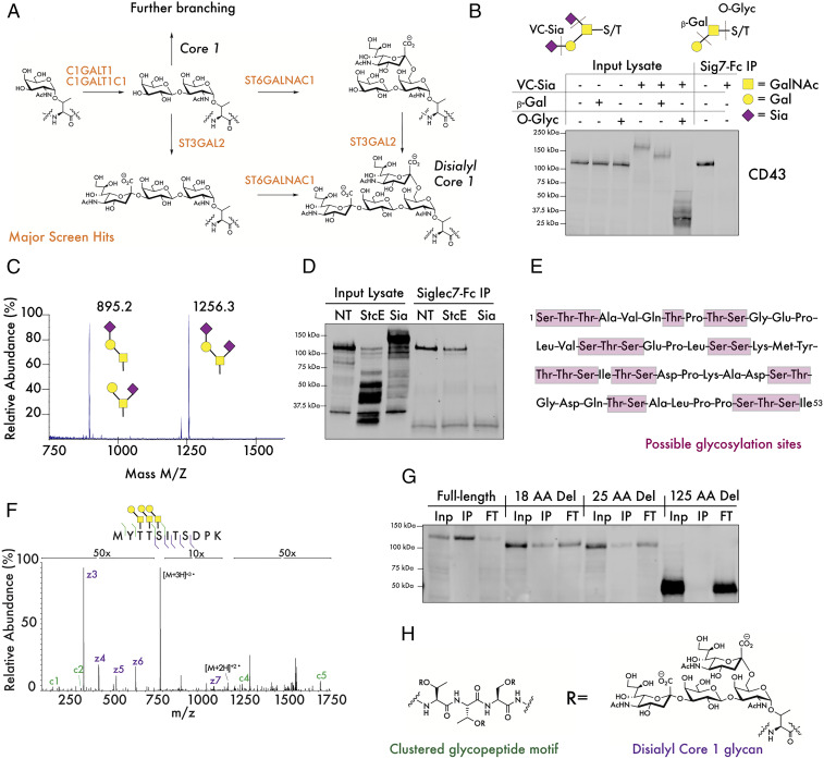
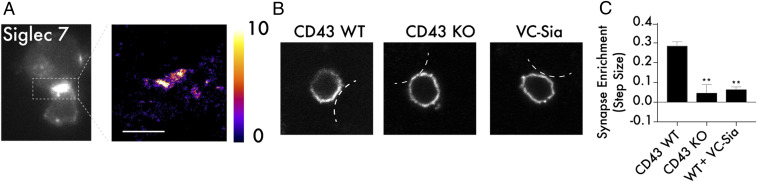
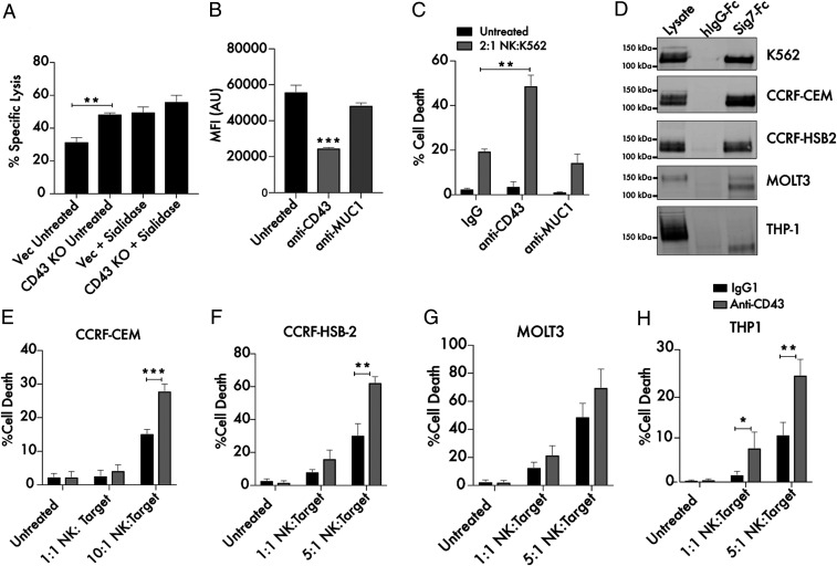
Glyco-immune checkpoint receptors, molecules that inhibit immune cell activity following binding to glycosylated cell-surface antigens, are emerging as attractive targets for cancer immunotherapy. Defining biologically relevant ligands that bind and activate such receptors, however, has historically been a significant challenge. Here, we present a CRISPRi genomic screening strategy that allowed unbiased identification of the key genes required for cell-surface presentation of glycan ligands on leukemia cells that bind the glyco-immune checkpoint receptors Siglec-7 and Siglec-9. This approach revealed a selective interaction between Siglec-7 and the mucin-type glycoprotein CD43. Further work identified a specific N-terminal glycopeptide region of CD43 containing clusters of disialylated O-glycan tetrasaccharides that form specific Siglec-7 binding motifs. Knockout or blockade of CD43 in leukemia cells relieves Siglec-7-mediated inhibition of immune killing activity. This work identifies a potential target for immune checkpoint blockade therapy and represents a generalizable approach to dissection of glycan-receptor interactions in living cells.

Keywords: Tumor Immunology; CRISPR Screening; Glycobiology.


Conflict of interest statement

Competing interest statement: C.R.B. is a cofounder and Scientific Advisory Board member of Palleon Pharmaceuticals, Enable Biosciences, Redwood Bioscience (a subsidiary of Catalent), and InterVenn Biosciences, and a member of the Board of Directors of Eli Lilly and Company. S.W., B.A.H.S., and C.R.B. are co-inventors on a patent application related to this work held by Stanford University (PCT/US2020/041603).

Figures

Fig. 1.
Fig. 1.
A genome-wide CRISPRi screen reveals core genes driving ligand expression for Siglec-7 and Siglec-9. (A) Siglecs possess an extracellular domain that binds to glycan ligands containing sialic acid. Ligand–receptor engagement initiates signaling through intracellular ITIM/ITSM domains, inhibiting immune activation. Sialic acid is incorporated into hundreds of glycan structures on thousands of individual proteins and lipids, making it challenging to define molecular details of ligand–receptor interactions. (B) K562 cells were incubated with 1 μg/mL Siglec-7–Fc and Siglec-9–Fc precomplexed to an Alexa Fluor 488 anti-huIgG secondary antibody and subjected to live cell flow cytometry. “Secondary” indicates noncomplexed Alexa Fluor 488 anti-huIgG. (C) A genome-wide library of ∼104,000 sgRNAs (five sgRNAs/gene) was transduced into K562-dCas9–KRAB cells and incubated with Siglec-7–Fc or Siglec-9–Fc as in B. FACS was performed to isolate a population of cells exhibiting at least a 10-fold reduction in ligand staining. Library amplification and sequencing was then performed to identify sgRNAs enriched in the low-staining population. (D) Hits for Siglec-7 and Siglec-9 were plotted and ranked by hit score (−log10[positive selection score]), where a higher value indicates a stronger enrichment of sgRNAs in the low-staining population. The screen identified a number of sialic acid biosynthesis genes (purple), glycotransferase genes specific for Siglec-7 and Siglec-9 (green and orange), as well as a single-cell surface glycoprotein, CD43, specific for Siglec-7 (red). (E) Enriched GO terms for hits in both screens show importance of Golgi and sialic acid biosynthesis genes. O-glycan biosynthesis is an enriched term in the Siglec-7 but not Siglec-9 screen. (F) The top hit of the Siglec-7 screen, CD43, is a mucin-type O-glycoprotein bearing many sialoglycan modification sites.
Fig. 2.
Fig. 2.
CD43 binds Siglec-7 with unusual selectivity and specificity. (A) K562 CD43 KO cells were generated by CRISPR/Cas9 editing, incubated with 1 μg/mL Siglec-7–Fc/Alexa Fluor 488 anti-huIgG as previously described and subjected to live cell flow cytometry. n = 3, **P < 0.01. MFI indicates mean fluorescence intensity and AU indicates arbitrary units. (B) K562 cells were lysed under nondenaturing conditions, and lysate was passed over magnetic beads functionalized with recombinant Siglec-Fc proteins. The bead-binding protein fraction was eluted and subjected to Western blot to assess the interaction of these Siglecs with the mucin-type glycoproteins CD43 and CD45. (C) K562 cell lysates were either left untreated or treated with 100 nM VC-Sialidase for 1 h and then incubated with magnetic beads functionalized with recombinant Siglec7-Fc. Tryptic digestion and MS/MS-based identification of Siglec-7–binding proteins was then performed, and the total spectral intensity for each interacting protein was calculated.
Fig. 3.
Fig. 3.
Super-resolution imaging reveals selective cell-surface colocalization between CD43 and Siglec-7 ligands. (A) K562 cells were plated onto slides coated with fibronectin and incubated with 1 μg/mL Siglec-7–Fc/Alexa Fluor 647 anti-huIgG (purple) and a CD43-CF568 antibody (blue). An analysis of colocalization was then performed using super-resolution microscopy. (B) A correlation analysis assessing colocalization of Siglec-7–Fc and CD43 signal on the cell surface was performed. (C) The same experiment as in A was performed with Siglec-9–Fc. (D) The same analysis as in B was performed for Siglec-9–Fc. The total edge length of the images is 40.96 microns. One px corresponds to the bin width of the two-dimensional histogram used for the reconstruction, that is, 32 nm.
Fig. 4.
Fig. 4.
Siglec-7 binds a specific glycopeptide motif at the N terminus of CD43. (A) The synthesis of the disialyl core 1 tetrasaccharide is catalyzed by enzymes that were significantly enriched hits in the Siglec-7 screen. (B) K562 lysates were digested with the indicated combinations of sialidase from Vibrio Cholerae (VC-Sia), bovine β1-3,4–Galactosidase (β-Gal), and O-Glycosidase from S. pneumoniae (O-Glyc). The digestion specificities of each of these enzymes are indicated, with the dotted lines indicating the preferred enzymatic cleavage point. GalNAc = N-Acetylgalactosamine, Gal = galactose, and Sia = sialic acid. (C) CD43 was isolated from CD43-myc–FLAG-expressing cells and subjected to glycomic analysis through β-elimination and liquid chromatography/MS. (D) K562 lysate was treated with 100 ng/mL StcE or 100 nM VC-Sia for 30 min at 37 °C, passed over magnetic beads functionalized with recombinant Siglec-7–Fc, and subjected to Western blot with an antibody binding the nonglycosylated CD43 intracellular domain. (E) The N terminus of CD43 contains repeated clusters of serine/threonine residues that can serve as points for glycan attachment. (F) CD43-myc–FLAG was isolated and digested with VC-Sia, GluC, and trypsin. Intact glycopeptides were subsequently analyzed by MS/MS. A representative electron transfer dissociation spectrum of a glycopeptide from the N terminus of CD43 with indicated glycans attached to adjacent residues is shown. (G) K562 cells expressing CD43-myc–FLAG constructs exhibiting truncations of the indicated number of amino acids (AA) from the N terminus of the CD43 extracellular domain were lysed, passed over Siglec-7–Fc-functionalized beads, and subjected to analysis by Western blot with an anti-myc antibody. Inp = input lysate, IP = bead-binding fraction, FT = flow-through. (H) The putative Siglec-7 binding structure on CD43.
Fig. 5.
Fig. 5.
CD43 drives Siglec-7 recruitment to the immunological synapse. (A) Primary NK cells and K562 cells were cocultured at a 2:1 ratio, fixed in 4% paraformaldehyde, and stained with a fluorescent antibody against Siglec-7. Super-resolution microscopy was performed to assess recruitment of Siglec-7 to the immune synapse. The scale bar corresponds to 2 microns and the color bar corresponds to number of localizations per bin. (B) Target cell expression of CD43 is required for recruitment of Siglec-7 to the immune synapse. Primary NK cells and K562 cells that were either CD43 WT, CD43 KO, or treated with VC-Sia at 100 nM for 30 min were prepared as in A, and normal resolution confocal microscopy was used to capture the distribution of Siglec-7 along the NK cell membrane at NK cell/target cell contact points. For simplicity, the membrane of the target cell is indicated with white lines in each image. (C) A synaptic enrichment algorithm was used to quantitate the level of Siglec-7 synaptic recruitment over a series of images. A value above 0 indicates higher abundance inside the synapse than outside, and a value below 0 indicates lower abundance inside the synapse than outside. n = 3, and error bars indicate SEM. **P < 0.01.
Fig. 6.
Fig. 6.
Targeting CD43 enhances killing of leukemia cells by primary NK cells. (A) Primary NK cells and K562 Vec (empty vector transfected) or CD43 KO target cells were cocultured for 4 h at a 2:1 ratio and subjected to flow cytometry using live cell (CellTrace Far Red) and dead cell (Sytox Green) stains to quantitate specific lysis of the target cells. The experiment was also performed following treatment of both Vec and KO target cells with 2 μM endotoxin-free sialidase from Salmonella Typhimurium. (B) A total of 104 K562 cells were treated with 10 μg/mL anti-CD43 (MEM-59) or anti-Muc1 (VU4H5) antibodies for 30 min, incubated with 100 ng/mL Siglec-7–Fc/Alexa Fluor 488, and subjected to flow cytometry. MFI indicates mean fluorescence intensity, and AU indicates arbitrary units. (C) K562 cells were treated for 30 min with 10 μg/mL isotype control, anti-CD43, and anti-MUC1 antibodies. Cells were then cocultured with primary NK cells at the indicated ratio as in A. The “Untreated” condition refers to target cells that have not been incubated with NK cells. (D) Lysates from the indicated leukemia and lymphoma cell lines were passed over Siglec-7–Fc functionalized beads. Bound proteins were eluted from the beads and subjected to Western blot to assess the presence of a Siglec-7–binding CD43 glycoform. Beads conjugated to huFc were used as a control. A total of 25 μg protein was loaded in the input lysate, while 500 μg protein was used for each IP. (E) CCRF-CEM cells, (F) CCRF-HSB2 cells, (G) MOLT3 cells, and (H) THP-1 cells were treated for 30 min with 10 μg/mL isotype control or anti-CD43 mouse IgG1 antibody and cocultured with primary NK cells at the indicated ratios as in A. n = 3 in all cases. *P < 0.05, **P < 0.01, ***P < 0.001. Error bars indicate SEM.

References

    1. Waldman A. D., Fritz J. M., Lenardo M. J., A guide to cancer immunotherapy: From T cell basic science to clinical practice. Nat. Rev. Immunol. 20, 651–668 (2020). - PMC - PubMed
    1. Seidel J. A., Otsuka A., Kabashima K., Anti-PD-1 and anti-CTLA-4 therapies in cancer: Mechanisms of action, efficacy, and limitations. Front. Oncol. 8, 86 (2018). - PMC - PubMed
    1. Tang J., et al. , Trial watch: The clinical trial landscape for PD1/PDL1 immune checkpoint inhibitors. Nat. Rev. Drug Discov. 17, 854–855 (2018). - PubMed
    1. Wolchok J. D., et al. , Overall survival with combined nivolumab and ipilimumab in advanced melanoma. N. Engl. J. Med. 377, 1345–1356 (2017). - PMC - PubMed
    1. Muñoz-Wolf N., Lavelle E. C., Innate immune receptors. Methods Mol. Biol. 1417, 1–43 (2016). - PubMed

Publication types

MeSH terms