. 2021 Jan 25;12(1):583.
doi: 10.1038/s41467-020-20825-6.

A conformation-selective monoclonal antibody against a small molecule-stabilised signalling-deficient form of TNF

Affiliations

A conformation-selective monoclonal antibody against a small molecule-stabilised signalling-deficient form of TNF

Daniel J Lightwood et al. Nat Commun. .

Abstract

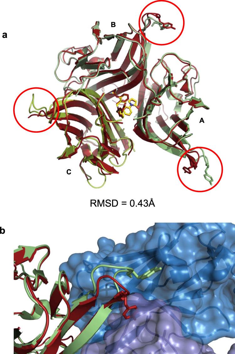
We have recently described the development of a series of small-molecule inhibitors of human tumour necrosis factor (TNF) that stabilise an open, asymmetric, signalling-deficient form of the soluble TNF trimer. Here, we describe the generation, characterisation, and utility of a monoclonal antibody that selectively binds with high affinity to the asymmetric TNF trimer-small molecule complex. The antibody helps to define the molecular dynamics of the apo TNF trimer, reveals the mode of action and specificity of the small molecule inhibitors, acts as a chaperone in solving the human TNF-TNFR1 complex crystal structure, and facilitates the measurement of small molecule target occupancy in complex biological samples. We believe this work defines a role for monoclonal antibodies as tools to facilitate the discovery and development of small-molecule inhibitors of protein-protein interactions.


Conflict of interest statement

D.J.L., J.P., D.M., B.C., A.T., A.M., T.C., T.B., J.O’C., and A.D.G.L. are/were all employees of UCB Pharma and may hold stock and/or stock options. A.S-T., E.S.H., and A.S. are/were employees of UCB Pharma.

Figures

Fig. 1
Fig. 1. CA1974 binding to human TNF in complex with small molecule inhibitors.
a TNF–small molecule (UCB-8733 and UCB-9260) complex or apo TNF (blue) were captured via TNFR1 which was pre-coated onto a 384-well ELISA plate. CA1974 Fab at a range of concentrations was then added to the plate to detect TNF–small molecule complex. Comparisons are presented as ratios of geometric means (points) and 95% confidence intervals for the ratios (error bars), i.e., Comparison of TNF-UCB-8733 complex and TNF-UCB-9260 (blue); Comparison of TNF-UCB-8733 complex and apo TNF (red); Comparison of TNF-UCB-9260 complex and apo TNF (green). b Human TNF-UCB-9260 complex at 25 ng/ml (blue), 250 ng/ml (green) or apo TNF (250 ng/ml) with DMSO (0.4%) (red) was added to HEK-293 Jump In cells expressing human TNFR1. CA1974 IgG at 10 µg/ml was used to detect TNF–small molecule complex bound to the surface of cells using flow cytometry. The binding was revealed using a goat anti-mouse IgG Alexa Fluor 488 secondary antibody. Data is shown for cells present in a gate which defines viable single cells and excludes doublets and other cell debris (Supplementary Fig. 1). Cell count (y-axis) is normalised to mode. Source data are provided as a Source Data file.
Fig. 2
Fig. 2. Stoichiometry assessment of the CA1974-TNF–small molecule complex.
A Fab fragment of CA1974 was incubated at various molar-ratios with TNF–UCB-9260 small molecule complex and then analysed by size-exclusion-HPLC. The appearance of only a single high-molecular-weight peak (at approx. 13.5 min) suggests the stoichiometry was 1 Fab: 1 TNF trimer. Source data are provided as a Source Data file.
Fig. 3
Fig. 3. Structure of CA1974 Fab-human TNF-UCB-8733-TNFR1 complex.
a Trimeric human TNF (green ribbons, monomers assigned A, B, & C) with UCB-8733 bound (yellow sticks), in complex with CA1974 Fab (blue surface rendered) and two copies of human TNFR1 (pink surface rendered), viewed from the top and side (left and right images respectively). b Detailed view of the CA1974 Fab-human TNF (with UCB-8733 bound) interface viewed from the top and side (left and right images respectively). c Detailed view of a structural model where compound-bound human TNF has been replaced by apo human TNF (brown ribbons) viewed from the top and side (left and right images respectively). This highlights side-chain clashes between the CA1974 Fab and symmetric apo TNF.
Fig. 4
Fig. 4. Comparison of human TNF-UCB-8733 free and antibody-complexed structures.
a Alignment of human TNF from the compound (UCB-8733)-only structure (red ribbons) with human TNF (plus UCB-8733) from the CA1974 Fab-human TNF-UCB-8733-TNFR1 complex structure (green ribbons) (Fab and receptors have been removed for clarity). Monomers A, B, and C are labelled. Red circles highlight a region of flexibility containing Tyr87 (sticks) on each monomer. b Detail showing the change in position of Tyr87 (sticks) in monomer A when Fab CA1974 is bound (green ribbon) compared to its position in the human TNF UCB-8733-only structure (red ribbon). Further details of residue movements are shown in Supplementary Table 2.
Fig. 5
Fig. 5. Detail of the epitope of CA1974 Fab.
a Complex of human TNF (green surface rendered), CA1974 Fab (blue ribbons), and human TNFR1 (pink ribbons) with epitope residues highlighted in red. b Head-on view of the CA1974 Fab epitope spanning monomers A & C on human TNF (highlighted in red). For clarity CA1974 Fab has been removed and monomers A and C have been labelled. c CA1974 Fab epitope with residues that vary in mouse TNF marked in blue. d Equivalent view with residues that vary in cynomolgus monkey TNF marked in blue. Residues that vary between the species are highlighted in Supplementary Table 2.
Fig. 6
Fig. 6. CA1974 represents a suitable reagent for measuring target occupancy.
CA1974 was used as a capture reagent to measure TNF complexed with UCB-8733 small-molecule inhibitor using a high-sensitivity ELISA, with a commercial anti-TNF antibody as the detection reagent. TNF pre-incubated with either excess compound or DMSO was spiked into neat human plasma (depleted of endogenous TNF) and diluted to various concentrations and used in the assay. TNF–small molecule complex signal (red) was significantly higher than apo–TNF background (blue), with probability of at least 0.999 at TNF concentrations of 25 pg/ml and above. Data from representative experiment. Source data from this and other experiments showing utility of CA1974 in measuring target occupancy are provided as a Source Data file.

References

    1. Kalliolias GD, Ivashkiv LB. TNF biology, pathogenic mechanisms and emerging therapeutic strategies. Nat. Rev. Rheumatol. 2016;12:49–62. doi: 10.1038/nrrheum.2015.169. - DOI - PMC - PubMed
    1. Locksley RM, Killeen N, Lenardo MJ. The TNF and TNF receptor superfamilies: integrating mammalian biology. Cell. 2001;104:487–501. doi: 10.1016/S0092-8674(01)00237-9. - DOI - PubMed
    1. Grell M, et al. The transmembrane form of tumor necrosis factor is the prime activating ligand of the 80 kDa tumor necrosis factor receptor. Cell. 1995;83:793–802. doi: 10.1016/0092-8674(95)90192-2. - DOI - PubMed
    1. Probert L. TNF and its receptors in the CNS: The essential, the desirable and the deleterious effects. Neuroscience. 2015;302:2–22. doi: 10.1016/j.neuroscience.2015.06.038. - DOI - PubMed
    1. O’Connell J, et al. Small molecules that inhibit TNF signalling by stabilising an asymmetric form of the trimer. Nat. Commun. 2019;10:5795. doi: 10.1038/s41467-019-13616-1. - DOI - PMC - PubMed

Publication types

MeSH terms