Review
. 2021 Jan 26;7(1):6.
doi: 10.1038/s41421-020-00237-y.

Emerging functions of branched ubiquitin chains

Affiliations
Review

Emerging functions of branched ubiquitin chains

Michael E French et al. Cell Discov. .

Abstract

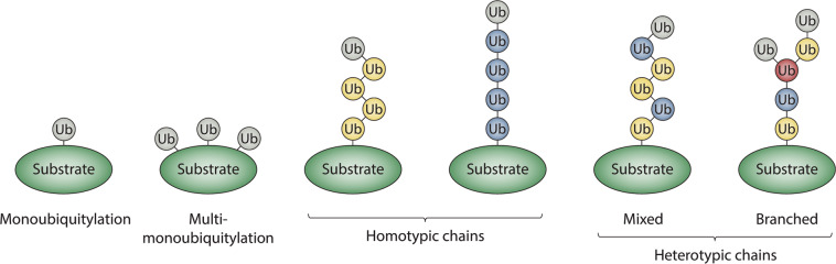
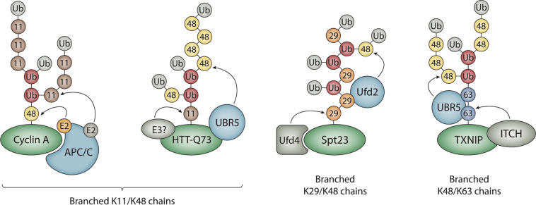
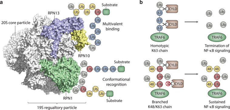
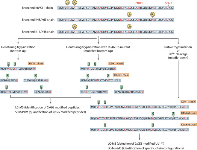
Ubiquitylation is a critical post-translational modification that controls a wide variety of processes in eukaryotes. Ubiquitin chains of different topologies are specialized for different cellular functions and control the stability, activity, interaction properties, and localization of many different proteins. Recent work has highlighted a role for branched ubiquitin chains in the regulation of cell signaling and protein degradation pathways. Similar to their unbranched counterparts, branched ubiquitin chains are remarkably diverse in terms of their chemical linkages, structures, and the biological information they transmit. In this review, we discuss emerging themes related to the architecture, synthesis, and functions of branched ubiquitin chains. We also describe methodologies that have recently been developed to identify and decode the functions of these branched polymers.


Conflict of interest statement

The authors declare that they have no conflict of interest.

Figures

Fig. 1
Fig. 1. Classification of ubiquitin modifications.
Protein substrates can be modified with ubiquitin monomers on one or more acceptor sites, referred to as monoubiquitylation or multi-monoubiquitylation, respectively. Alternatively, ubiquitin monomers can be joined to each other via isopeptide bonds to form chains of varying lengths, linkages, and structures. Homotypic chains are linked uniformly through the same acceptor site of ubiquitin (e.g., K48-linked chains), whereas heterotypic chains contain multiple types of linkages and can be further classified as either mixed or branched. Mixed chains consist of ubiquitin subunits that are modified on only a single acceptor site. Branched chains contain at least one ubiquitin subunit that is simultaneously modified on multiple acceptor sites. Ubiquitins modified on one acceptor site are colored in blue or yellow; the branch point ubiquitin is colored in red; unmodified or “terminal” ubiquitins are colored gray.
Fig. 2
Fig. 2. Architecture and synthesis of branched ubiquitin chains.
The APC/C collaborates with two different E2s, UBE2C and UBE2S, to assemble branched K11/K48 chains on cyclin A and other mitotic substrates. UBR5 collaborates with an unknown K11-specific E3 to form branched K11/K48 chains of a different architecture on a pathological mutant version of the Huntingtin protein (HTT-Q73). UBR5 has also been reported to synthesize branched K11/K48 chains on newly synthesized misfolded polypeptides. Spt23 and substrates of the ubiquitin fusion degradation pathway are modified with branched K29/K48 chains synthesized by Ufd4 and Ufd2. ITCH cooperates with UBR5 to assemble branched K48/K63 chains on the pro-apoptotic regulator TXNIP. Substrates are colored in green; chain branching E3s are colored in light blue; the color coding for ubiquitins is as described in Fig. 1.
Fig. 3
Fig. 3. Models for the recognition and functions of branched ubiquitin chains.
a The binding of branched chains to the proteasome (PDB ID: 5T0J) is illustrated schematically. The ubiquitin-binding subunits of the 19S regulatory particle are colored in blue, yellow, and green; all other proteasome subunits are colored in white. The enhanced binding of branched chains to the proteasome as a result of an increase in the local concentration or “density” of ubiquitin subunits surrounding the substrate is illustrated by the multivalent-binding model. Enhanced binding due to the recognition of novel interaction surfaces created by branching or recognition of the branch point itself is represented by the conformational recognition model. Non-covalent interactions between ubiquitin and proteasome subunits are represented by arcs. The positions of the ubiquitin-binding sites on the proteasome are shown for schematic purposes only. b Model for the role of branched K48/K63 chains in the activation of NF-κB signaling. Homotypic K63-linked chains are efficiently disassembled by CYLD, resulting in the removal of K63 linkages from TRAF6 and the termination of NF-κB signaling (top). Branched K48/K63 chains are resistant to CYLD cleavage, resulting in the persistence of K63 linkages on TRAF6 and sustained activation of NF-κB signaling (bottom). Branched ubiquitin subunits modified at both K48 and K63 are colored in red.
Fig. 4
Fig. 4. Mass spectrometry-based workflows to detect branched ubiquitin chains.
Three different approaches to detect branched chains are illustrated. In the classical bottom-up approach, branched chains involving neighboring lysines can be detected, but all other types of branched chains are invisible due to the cleavage of branched peptides at intervening lysines or arginines (left). Mutation of the single arginine located between K48 and K63 of ubiquitin (Arg54) allows the detection of branched K48/K63 chains using classical bottom-up methods (middle). In principle, other types of branched chains can be detected in this manner. In the middle-down approach, ubiquitin chains are digested with trypsin under native conditions or cleaved after Arg74 with a site-specific protease (right). This approach leaves all possible combinations of branch points intact. Specific chain configurations can then be identified by tandem mass spectrometry. LC–MS liquid chromatography-mass spectrometry, SRM selected reaction monitoring, PRM parallel reaction monitoring, LC–MS/MS liquid chromatography-tandem mass spectrometry.

References

    1. Rape M. Ubiquitylation at the crossroads of development and disease. Nat. Rev. Mol. Cell Biol. 2018;19:59–70. doi: 10.1038/nrm.2017.83. - DOI - PubMed
    1. Pohl C, Dikic I. Cellular quality control by the ubiquitin-proteasome system and autophagy. Science. 2019;366:818–822. doi: 10.1126/science.aax3769. - DOI - PubMed
    1. Oh E, Akopian D, Rape M. Principles of ubiquitin-dependent signaling. Annu. Rev. Cell Dev. Biol. 2018;34:137–162. doi: 10.1146/annurev-cellbio-100617-062802. - DOI - PubMed
    1. Schwertman P, Bekker-Jensen S, Mailand N. Regulation of DNA double-strand break repair by ubiquitin and ubiquitin-like modifiers. Nat. Rev. Mol. Cell Biol. 2016;17:379–394. doi: 10.1038/nrm.2016.58. - DOI - PubMed
    1. Foot N, Henshall T, Kumar S. Ubiquitination and the regulation of membrane proteins. Physiol. Rev. 2017;97:253–281. doi: 10.1152/physrev.00012.2016. - DOI - PubMed

LinkOut - more resources