. 2021 Jan 25;12(1):562.
doi: 10.1038/s41467-020-20858-x.

Dual mechanism β-amino acid polymers promoting cell adhesion

Affiliations

Dual mechanism β-amino acid polymers promoting cell adhesion

Qi Chen et al. Nat Commun. .

Abstract

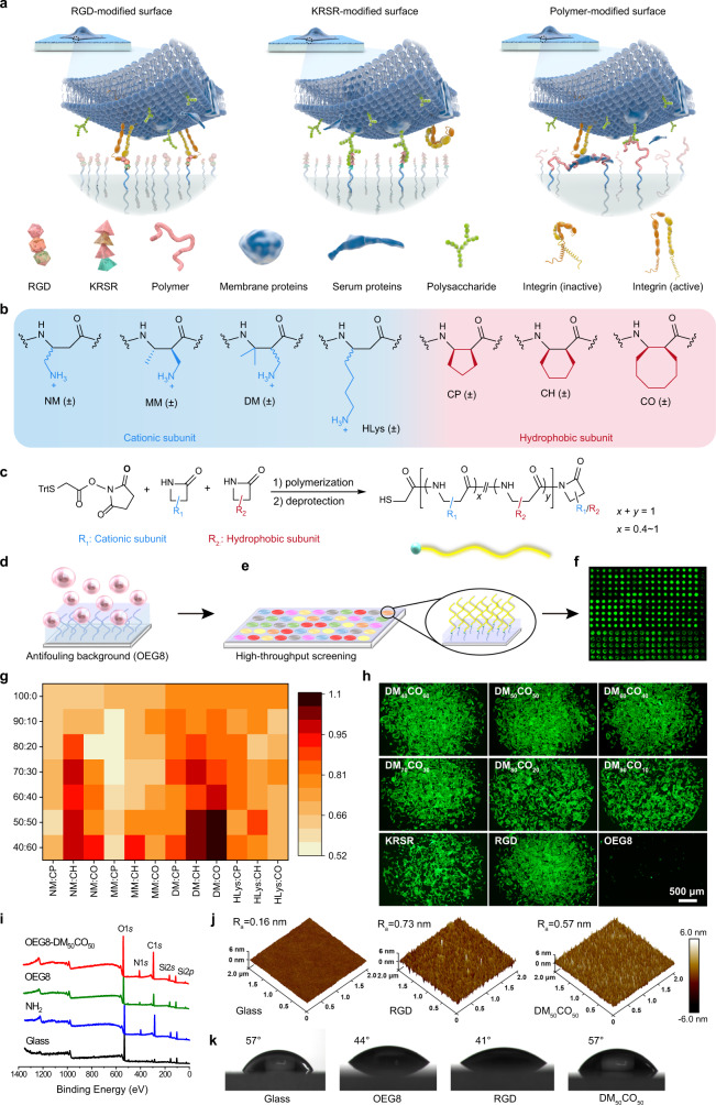
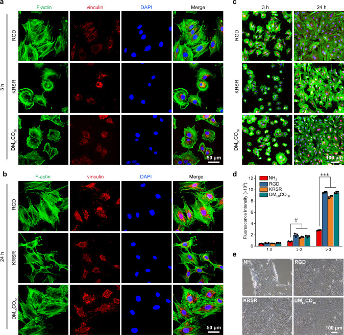
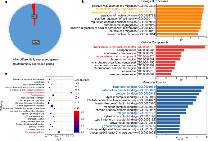
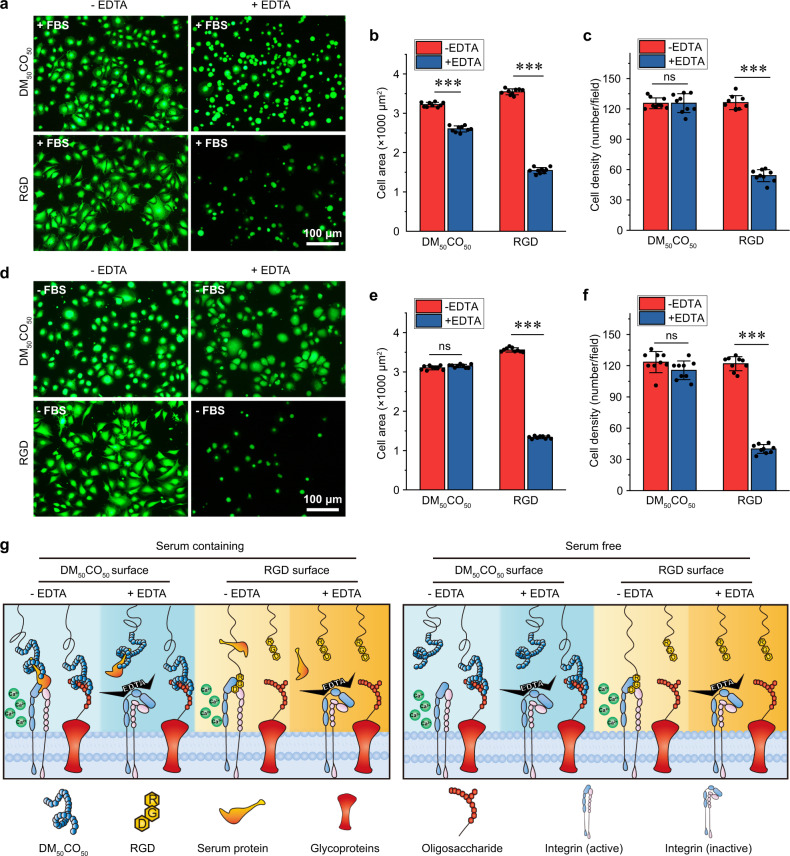
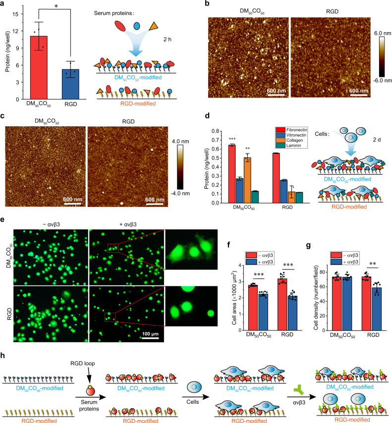
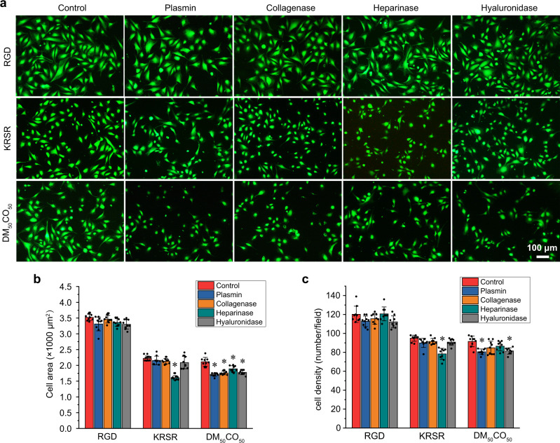
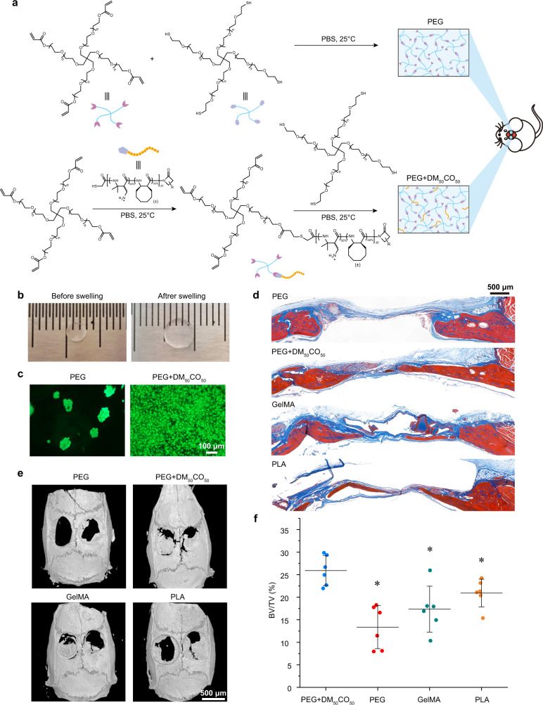
Cell adhesion has tremendous impact on the function of culture platforms and implants. Cell-adhesive proteins and peptides have been extensively used for decades to promote cell adhesion, however, their application suffers from their easy enzymatic degradation, difficulty in large-scale preparation and expensiveness. To develop the next-generation cell-adhesive materials, we mimic the cell adhesion functions and mechanisms of RGD and KRSR peptides and design cell-adhesive cationic-hydrophobic amphiphilic β-amino acid polymers that are stable upon proteolysis and easily prepared in large scale at low cost. The optimal polymer strongly promotes cell adhesion, using preosteoblast cell as a model, by following dual mechanisms that are independent of sequence and chirality of the statistic copolymer. Our strategy opens avenues in designing the next-generation cell-adhesive materials and may guide future studies and applications.


Conflict of interest statement

R.L. and Q.C. are co-inventors on a patent covering the function of β-amino acid polymers presented in this report. The remaining authors declare no competing interests.

Figures

Fig. 1
Fig. 1. High-throughput screening of β-amino acid polymers for preosteoblast cell adhesion.
a Cell adhesion mechanisms of RGD, KRSR, and β-amino acid polymer-modified surfaces. b Subunits to compose β-amino acid polymers. All subunits are racemic and the resulting polymers are heterochiral. c The general synthesis of the 76 β-peptide polymers with a chain length of 20 amino acid residue. d An OEG8 antifouling layer was used to resist protein adsorption and non-specific cell adhesion to the substrate. e Thiol-terminated polymers were covalently attached to maleimide-functionalized glass surface for high-throughput screening of cell adhesion, using RGD (RGDSPC) and KRSR (KRSRGYC) peptide-modified surfaces for comparison. f High-throughput screening of cell adhesion evaluated by fluorescence scanning and quantification on live/dead stained cells, calcein AM (green) for live cells. g Heat map showing the relationship of preosteoblast adhesion vs. polymer composition. The fluorescence intensity was normalized with RGD-modified surface. h Micrograph of F-actin (green) stained preosteoblast cell adhesion to surfaces modified with DMxCOy (x + y = 100, x = 40, 50, 60, 70, 80, 90), RGD, KRSR, and OEG8, respectively. Scale bar: 500 μm. i XPS spectra of bare glass, NH2-glass, OEG8, and DM50CO50-modified surfaces. j 3D AFM images of bare glass, RGD, and DM50CO50-modified surfaces. k Water contact angle of bare glass, OEG8, RGD, and DM50CO50-modified surfaces.
Fig. 2
Fig. 2. Adhesion and proliferation of preosteoblast cells.
ac Fluorescence confocal microscope images (green, actin; red, vinculin; blue, nucleus) of preosteoblast cells on RGD, KRSR, and DM50CO50-modified surfaces after cell seeding for 3 h (a) and 24 h (b), Scale bar: 50 μm. Lower magnification images (c) after cell seeding for 3 and 24 h, Scale bar: 100 μm. d Cell proliferation after 1, 3, and 5 days of culture. Quantification of cell density was done with Alamar Blue assay. e Bright field images of preosteoblast cells cultured on NH2, RGD, KRSR, and DM50CO50-modified surfaces for 5 days. Scale bar: 100 μm. Data (in d) represent mean ± s.d. (n = 3). Statistical analysis: one-way ANOVA with Tukey post-test, #p < 0.05, ***p < 0.0001.
Fig. 3
Fig. 3. RNA-seq analysis on DM50CO50-modified surface compared to RGD-modified surface.
a From 17,267 gene transcripts in analysis, 305 differentially expressed genes showed greater than 50% change in expression level after 2 days of culture. b Significant GO terms of associated biological processes, cellular components, and molecular functions from differentially regulated genes. c KEGG pathway enrichment. The y-axis label represents KEGG pathway, and the x-axis shows -lgP. The size of the dots represents the number of gene enrichment.
Fig. 4
Fig. 4. Cells adhesion via both integrin-dependent and -independent mechanisms.
a Fluorescent micrographs of live/dead stained cells, b cell area analysis, and c cell density analysis, of cell adhesion to DM50CO50 and RGD-modified surfaces in serum-containing media for 3 h, with or without EDTA treatment. d Fluorescent micrographs, e cell area analysis, and f cell density analysis, of cell adhesion to DM50CO50 and RGD-modified surfaces in serum-free media for 3 h, with or without EDTA treatment. g Cell adhesion mechanisms of DM50CO50 and RGD-modified surfaces in different conditions. Data in (b, c, e, f) represent mean ± s.d. (n = 9). Statistical analysis: two-tailed t-test, ***p < 0.0001, ns = not significant.
Fig. 5
Fig. 5. Effect of adsorbed serum proteins on preosteoblast cell adhesion to DM50CO50-modified surfaces.
a The amount of surface-adsorbed total protein after variable surfaces were incubated with a serum-containing cell culture medium for 2 h. b, c AFM images of DM50CO50 and RGD-modified surfaces after incubation with serum-containing cell culture medium (b) and pure fibronectin (c) for 2 h. d The amount of surface-adsorbed individual protein (fibronectin, vitronectin, collagen, laminin) on DM50CO50 and RGD-modified surfaces after cells were cultured on the surfaces for 2 days. Fluorescence micrographs of: e live/dead stained cells, f spreading area, and g density analysis, of cells 2 h after seeding on DM50CO50 and RGD-modified surfaces, which were first incubated with serum-containing medium and then treated with or without integrin αvβ3 before cell seeding. h Carton illustration of results in (eg). Data in (a, d, f, g) represent mean ± s.d. (n = 3 for a, d and n = 9 for f, g). Statistical analysis: two-tailed t-test, **p < 0.001, ***p < 0.0001.
Fig. 6
Fig. 6. Effect of membrane proteins and polysaccharides on preosteoblast cell adhesion.
Fluorescent micrographs of: a live/dead stained cells, b spreading area, and c density analysis, of cells adhered to RGD, KRSR, and DM50CO50-modified surfaces for 1 day in a serum-free medium. Cells were treated with plasmin, collagenase, heparinase, and hyaluronidase, respectively, before seeded on the surfaces. Untreated cells were used as the control. Data in (b, c) represent mean ± s.d. (n = 9). Statistical analysis: one-way ANOVA with Tukey post-test, *p < 0.01.
Fig. 7
Fig. 7. Demonstration of cell adhesion impact of DM50CO50-incorporated hydrogel on in vivo bone regeneration using a critical-size rat calvarial defect model.
a Synthesis of bare PEG hydrogel and DM50CO50-modified PEG hydrogel. b Comparison before and after PEG hydrogel swelling. c Preosteoblast cells adhesion to bare PEG hydrogel and DM50CO50-modified PEG hydrogel after cell seeding for 2 days. Cells were subjected to live/dead staining. d Masson’s trichrome stain, e 3D micro-CT analysis, and f bone volume analysis, of the cranial bone samples from bare PEG hydrogels, DM50CO50-modified PEG hydrogels, GelMA, and PLA after 8 weeks of implantation. Data in (f) represent mean ± s.d. (n = 6). Statistical analysis: two-tailed t-test, *p < 0.05.

References

    1. Changede R, Cai H, Wind SJ, Sheetz MP. Integrin nanoclusters can bridge thin matrix fibres to form cell-matrix adhesions. Nat. Mater. 2019;18:1366–1375. doi: 10.1038/s41563-019-0460-y. - DOI - PMC - PubMed
    1. Kanchanawong P, et al. Nanoscale architecture of integrin-based cell adhesions. Nature. 2010;468:580–584. doi: 10.1038/nature09621. - DOI - PMC - PubMed
    1. Li W, Yan Z, Ren J, Qu X. Manipulating cell fate: dynamic control of cell behaviors on functional platforms. Chem. Soc. Rev. 2018;47:8639–8684. doi: 10.1039/C8CS00053K. - DOI - PubMed
    1. Bauer S, Schmuki P, von der Mark K, Park J. Engineering biocompatible implant surfaces. Prog. Mater. Sci. 2013;58:261–326. doi: 10.1016/j.pmatsci.2012.09.001. - DOI
    1. Chen S, et al. Tuning surface properties of bone biomaterials to manipulate osteoblastic cell adhesion and the signaling pathways for the enhancement of early osseointegration. Colloids Surf. B. 2018;164:58–69. doi: 10.1016/j.colsurfb.2018.01.022. - DOI - PubMed

Publication types

MeSH terms