. 2021 Jan 25;11(1):2160.
doi: 10.1038/s41598-021-81669-8.

Novel myostatin-specific antibody enhances muscle strength in muscle disease models

Affiliations

Novel myostatin-specific antibody enhances muscle strength in muscle disease models

Hiroyasu Muramatsu et al. Sci Rep. .

Abstract

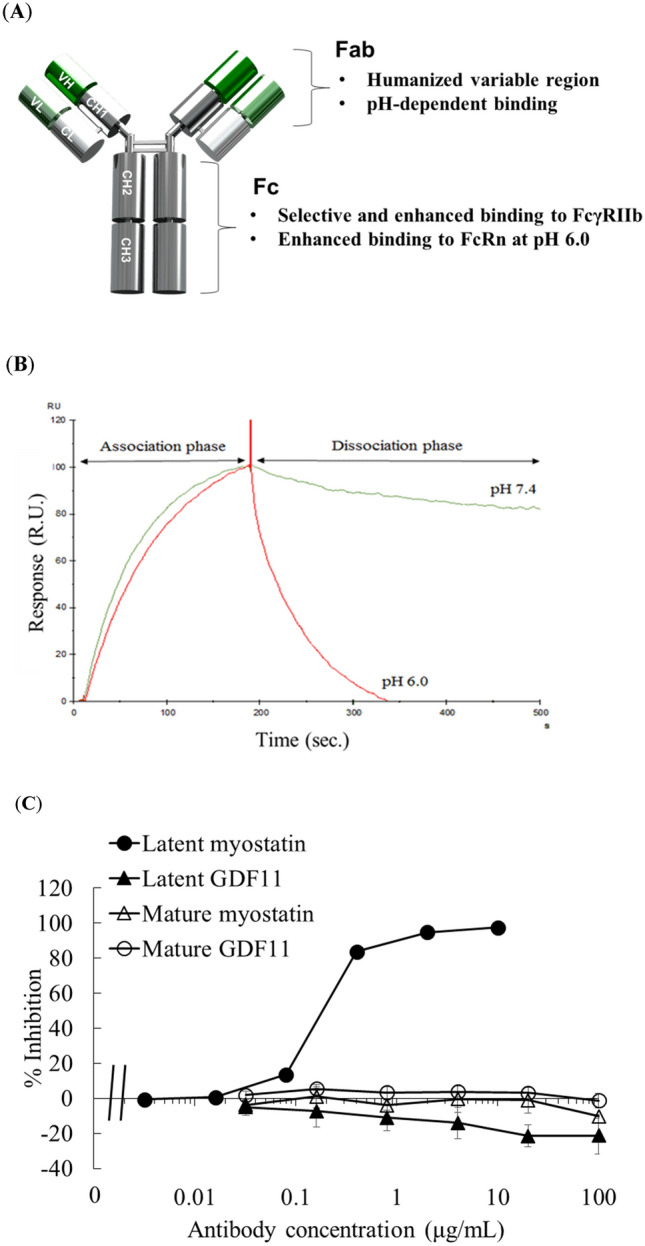
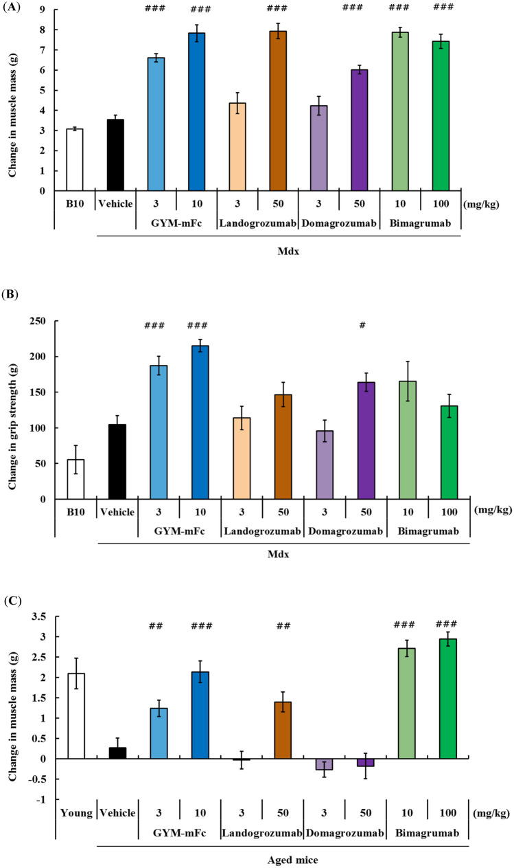
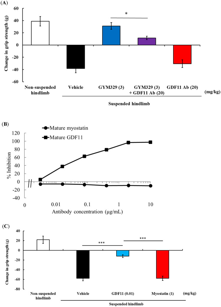
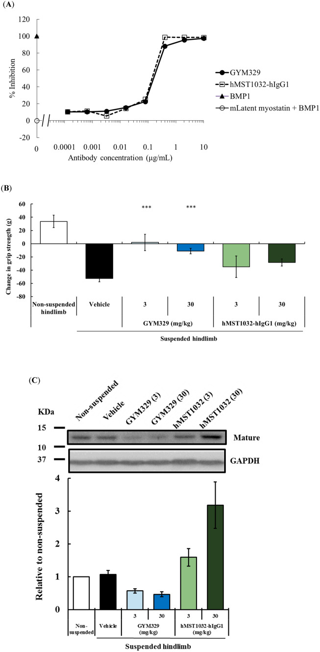
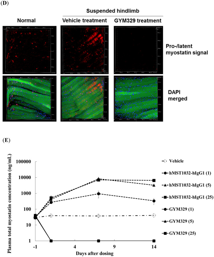
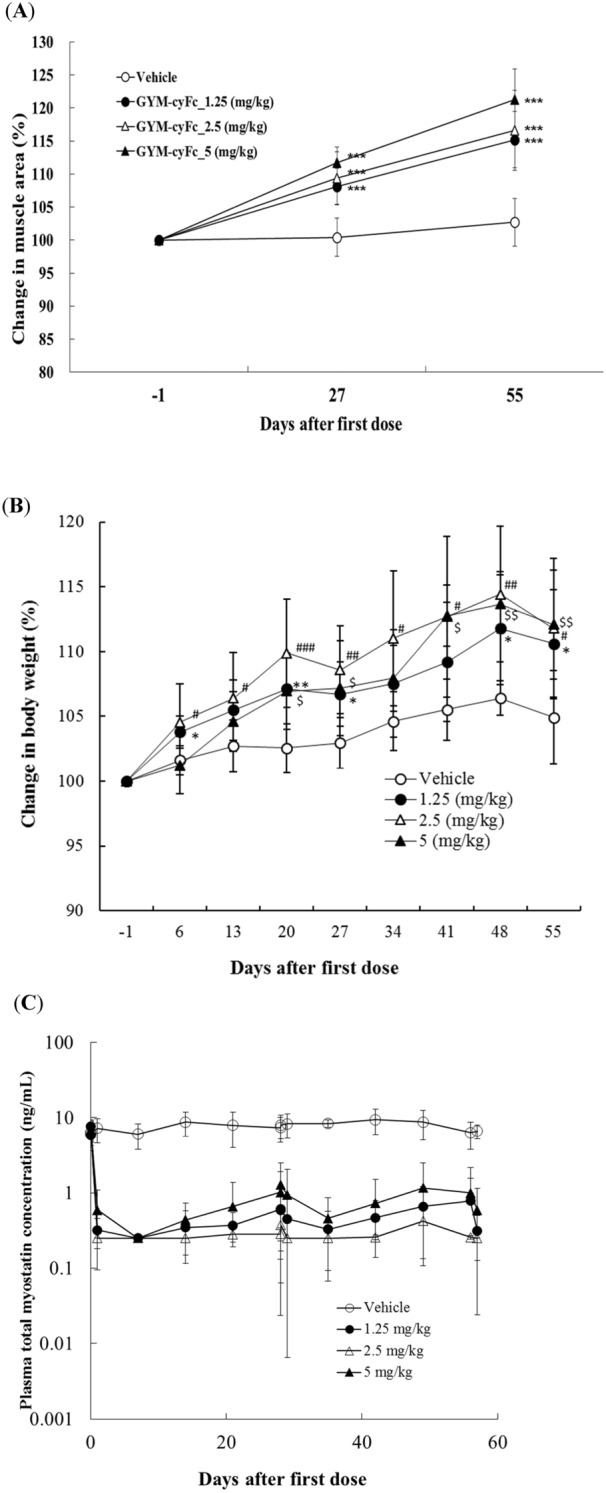
Myostatin, a member of the transforming growth factor-β superfamily, is an attractive target for muscle disease therapy because of its role as a negative regulator of muscle growth and strength. Here, we describe a novel antibody therapeutic approach that maximizes the potential of myostatin-targeted therapy. We generated an antibody, GYM329, that specifically binds the latent form of myostatin and inhibits its activation. Additionally, via "sweeping antibody technology", GYM329 reduces or "sweeps" myostatin in the muscle and plasma. Compared with conventional anti-myostatin agents, GYM329 and its surrogate antibody exhibit superior muscle strength-improvement effects in three different mouse disease models. We also demonstrate that the superior efficacy of GYM329 is due to its myostatin specificity and sweeping capability. Furthermore, we show that a GYM329 surrogate increases muscle mass in normal cynomolgus monkeys without any obvious toxicity. Our findings indicate the potential of GYM329 to improve muscle strength in patients with muscular disorders.


Conflict of interest statement

All authors are employees of Chugai Pharmaceutical Co., Ltd. Chugai Pharmaceutical Co., Ltd. is clinically developing GYM329.

Figures

Figure 1
Figure 1
Generation of the anti-latent myostatin sweeping antibody, GYM329. (A) Summary of the characteristics engineered into GYM329. (B) Surface plasmon resonance analysis illustrating pH-dependent binding of GYM329 to latent myostatin. Binding of GYM329 to human latent myostatin was monitored at pH 7.4 in the association phase (0 to 180 s) and dissociation of GYM329 from latent myostatin at either pH 6.0 or 7.4 were assessed in the dissociation phase (from 180 s). The sensorgram was normalized by adjusting the latent myostatin binding response to ‘100.’ RU: resonance unit. (C) Inhibitory effects of GYM329 on latent myostatin, latent GDF11, mature myostatin, and mature GDF11. Latent myostatin or latent GDF11: 3 nmol/L; mature GDF11 or mature myostatin: 5 ng/mL. Mean ± SD (n = 3).
Figure 2
Figure 2
The mouse surrogate of GYM329 (GYM-mFc) strongly increases muscle strength and muscle mass in three mouse models of muscle disease. Changes in (A) whole-body muscle mass and (B) appendicular grip strength 4 weeks after antibody injection in mdx mice (n = 5–6 per group). Changes in (C) whole-body muscle mass and (D) appendicular grip strength 4 weeks after antibody injection in aged mice (n = 9–10 per group). (E) Hindlimb muscle weights after 2 weeks of hindlimb suspension in the muscular atrophy model. Hindlimb muscles (quadriceps, gastrocnemius, tibialis anterior, EDL, and soleus) were collected and weighed 2 weeks after treatment. (F) Changes in hindlimb grip strength after pre-treatment with the antibodies in the muscular atrophy model. Data represent the changes in hindlimb grip strength during 2 weeks of the hindlimb suspension period. Data represent mean ± SEM (n = 5–6 per group). #P < 0.025, ##P < 0.005, and ###P < 0.0005, Williams’ test compared with the vehicle (150 mmol/L NaCl, 20 mmol/L L-Histidine, pH 6.0) group.
Figure 2
Figure 2
The mouse surrogate of GYM329 (GYM-mFc) strongly increases muscle strength and muscle mass in three mouse models of muscle disease. Changes in (A) whole-body muscle mass and (B) appendicular grip strength 4 weeks after antibody injection in mdx mice (n = 5–6 per group). Changes in (C) whole-body muscle mass and (D) appendicular grip strength 4 weeks after antibody injection in aged mice (n = 9–10 per group). (E) Hindlimb muscle weights after 2 weeks of hindlimb suspension in the muscular atrophy model. Hindlimb muscles (quadriceps, gastrocnemius, tibialis anterior, EDL, and soleus) were collected and weighed 2 weeks after treatment. (F) Changes in hindlimb grip strength after pre-treatment with the antibodies in the muscular atrophy model. Data represent the changes in hindlimb grip strength during 2 weeks of the hindlimb suspension period. Data represent mean ± SEM (n = 5–6 per group). #P < 0.025, ##P < 0.005, and ###P < 0.0005, Williams’ test compared with the vehicle (150 mmol/L NaCl, 20 mmol/L L-Histidine, pH 6.0) group.
Figure 3
Figure 3
GDF11 signaling blockade negatively affects muscle strength in the muscular atrophy model. (A) Changes in hindlimb grip strength 2 weeks after treatment (n = 5–6 per group). Antibodies were administered (i.v.), followed by hindlimb suspension on day 0. Two weeks after hindlimb suspension, hindlimb muscles (quadriceps, TA, EDL, soleus, and gastrocnemius) were isolated and weighed (n = 5–6 per group). Data represent mean ± SEM. *P < 0.05 with Student’s t-test. (B) Inhibitory activities of antibodies against mature GDF11 or mature myostatin; 5 ng/mL of mature GDF11 (square) or mature myostatin (circle) was used for the reporter gene assay. Data are presented as mean ± SD (n = 3). (C) Changes in hindlimb grip strength over three days. (n = 6 per group). Recombinant GDF11 or myostatin were administered (i.p.) on day 0, 1, and 2. Data represent mean ± SEM (n = 6 per group). ***P < 0.001 with Tukey test performed without the non-suspension group.
Figure 4
Figure 4
The sweeping function of GYM329 positively contributes to muscle strength improvement in the muscular atrophy model. (A) Inhibition of BMP1-mediated activation of mouse latent myostatin (3 nmol/L) by the anti-latent myostatin sweeping antibody, GYM329, and the non-sweeping antibody, hMST1032-hIgG1, determined by the Smad reporter gene assay. Data represent mean ± SD (n = 3). (B) Changes in hindlimb grip strength one week after injection (i.v.) with GYM329 or hMST1032-hIgG1. ***P < 0.0005 using Williams’ test compared with the vehicle group. (C) Mature myostatin levels in isolated mouse quadriceps after the in vivo study in (B). A representative western blotting image and quantified results are shown (the non-suspension group was assigned the value of 1; n = 6; mean ± SEM). Full length blots are presented in Supplementary Information (Fig. S7). (D) Confocal imaging of whole mounts of the extensor digitorum longus (EDL) in the muscular atrophy model. Pro-/latent myostatin localized in the extracellular space of the skeletal muscles was labeled by the anti-pro-myostatin/latent myostatin antibody, MST1098-rabbit IgG, followed by the secondary anti-rabbit-IgG Alexa Fluor 568 (red). Representative confocal images of muscles from the non-suspension (left) and hindlimb suspension group (vehicle treatment, middle; GYM329 treatment, right) are shown. (E) Plasma concentration–time curve of total myostatin in normal mice after administration of GYM329 or hMST1032-hIgG1. GYM329 or hMST1032-hIgG1 was intravenously injected into normal mice on day 0. Total plasma myostatin concentration was measured by the electrochemiluminescence immunoassay (n = 6, mean ± SD). Total myostatin is the C-terminal domain of myostatin including both antibody-bound and unbound, or other protein-bound myostatin forms. Values < 1 ng/mL were considered to be below the limit of quantification (BLQ).
Figure 4
Figure 4
The sweeping function of GYM329 positively contributes to muscle strength improvement in the muscular atrophy model. (A) Inhibition of BMP1-mediated activation of mouse latent myostatin (3 nmol/L) by the anti-latent myostatin sweeping antibody, GYM329, and the non-sweeping antibody, hMST1032-hIgG1, determined by the Smad reporter gene assay. Data represent mean ± SD (n = 3). (B) Changes in hindlimb grip strength one week after injection (i.v.) with GYM329 or hMST1032-hIgG1. ***P < 0.0005 using Williams’ test compared with the vehicle group. (C) Mature myostatin levels in isolated mouse quadriceps after the in vivo study in (B). A representative western blotting image and quantified results are shown (the non-suspension group was assigned the value of 1; n = 6; mean ± SEM). Full length blots are presented in Supplementary Information (Fig. S7). (D) Confocal imaging of whole mounts of the extensor digitorum longus (EDL) in the muscular atrophy model. Pro-/latent myostatin localized in the extracellular space of the skeletal muscles was labeled by the anti-pro-myostatin/latent myostatin antibody, MST1098-rabbit IgG, followed by the secondary anti-rabbit-IgG Alexa Fluor 568 (red). Representative confocal images of muscles from the non-suspension (left) and hindlimb suspension group (vehicle treatment, middle; GYM329 treatment, right) are shown. (E) Plasma concentration–time curve of total myostatin in normal mice after administration of GYM329 or hMST1032-hIgG1. GYM329 or hMST1032-hIgG1 was intravenously injected into normal mice on day 0. Total plasma myostatin concentration was measured by the electrochemiluminescence immunoassay (n = 6, mean ± SD). Total myostatin is the C-terminal domain of myostatin including both antibody-bound and unbound, or other protein-bound myostatin forms. Values < 1 ng/mL were considered to be below the limit of quantification (BLQ).
Figure 5
Figure 5
The monkey GYM329 surrogate antibody, GYM-cyFc, increases muscle area and bodyweight, and reduces total myostatin levels in cynomolgus monkeys. (A) Muscle section area determined by MRI are presented as the sum of the area of the quadriceps femoris, brachialis, and elector spinae on days − 1 (Pre), 27 (4 weeks), and 55 (8 weeks) from the administration (i.v.) of the first dose of GYM-cyFc. GYM-cyFc was administered every 4 weeks for 2 months. From the results of anti-drug antibody (ADA) and pharmacokinetic analyses, ADA-positive animals were excluded (vehicle group: n = 6; 1.25 mg/kg dose: n = 9; 2.5 mg/kg dose: n = 6; and 5 mg/kg dose: n = 5). Data represent mean ± SD. ***P < 0.0005, Williams’ test for multiple comparisons to the vehicle group. (B) The rates of increase in bodyweight relative to baseline after the 1st administration of GYM-cyFc into cynomolgus monkeys. Data represent mean ± SD. */#/$P < 0.025, **/##/$$P < 0.005, ***/###/$$$P < 0.0005, Williams test for multiple comparisons to the vehicle group (*: 1.25 mg/kg; #: 2.5 mg/kg; and $: 5 mg/kg). (C) Plasma concentration–time curve of total myostatin. Total myostatin is the C-terminal domain of myostatin including both antibody-bound and unbound, or other protein-bound myostatin forms. Values under 0.25 ng/mL were considered to be below the limit of quantification (BLQ). Data represent mean ± SD.

References

    1. McPherron AC, Lawler AM, Lee SJ. Regulation of skeletal muscle mass in mice by a new TGF-beta superfamily member. Nature. 1997;387:83–90. doi: 10.1038/387083a0. - DOI - PubMed
    1. Szabó G, et al. A deletion in the myostatin gene causes the compact (Cmpt) hypermuscular mutation in mice. Mamm. Genome. 1998;9:671–672. doi: 10.1007/s003359900843. - DOI - PubMed
    1. Zhu X, Hadhazy M, Wehling M, Tidball JG, McNally EM. Dominant negative myostatin produces hypertrophy without hyperplasia in muscle. FEBS Lett. 2000;474:71–75. doi: 10.1016/s0014-5793(00)01570-2. - DOI - PubMed
    1. Grobet L, et al. A deletion in the bovine myostatin gene causes the double-muscled phenotype in cattle. Nat. Genet. 1997;17:71–74. doi: 10.1038/ng0997-71. - DOI - PubMed
    1. Kambadur R, Sharma M, Smith TP, Bass JJ. Mutations in myostatin (GDF8) in double-muscled Belgian Blue and Piedmontese cattle. Genome Res. 1997;7:910–916. doi: 10.1101/gr.7.9.910. - DOI - PubMed

Publication types

MeSH terms