Review
. 2021 May;19(5):303-318.
doi: 10.1038/s41579-020-00501-8. Epub 2021 Jan 25.

Structural insights into RNA polymerases of negative-sense RNA viruses

Affiliations
Review

Structural insights into RNA polymerases of negative-sense RNA viruses

Aartjan J W Te Velthuis et al. Nat Rev Microbiol. 2021 May.

Erratum in

Abstract

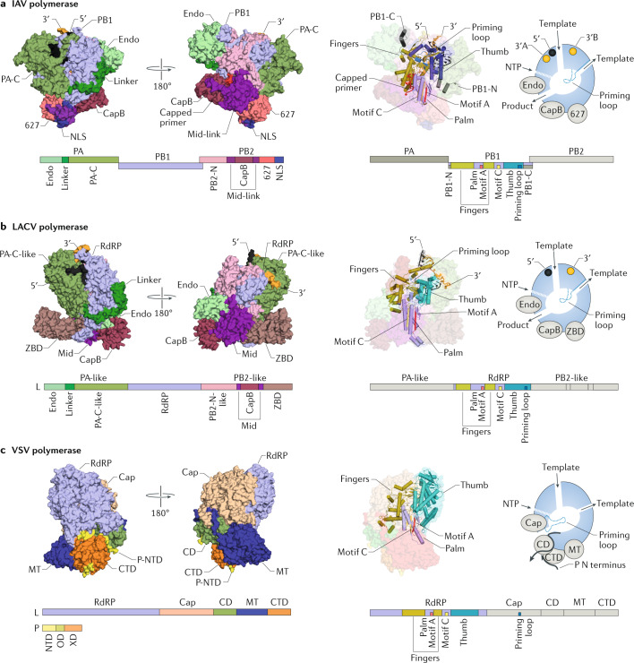
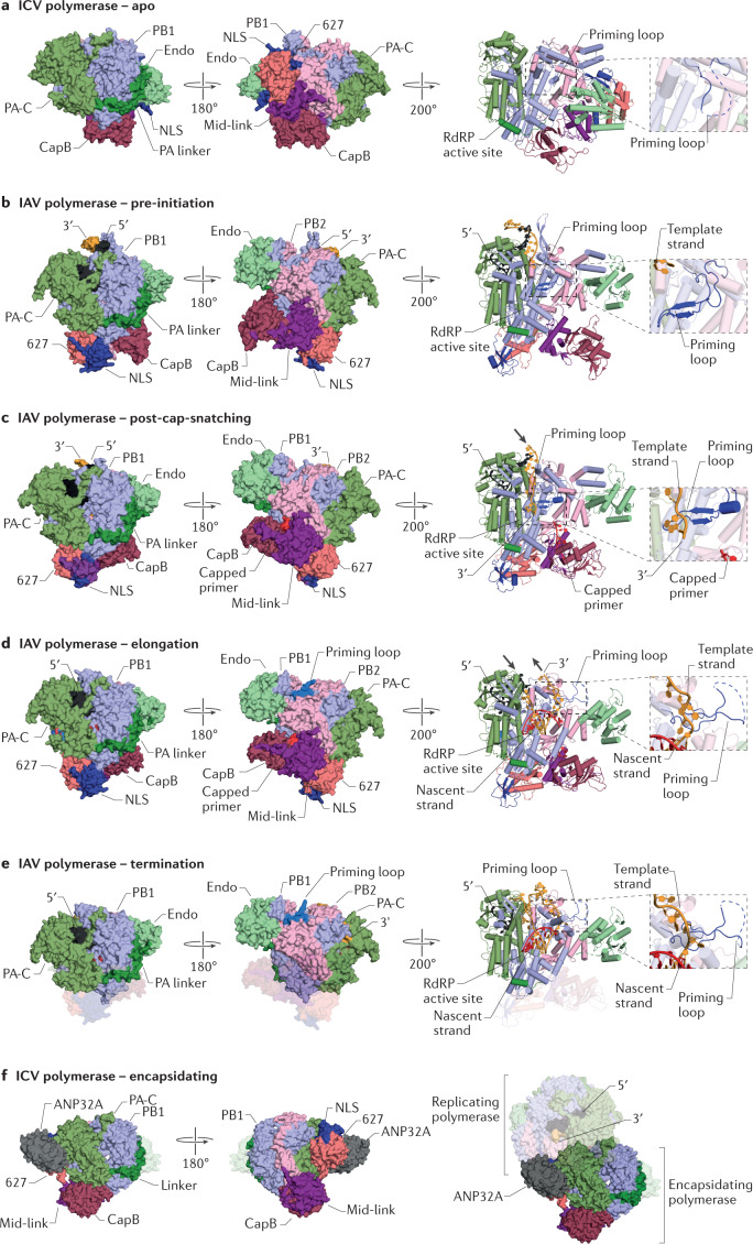
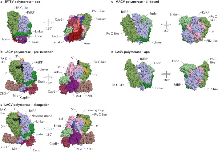
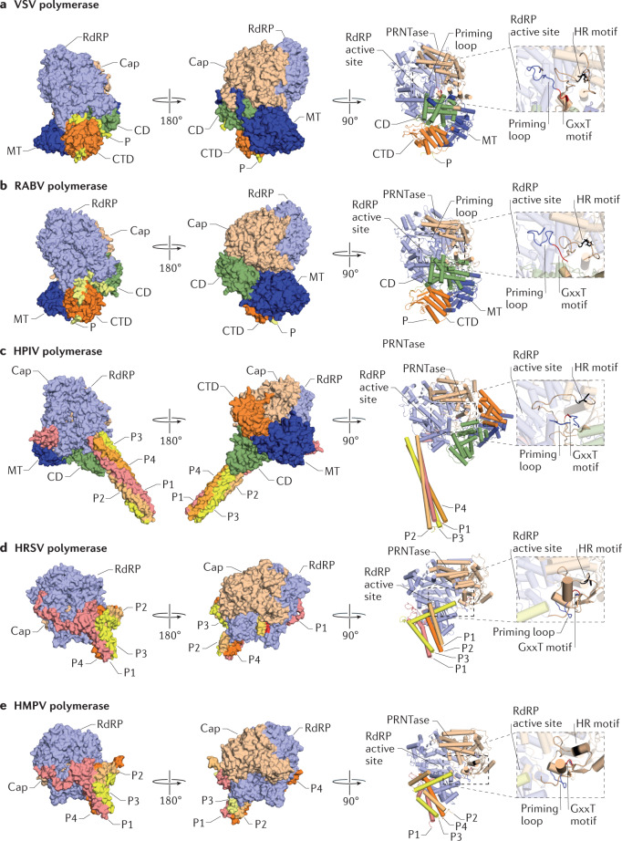
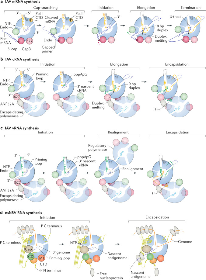
RNA viruses include many important human and animal pathogens, such as the influenza viruses, respiratory syncytial virus, Ebola virus, measles virus and rabies virus. The genomes of these viruses consist of single or multiple RNA segments that assemble with oligomeric viral nucleoprotein into ribonucleoprotein complexes. Replication and transcription of the viral genome is performed by ~250-450 kDa viral RNA-dependent RNA polymerases that also contain capping or cap-snatching activity. In this Review, we compare recent high-resolution X-ray and cryoelectron microscopy structures of RNA polymerases of negative-sense RNA viruses with segmented and non-segmented genomes, including orthomyxoviruses, peribunyaviruses, phenuiviruses, arenaviruses, rhabdoviruses, pneumoviruses and paramyxoviruses. In addition, we discuss how structural insights into these enzymes contribute to our understanding of the molecular mechanisms of viral transcription and replication, and how we can use these insights to identify targets for antiviral drug design.


Conflict of interest statement

The authors declare no competing interests.

Figures

Fig. 1
Fig. 1. Domain organization and overall structure of NSV RNA polymerases.
a | Structure of the influenza A virus (IAV) heterotrimeric RNA polymerase bound to viral RNA (vRNA) and a capped RNA primer (Protein Data Bank (PDB) identifier (ID) 6RR7). b | Structure of the La Crosse virus (LACV) L protein with bound vRNA (PDB ID 6Z6B). c | Structure of the vesicular stomatitis virus (VSV) L protein and phosphoprotein (P) monomer complex (PDB ID 6U1X). The structures are rendered in the same orientation after superimposing motif C of the RNA-dependent RNA polymerase (RdRP) domain. CD, connector domain; CTD, C-terminal domain; Endo, endonuclease; Mid-link, middle and linker regions; MT, methyltransferase; NLS, nuclear localization signal; NSV, negative-sense RNA virus; NTD, N-terminal domain; NTP, nucleoside triphosphate; PA, polymerase acidic; PA-C, polymerase acidic C-terminal domain; PB, polymerase basic; P-NTD, P protein N-terminal domain; P-OD, P protein oligomerization domain; P-XD, P protein X domain; ZBD, zinc-binding domain.
Fig. 2
Fig. 2. Structures of influenza virus RNA polymerase conformations.
a | Structure of the apo form of the influenza C virus (ICV) RNA polymerase (Protein Data Bank (PDB) identifier (ID) 5D9A). b | Structure of the pre-initiation conformation of the influenza A virus (IAV) RNA polymerase (PDB ID 4WSB). c | Structure of the post-cap-snatching conformation of the IAV RNA polymerase (PDB ID 6RR7). d | Structure of the elongation conformation of the IAV RNA polymerase (PDB ID 6T0V). e | Structure of the termination conformation of the IAV RNA polymerase (PDB ID 6SZU). f | Structure of the encapsidating polymerase of the ICV RNA polymerase bound to ANP32A (PDB ID 6XZR) and the putative replicating–encapsidating polymerase dimer bound to ANP32A. Magnified views show the priming loop. Finger subdomain residues are not shown so that the priming loop is visible. Structures in surface representation in each panel are shown in the same orientation after superimposing motif C of the RNA-dependent RNA polymerase (RdRP) domain. Structures in cartoon representation have been rotated relative to the structures in surface representation to focus on the priming loop. Endo, endonuclease; Mid-link, middle and linker regions; NLS, nuclear localization signal; PA, polymerase acidic; PA-C, polymerase acidic C-terminal domain; PB, polymerase basic.
Fig. 3
Fig. 3. Structures of sNSV L protein conformations.
a | Structure of the apo form of the severe fever with thrombocytopenia syndrome virus (SFTSV) L protein (Protein Data Bank (PDB) identifier (ID) 6L42). b | Structure of the pre-initiation conformation of the La Crosse virus (LACV) L protein (PDB ID 6Z6B). c | Structure of the elongation conformation of the LACV L protein (PDB ID 6Z8K). d | Structure of the Machupo virus (MACV) L protein bound to the viral RNA 3′ terminus (PDB ID 6KLE). e | Structure of the apo form of the Lassa mammarenavirus (LASV) L protein (PDB ID 6KLC). Structures in each panel are shown in the same orientation after superimposing motif C of the RNA-dependent RNA polymerase (RdRP) domain. Endo, endonuclease; PA, polymerase acidic; PA-C, polymerase acidic C-terminal domain; PB, polymerase basic; sNSV, segmented negative-sense RNA virus; ZBD, zinc-binding domain.
Fig. 4
Fig. 4. Structures of nsNSV L–P complex conformations.
a | Structure of the vesicular stomatitis virus (VSV) L protein–phosphoprotein (P) monomer complex (Protein Data Bank (PDB) identifier (ID) 6U1X). b | Structure of the rabies lyssavirus (RABV) L protein–P monomer complex (PDB ID 6UEB). c | Structure of the human parainfluenza virus type 5 (HPIV) L protein–P tetramer complex (PDB ID 6V86). d | Structure of the human respiratory syncytial virus (HRSV) L protein–P tetramer complex (PDB ID 6UEN). e | Structure of the human metapneumovirus (HMPV) L protein–P tetramer complex (PDB ID 6U5O). Insets show the priming loop and histidine–arginine (HR) and GxxT motifs of the Cap domain. Structures in surface representation in each panel are shown in the same orientation after superimposing motif C of the RNA-dependent RNA polymerase (RdRP) domain. Structures in cartoon representation have been rotated relative to the structures in surface representation to focus on the priming loop. Finger subdomain residues are not shown so that the priming loop and Cap domain active site are visible. CD, connector domain; CTD, C-terminal domain; MT, methyltransferase; nsNSV, non-segmented negative-sense RNA virus; P1–P4, phosphoprotein subunits; PRNTase, polyribonucleotidyl transferase.
Fig. 5
Fig. 5. Mechanisms of RNA synthesis by NSV RNA polymerases.
a | Synthesis of mRNA in influenza virus. b | Synthesis of complementary RNA (cRNA) in influenza virus. c | Synthesis of viral RNA (vRNA) in influenza virus. d | RNA synthesis by a model non-segmented negative-sense RNA virus (nsNSV) RNA polymerase. Arrows indicate direction of C terminus in partial structures of the phosphoprotein (P). CD, connector domain; CTD, C-terminal domain; Endo, endonuclease; MT, methyltransferase; NTP, nucleoside triphosphate; PA, polymerase acidic; Pol II, RNA polymerase II.

References

    1. Luo M, Terrell JR, McManus SA. Nucleocapsid structure of negative strand RNA virus. Viruses. 2020;12:835. doi: 10.3390/v12080835. - DOI - PMC - PubMed
    1. Ruigrok RW, Crepin T, Kolakofsky D. Nucleoproteins and nucleocapsids of negative-strand RNA viruses. Curr. Opin. Microbiol. 2011;14:504–510. doi: 10.1016/j.mib.2011.07.011. - DOI - PubMed
    1. Gallagher JR, Torian U, McCraw DM, Harris AK. Structural studies of influenza virus RNPs by electron microscopy indicate molecular contortions within NP supra-structures. J. Struct. Biol. 2017;197:294–307. doi: 10.1016/j.jsb.2016.12.007. - DOI - PMC - PubMed
    1. Coloma R, et al. Structural insights into influenza A virus ribonucleoproteins reveal a processive helical track as transcription mechanism. Nat. Microbiol. 2020;5:727–734. doi: 10.1038/s41564-020-0675-3. - DOI - PubMed
    1. Te Velthuis AJ, Fodor E. Influenza virus RNA polymerase: insights into the mechanisms of viral RNA synthesis. Nat. Rev. Microbiol. 2016;14:479–493. doi: 10.1038/nrmicro.2016.87. - DOI - PMC - PubMed

Publication types

MeSH terms

Substances

LinkOut - more resources