. 2021 Apr;6(4):425-434.
doi: 10.1038/s41564-020-00854-z. Epub 2021 Jan 25.

An Alveolata secretory machinery adapted to parasite host cell invasion

Affiliations

An Alveolata secretory machinery adapted to parasite host cell invasion

Eleonora Aquilini et al. Nat Microbiol. 2021 Apr.

Abstract

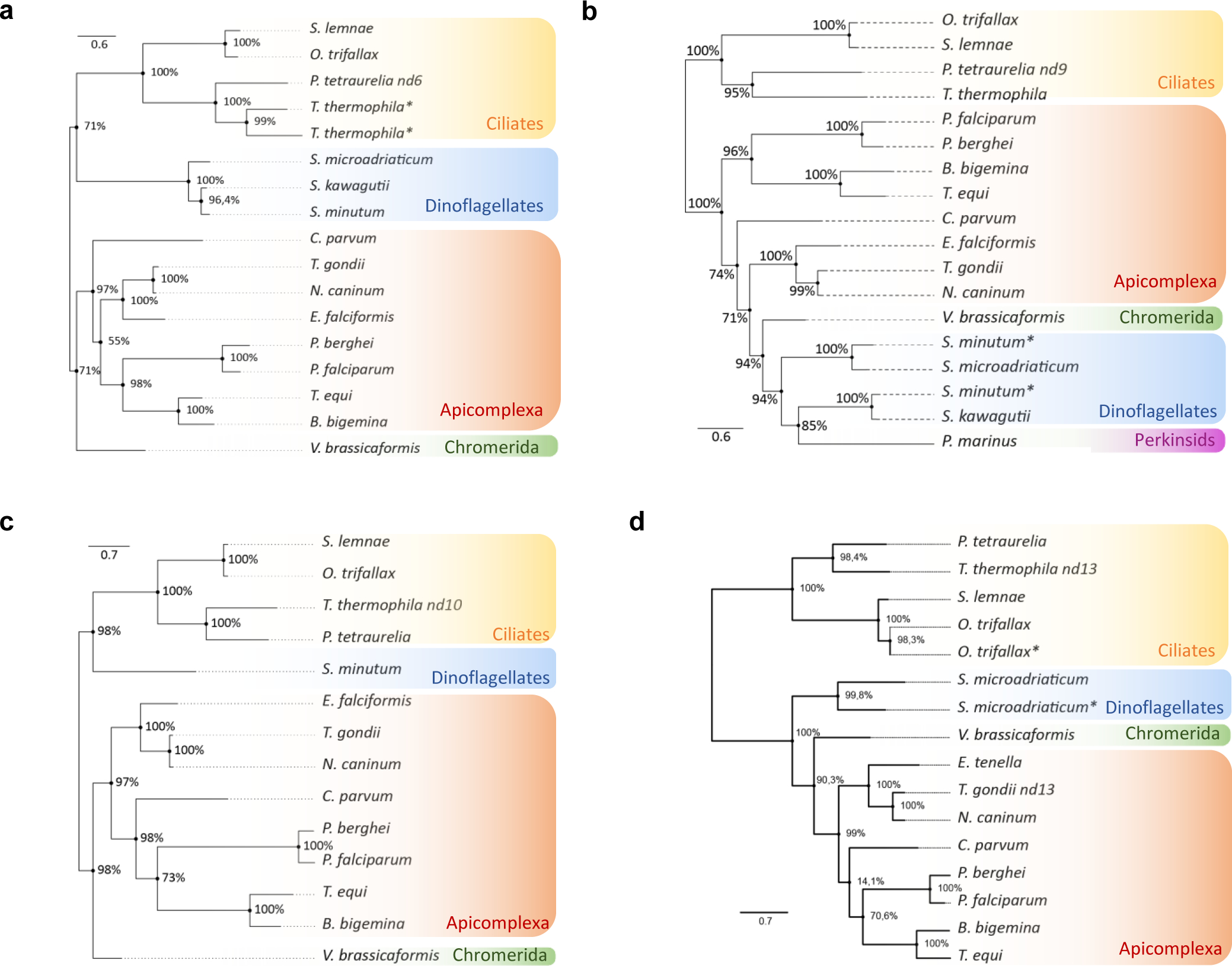
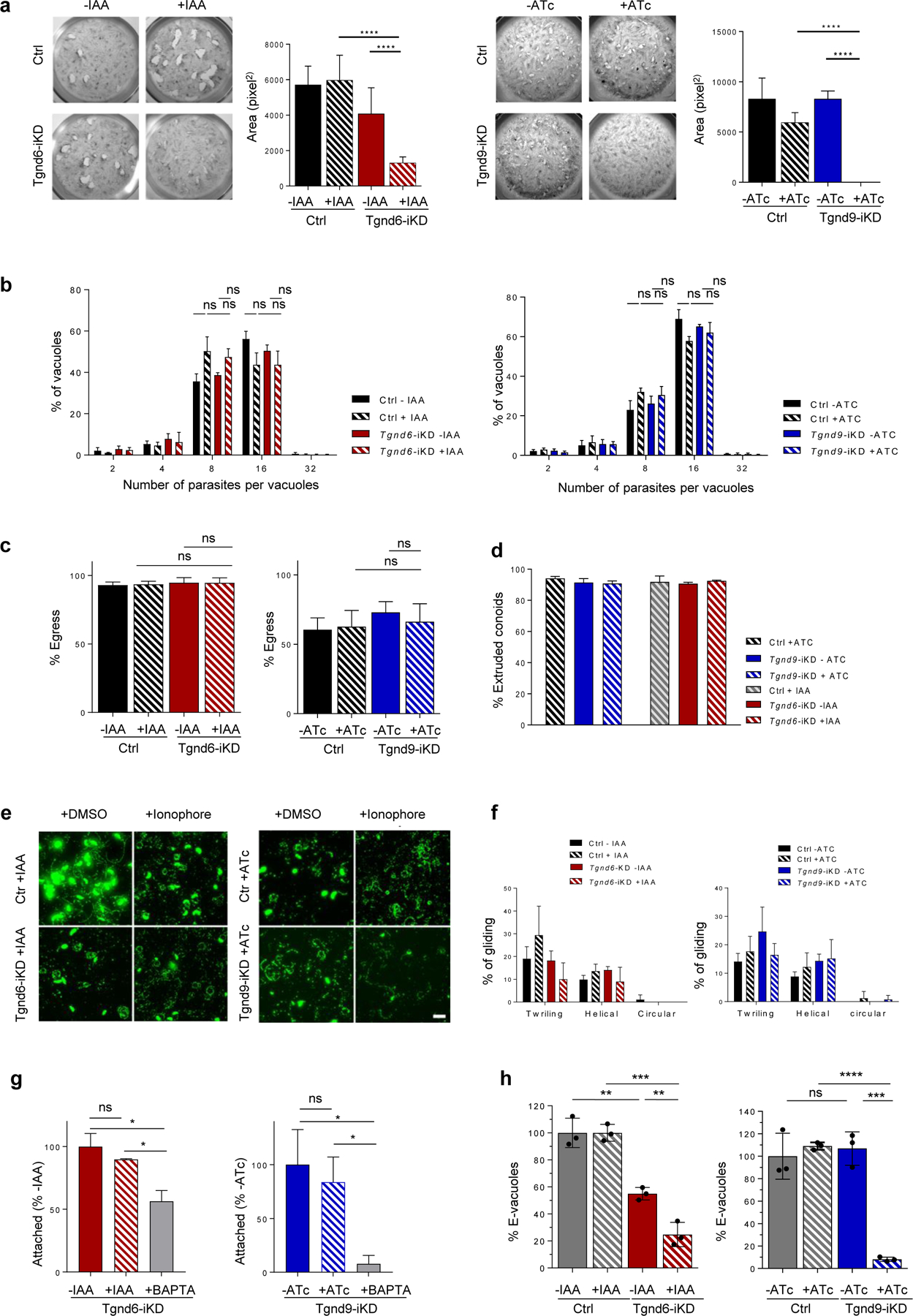
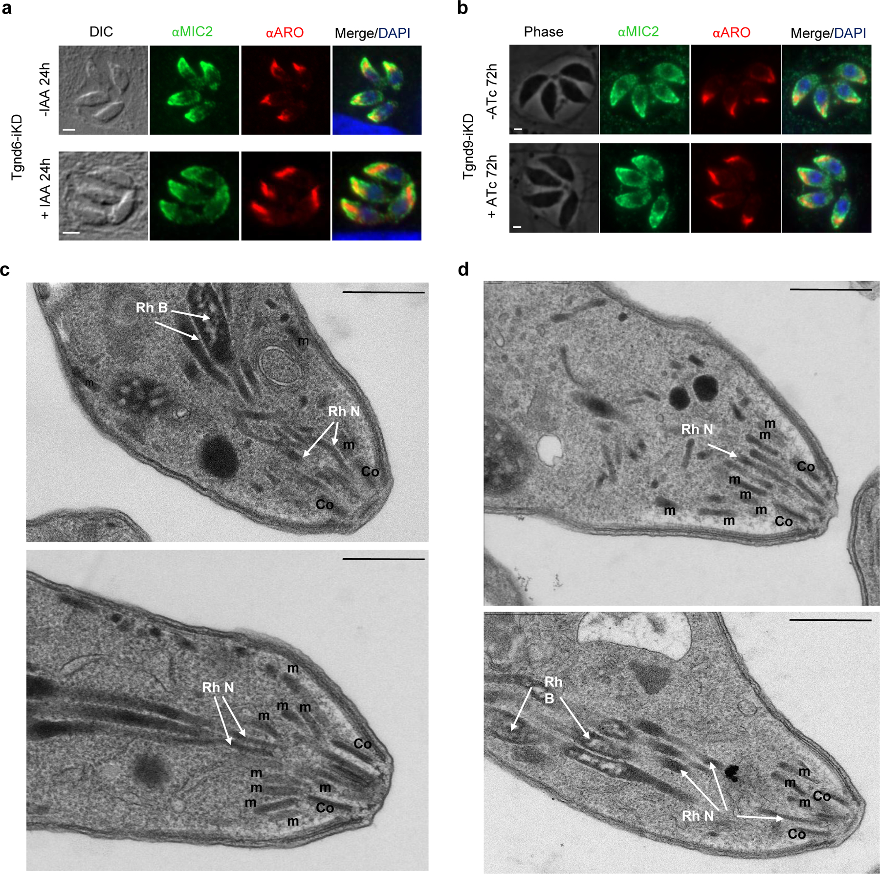
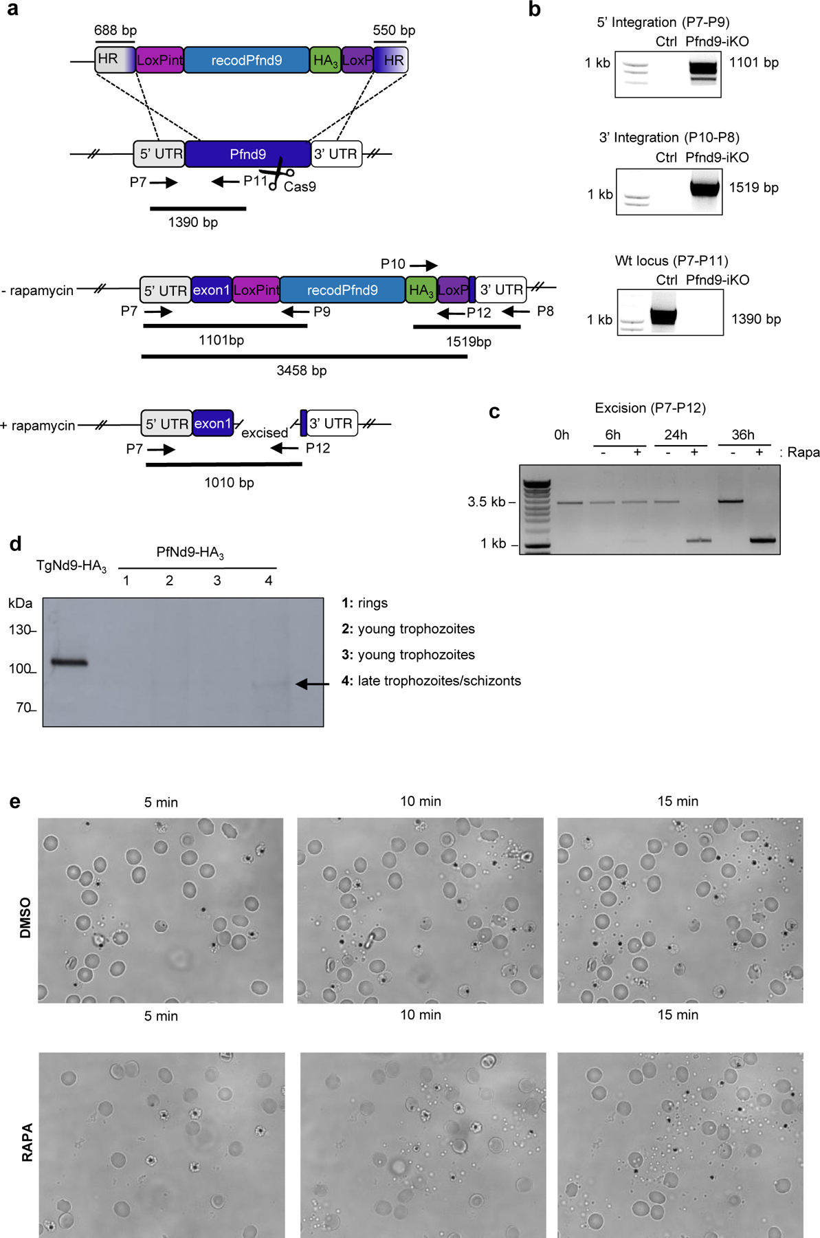
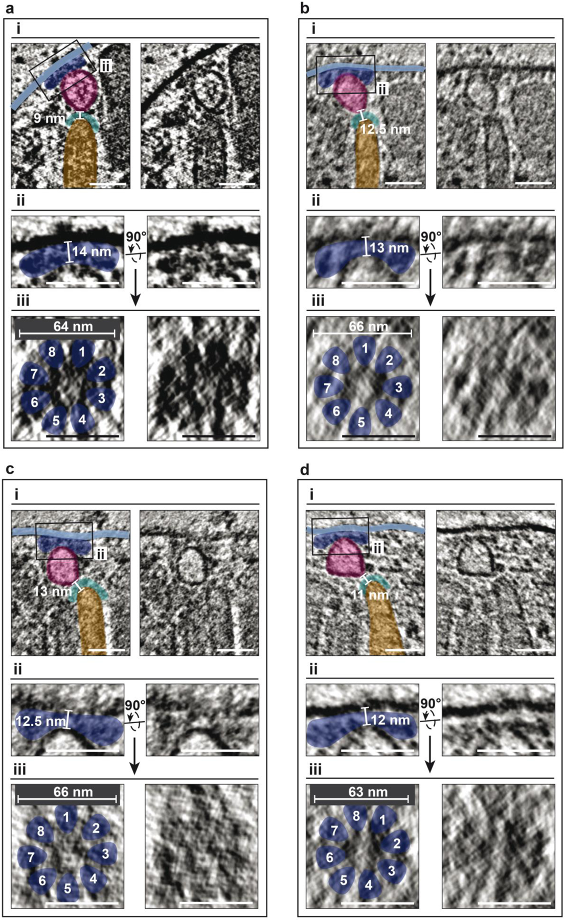
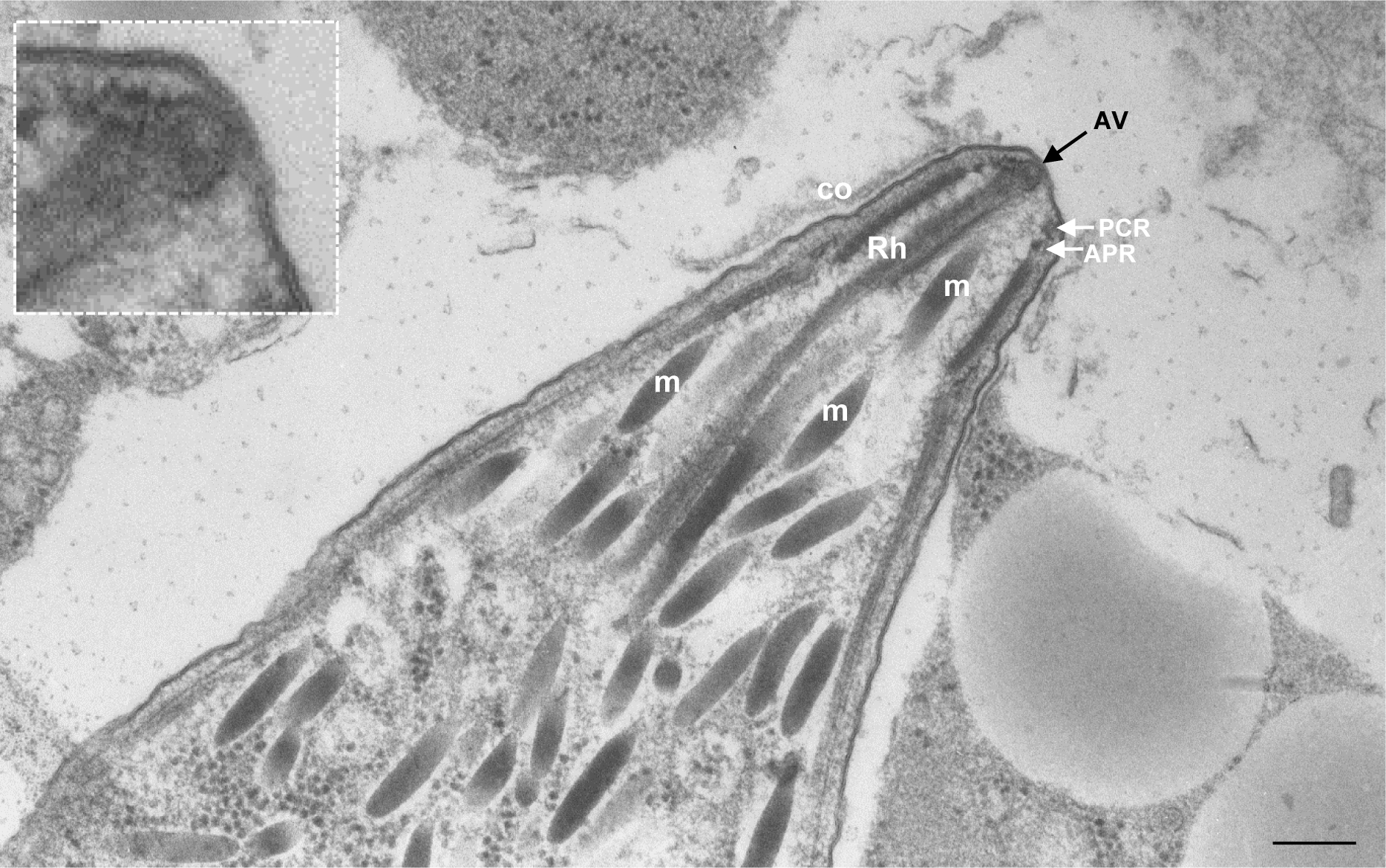
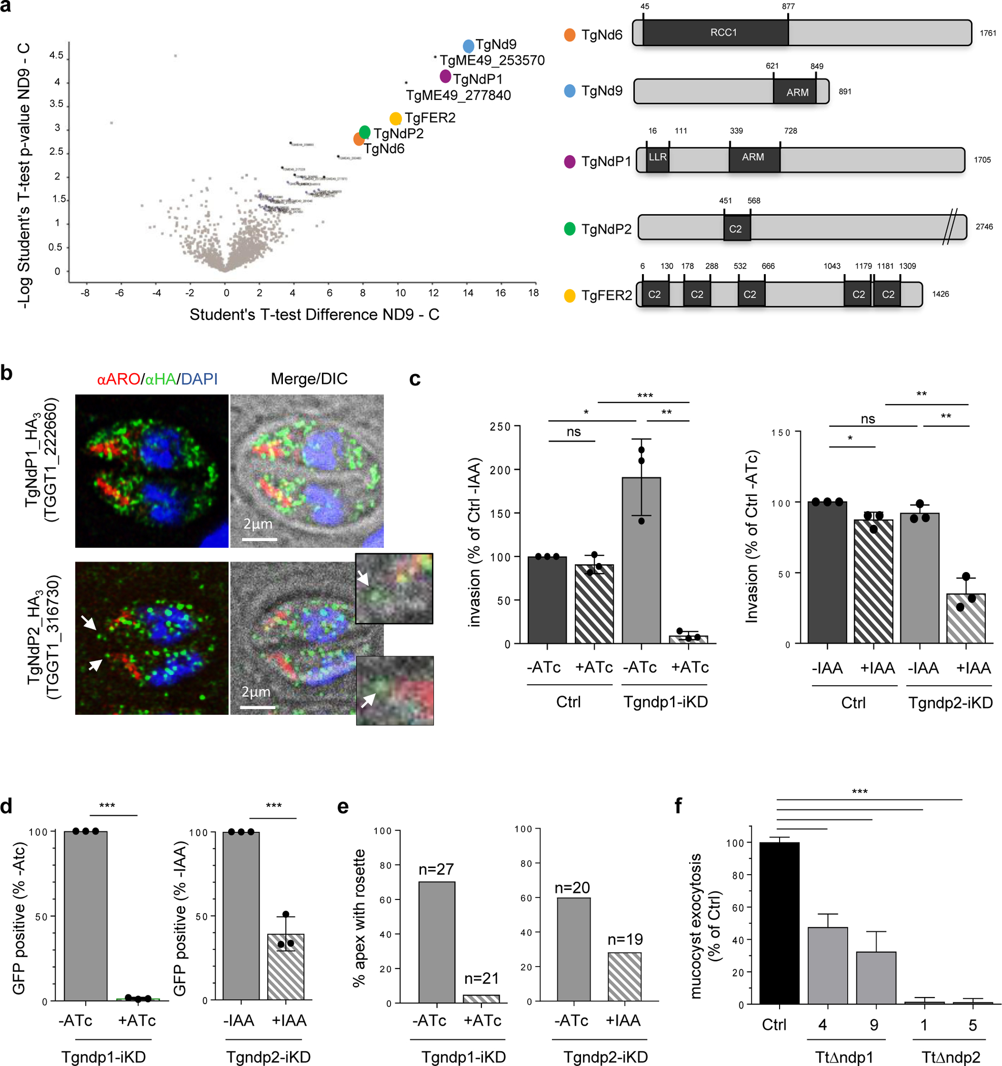
Apicomplexa are unicellular eukaryotes and obligate intracellular parasites, including Plasmodium (the causative agent of malaria) and Toxoplasma (one of the most widespread zoonotic pathogens). Rhoptries, one of their specialized secretory organelles, undergo regulated exocytosis during invasion1. Rhoptry proteins are injected directly into the host cell to support invasion and subversion of host immune function2. The mechanism by which they are discharged is unclear and appears distinct from those in bacteria, yeast, animals and plants. Here, we show that rhoptry secretion in Apicomplexa shares structural and genetic elements with the exocytic machinery of ciliates, their free-living relatives. Rhoptry exocytosis depends on intramembranous particles in the shape of a rosette embedded into the plasma membrane of the parasite apex. Formation of this rosette requires multiple non-discharge (Nd) proteins conserved and restricted to Ciliata, Dinoflagellata and Apicomplexa that together constitute the superphylum Alveolata. We identified Nd6 at the site of exocytosis in association with an apical vesicle. Sandwiched between the rosette and the tip of the rhoptry, this vesicle appears as a central element of the rhoptry secretion machine. Our results describe a conserved secretion system that was adapted to provide defence for free-living unicellular eukaryotes and host cell injection in intracellular parasites.


Conflict of interest statement

Competing interests

The authors declare no competing financial interests.

Figures

Extended Data Fig. 1 |
Amino acid sequence alignments and phylogenetic analysis
Extended Data Fig. 2 |
Generation of TgNd6-KD and TgNd9-KD
Extended Data Fig. 3 |
Depletion of TgNd6 or TgNd9 in T.gondii parasites does not affect replication, egress, nor gliding motility.
Extended Data Fig. 4 |
Secretory organelles biogenesis and positioning is not affected in Tgnd6iKO and Tgnd9iKO.
Extended Data Fig. 5 |
Generation of Pfnd9iKO
Extended Data Fig. 6 |
Generation of Tgnd6-iKD and Tgnd9-iKD
Extended Data Fig. 7 |
Depletion of NdP1 or NdP2 in T. gondii parasites does not affect replication, egress, gliding motility, nor attachment
Extended Data Fig. 7 |
Depletion of NdP1 or NdP2 in T. gondii parasites does not affect replication, egress, gliding motility, nor attachment
Extended Data Fig. 8 |
Mucocyst biogenesis and positioning is not affected in TtΔnd10
Extended Data Fig. 9 |
Cryo-ET of rhoptry secretion system.
Extended Data Fig. 10 |
Apical area of a Sarcocystis tenella cystozoite.
Extended Data Fig. 11 |
Unedited full-length gels/blots.
Extended Data Fig. 11 |
Unedited full-length gels/blots.
Extended Data Fig. 11 |
Unedited full-length gels/blots.
Extended Data Fig. 11 |
Unedited full-length gels/blots.
Fig. 1 |
Fig. 1 |. Rhoptry secretion is dependent on rosette formation.
a, Immunofluorescence assay (IFA) of control untagged line and endogenously HA3-tagged TgNd6 and HA3-tagged TgNd9 tachyzoites. The green arrow points to TgNd6-HA3 apical puncta. ARO is a marker for rhoptries. b, Super-resolution microscopy of the apical dot of TgNd6. Schematic of apical end of a tachyzoite. Maximum intensity projections of z-stacks of TgNd6-HA3 parasites transiently expressing RNG1_GFP (top) and of TgNd6-HA3 parasites expressing centrin 2-Ty3 (bottom). The protein RNG1_GFP marks the apical polar ring (APR) and CEN2_GFP marks pre-conoidal rings (PCR). Higher magnifications show that TgNd6-HA3 (red arrow) localizes above the apical polar ring (green arrow) and co-localizes partially with CEN2. DIC: differential interference contrast. c, Immunogold labelling of TgNd6-HA3. Right panel shows TgNd6-HA3 on the apical vesicle. Insert panel: higher magnification of the apical vesicle. Micronemes (m) and rhoptries (Rh) are visible in transverse section of the conoid (Co). Bar is 200 nm. d, Quantification of invasion after depletion of TgNd6 and TgNd9. Mean ± SD of n=3 independent experiments. e, Immunoblot showing microneme secretion (arrow=processed/secreted TgAMA1) in Tgnd6-iKD ± IAA 24h (top) and Tgnd9-iKD ± ATC 72h (bottom). P = pellet, Sup: supernatant, Ind: propanolol-induced secretion. GRA3, loading control. f, Rhoptry secretion assay by Secreted Cre, epitope-tagged (SeCrEt). Rhoptry secretion quantification of Tgnd6i-KD (left) ± IAA and Tgnd9-iKD (right) ± ATC. Mean ± SD of n=3 independent experiments. f, Left: Freeze-fracture electron microscopy of a T. gondii tachyzoite (P face) showing a rosette of intramembranous particles (white arrow). Middle: Higher magnification of the right panel. The white arrows point to the eight IMPs of the rosette. Right: Quantification of rosettes of IMPs in Tgnd9-iKD ± ATc 72h and Tgnd6-iKD ± IAA 24h using freeze fracture. (d, f) Unpaired two tail student’s t test: **** p-value < 0.0001, *** p-value < 0.001, ** p-value < 0.01, * p-value < 0.05.
Fig. 2 |
Fig. 2 |. PfNd9 is essential for rhoptry secretion in P. falciparum.
a, Freeze-fracture electron microscopy of a P. falciparum merozoite (P face) showing a rosette of intramembranous particles (white arrow). Higher magnification at the bottom. Bar is 100 nm. b, Growth curves (parasitaemias) of p230p DiCre (Ctrl) and Pfnd9-iKO mutant ± rapamycin shows that PfNd9-depleted parasites have a growth defect. On the right: Giemsa staining of the growth experiment illustrating development and reinvasion of p230p DiCre (Ctrl) and Pfnd9-iKO merozoites (along 2 cycles) ± rapamycin treatment. c, Quantification of egress of Pfnd9iKO ± rapamycin schizonts. Data collected from 8 movies of Pfnd9-iKO ± rapamycin. d, Left: IFA illustrating AMA1 protein stored in micronemes (top) or secreted and translocated at the surface of the parasite prior to egress (bottom). MSP1: surface marker. Right: Proportion of infected cells (± rapamycin) exhibiting AMA1 secretion.. e, Quantification of rhoptry secretion events in Pfnd9-iKO ± rapamycin-treated schizonts using anti-PfRAP2 antibodies to visualise rhoptry secretion events (‘spits’ of RAP2 export into the RBC).
Fig. 3 |
Fig. 3 |. Nd6 and Nd9 are part of an Alveolate complex essential for organellar secretion in T. gondii (Apicomplexa) and T. thermophila (Ciliate)
a, Mass spectrometry analysis of immuno-isolated Nd9-HA. Left: Volcano Plot of proteins differentially enriched in Nd9 vs control IP. This plot presents the fold change (Difference) and significance (-Log p) obtained from a t-test of three independent IPs using LFQ intensity values. Right: Schematic representation of TgNds proteins using SUPERFAMILY. RCC1: regulator of chromosome condensation 1-like domains (RLDs), a versatile domain that performs many different functions, including guanine nucleotide exchange on small GTP-binding proteins. LRR: Leucine Rich Repeat domain. ARM: Armadillo Repeat domain. C2: lipid-calcium binding domain. b, Immunofluorescence (IF) of endogenously HA3-tagged TgNdP1 and TgNdP2 tachyzoites. The white arrow points to TgNdP1-HA3 apical dots of the two parasites, which are magnified on the right. ARO: rhoptry marker. c, Quantification of invasion after depletion of TgNdP1 (left) and TgNdP2 (right). Mean ± SD of n=3 independent experiments. d, Rhoptry secretion quantification of TgndP1i-KD (left) ± ATc and TgndP2-iKD (right) ± IAA. Mean ± SD of n=3 independent experiments. e, Quantification of rosettes of IMPs in TgndP1-iKD ± ATC 72h and in TgndP2-iKD ± IAA 24h. f, Quantification of mucocyst exocytosis by dibucaine assay. Data collected from three experiments. (c, e) Unpaired two tail student’s t test: **** p-value < 0.0001, *** p-value < 0.001, ** p-value < 0.01, * p-value < 0.05.
Fig. 4 |
Fig. 4 |. The rhoptry secretion machine includes an apical vesicle tightly connected with the rosette
a, A slice through a tomogram showing a side view of the apical complex – conoid (brown), micronemes (yellow), plasma membrane (PM; light blue) and the rhoptry secretion system consisting of the rosette (dark blue), apical vesicle (AV; magenta), rhoptry (orange) and rhoptry tip density (cyan). Original image (right) is annotated with color overlays (left). b, Left: magnified image of the boxed region in (a) showing the connections between the rhoptry, tip density, AV, rosette and the PM. The rhoptry tip is 9 nm distant from the AV. Right: 3-dimensional (3D) segmentation of the scene above. The PM is rendered transparent on the right to reveal the rosette. c, Magnified image of the boxed region in (b) showing the side view of the rosette. The AV is 14 nm distant from the PM. d) Top view of the rosette from a horizontal tomogram section, perpendicular to the plane in (c), showing 8-fold rotational symmetry and a diameter of ~67 nm. e) AV connected with the PM via a rosette in the absence of a rhoptry. All measurements are made in 3D. Images in (b-e) are low pass filtered to boost contrast. The images in (c) and (d) are from two different cells that were initially oriented perpendicular to each other on the EM grid. f, Quantification of apical rosettes in TgARO-iKD mutants ± ATc 72h. g, Left: Ultrastructure of wild type (RH strain type I) tachyzoites with the apical vesicle positioned beneath the plasma membrane, where the rosette is located, and above the tip of the rhoptry neck. Right: In TgARO-iKD ATc-treated tachyzoites (72h), the apical vesicle is still properly positioned under the rosette, while rhoptries are not engaged in the conoid. co: conoid; m: micronemes; Rh: rhoptry; APR: apical polar ring; PCR: pre-conoidal apical rings; AV: apical vesicle; CM: pair of intraconoidal microtubules. h, Schematic of similarities and differences of the exocytic machinery between ciliates and Apicomplexa. Exocytosis in Alveolata (Ciliate, Dinoflagellate and Apicomplexa) is outlined by the presence of a rosette of particles embedded in the outer membrane, defining the site of exocytosis. In ciliates, organelles discharge can have a defensive or predatory function. Rhoptry exocytosis in Apicomplexa is one of the critical steps of host cell invasion and therefore fundamental for parasitism. In Apicomplexa, but not in ciliates, an apical vesicle (Av) of unknown function is present between the tip of the rhoptry and the plasmalemma, plausibly involved in the injection of rhoptry proteins into the host cell. PM, plasma membrane, PVM, parasitophorous vacuole membrane, AS, alveolar sac, which is homologous to the IMC (inner membrane complex) of Apicomplexa.

Comment in

  • The art of the steal.
    Hu K. Hu K. Nat Microbiol. 2021 Apr;6(4):421-422. doi: 10.1038/s41564-021-00886-z. Nat Microbiol. 2021. PMID: 33782557 No abstract available.

References

    1. Dubremetz JF Rhoptries are major players in Toxoplasma gondii invasion and host cell interaction. Cell Microbiol 9, 841–8 (2007). - PubMed
    1. Boothroyd JC & Dubremetz JF Kiss and spit: the dual roles of Toxoplasma rhoptries. Nat Rev Microbiol 6, 79–88 (2008). - PubMed
    1. Carruthers VB & Sibley LD Sequential protein secretion from three distinct organelles of Toxoplasma gondii accompanies invasion of human fibroblasts. Eur J Cell Biol 73, 114–23 (1997). - PubMed
    1. Frenal K, Dubremetz JF, Lebrun M & Soldati-Favre D Gliding motility powers invasion and egress in Apicomplexa. Nat Rev Microbiol 15, 645–660 (2017). - PubMed
    1. Besteiro S, Dubremetz JF & Lebrun M The moving junction of apicomplexan parasites: a key structure for invasion. Cell Microbiol 13, 797–805 (2011). - PubMed

Publication types

Substances

LinkOut - more resources