Review
. 2021 Apr;22(4):266-282.
doi: 10.1038/s41580-020-00324-8. Epub 2021 Jan 25.

Ferroptosis: mechanisms, biology and role in disease

Affiliations
Review

Ferroptosis: mechanisms, biology and role in disease

Xuejun Jiang et al. Nat Rev Mol Cell Biol. 2021 Apr.

Abstract

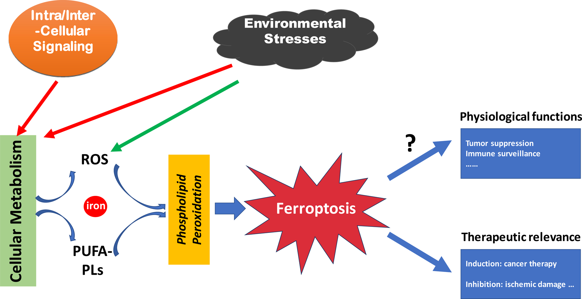
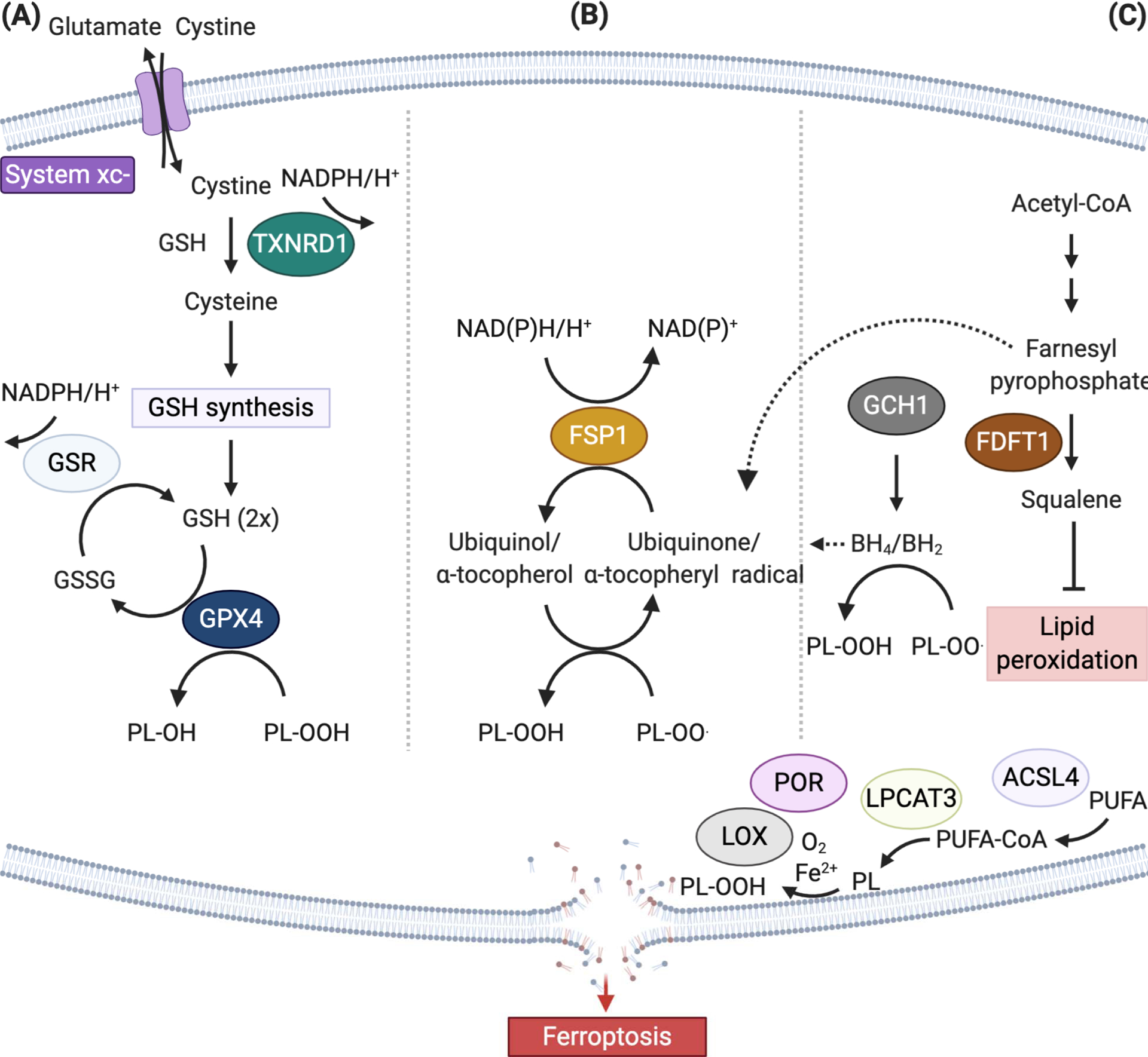
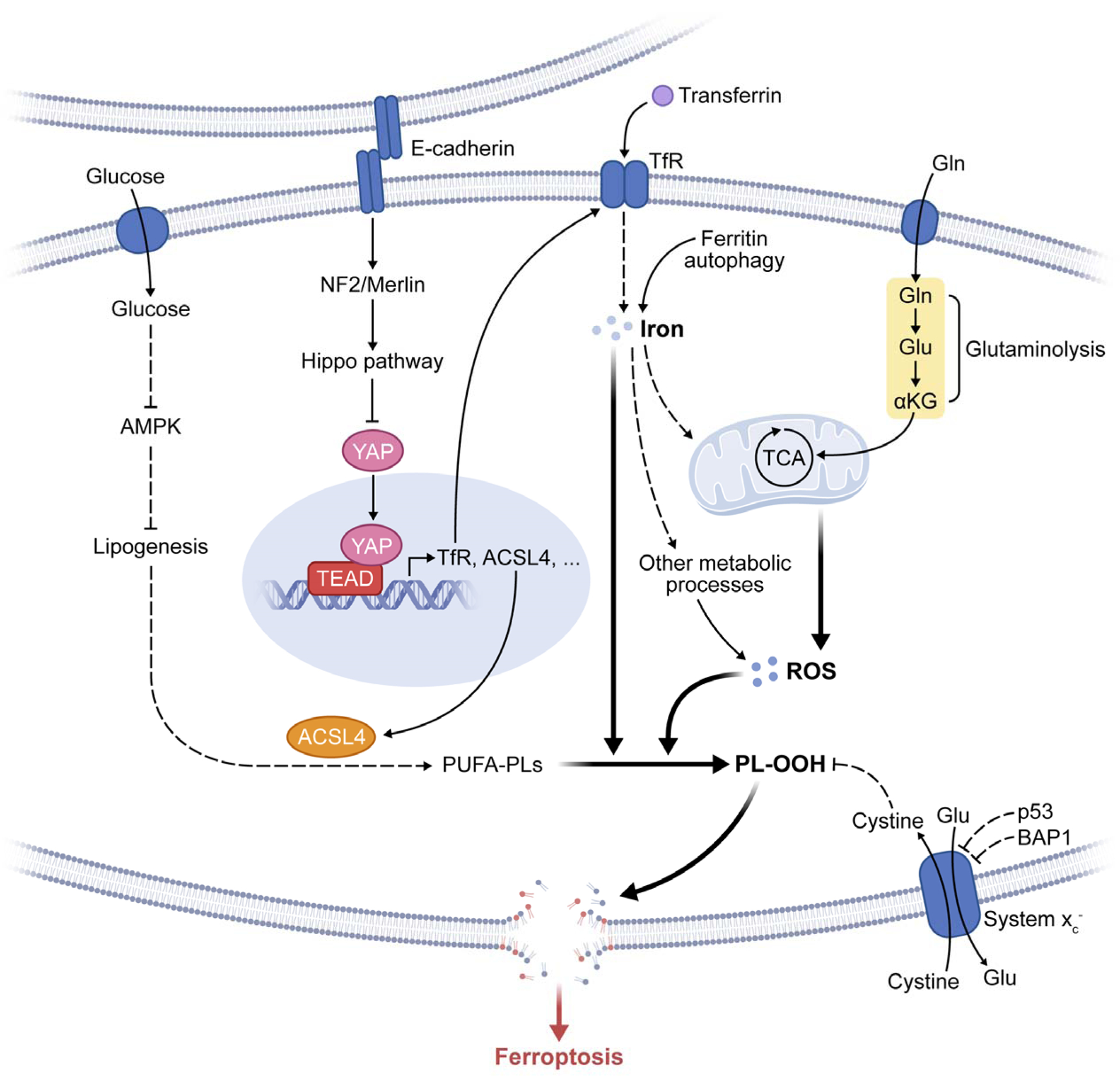
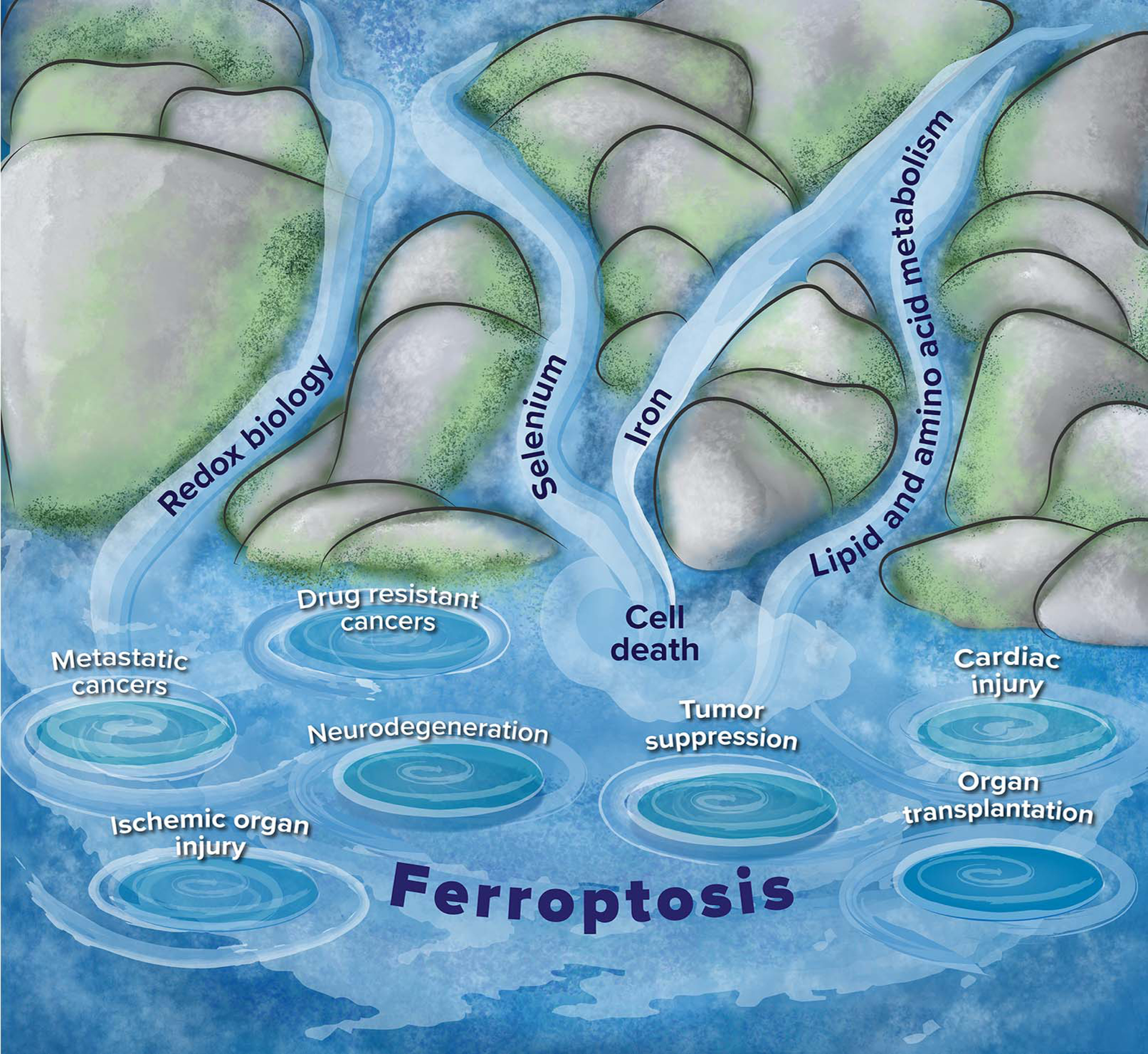
The research field of ferroptosis has seen exponential growth over the past few years, since the term was coined in 2012. This unique modality of cell death, driven by iron-dependent phospholipid peroxidation, is regulated by multiple cellular metabolic pathways, including redox homeostasis, iron handling, mitochondrial activity and metabolism of amino acids, lipids and sugars, in addition to various signalling pathways relevant to disease. Numerous organ injuries and degenerative pathologies are driven by ferroptosis. Intriguingly, therapy-resistant cancer cells, particularly those in the mesenchymal state and prone to metastasis, are exquisitely vulnerable to ferroptosis. As such, pharmacological modulation of ferroptosis, via both its induction and its inhibition, holds great potential for the treatment of drug-resistant cancers, ischaemic organ injuries and other degenerative diseases linked to extensive lipid peroxidation. In this Review, we provide a critical analysis of the current molecular mechanisms and regulatory networks of ferroptosis, the potential physiological functions of ferroptosis in tumour suppression and immune surveillance, and its pathological roles, together with a potential for therapeutic targeting. Importantly, as in all rapidly evolving research areas, challenges exist due to misconceptions and inappropriate experimental methods. This Review also aims to address these issues and to provide practical guidelines for enhancing reproducibility and reliability in studies of ferroptosis. Finally, we discuss important concepts and pressing questions that should be the focus of future ferroptosis research.


Figures

Figure 1.
Figure 1.. An overview of ferroptosis.
A schematic chart showing that ferroptosis is executed by phospholipid peroxidation, a process relying on metabolic products reactive oxygen species (ROS), phospholipid containing polyunsaturated fatty acid chain(s) (PUFA-PL), and transition metal iron, and that intra- and intercellular signaling events and environmental stresses can impact ferroptosis by regulating cellular metabolism and ROS level. The figure also shows the role of ferroptosis in disease and its potential physiological functions.
Figure 2.
Figure 2.. Ferroptosis-suppressing pathways.
(A) The canonical ferroptosis controlling axis entails uptake of cystine via the cystine-glutamate antiporter, designated system xc, glutathione (GSH)- and/or thioredoxin reductase 1 (TXNRD1)-dependent reduction of cystine to cysteine, GSH biosynthesis, and glutathione peroxidase 4 (GPX4)-mediated reduction of phospholipid hydroperoxides (PL-OOH) yielding the corresponding alcohols (P-LOH). Recycling of oxidized glutathione (GSSG) is achieved via glutathione-disulfide reductase (GSR) using electrons provided by NADPH/H+. (B) In two independent genetics screens the FSP1/ubiquinone (CoQ10) system has been recently identified that completely protects against ferroptosis induced by pharmacological inhibition or genetic deletion of GPX4. FSP1 prevents lipid peroxidation and associated ferroptosis via reduction of ubiquinol/α-tocopherol on the level of lipid radicals unlike GPX4/GSH. (C) Alternate ferroptosis-suppressive mechanisms include squalene- and di-/tetrahydrobiopterin (BH2/BH4)-mediated inhibition of lipid peroxidation, although the chemical mechanisms how this is achieved remains to be shown (Abbreviations: ACSL4, acyl-CoA synthetase long chain family member 4; FDFT1, Farnesyl-diphosphate farnesyltransferase 1; GCH1, GTP cyclohydrolase 1, LOX, lipoxygenase; LPCAT3 lysophosphatidylcholine acyltransferase 3, POR, cytochrome P450 oxidoreductase, PUFA, polyunsaturated fatty acid).
Figure 3.
Figure 3.. Mechanisms of phospholipid peroxidation.
Lipid peroxidation, the hallmark of ferroptosis, occurs in both non-enzymatic and enzymatic ways (marked by a dashed box). For the latter, lipoxygenases (LOXs) and/or cytochrome P450 oxidoreductase (POR) have been implied in initiating the process of lipid peroxidation by dioxygenation of lipids, although definitive genetic evidence for an involvement of lipoxygenases in the ferroptotic process is lacking. Lipid peroxidation can be divided in three phases, i.e. initiation, propagation and termination, as indicated with the differently colored arrows. The lipid peroxidation-inhibiting systems – involving enzymes and small molecules – act on the different levels of the lipid peroxidation cascade (Abbreviations: ETC, electron transport chain; FSP1, ferroptosis suppressor protein 1; Fe2+, ferrous iron, Fe3+, ferric iron, GPX4, glutathione peroxidase; LOX, lipoxygenase; POR, cytochrome P450 oxidoreductase; L•, lipid radical; L-H, lipid; L-O•, alkoxyl radical; L-OO•, peroxyl radical; L-OH, lipid alcohol; L=O, lipid carbonyl, NOX, NADPH oxidase; OH hydroxide ion; O2·, superoxide anion; RTA, radical trapping antioxidant).
Figure 4.
Figure 4.. Metabolism and cell signaling in ferroptosis.
The figure depicts the regulation of ferroptosis by multiple metabolic events (such as lipogenesis, autophagy, and mitochondrial TCA cycle) and signaling pathways (such as E-cadherin-NF2-Hippo-YAP pathway, glucose-regulated AMPK signaling, and p53 and BAP1 tumor suppressor function). See text for detail. Abbreviations: TfR, transferrin receptor; PL-OOH, phospholipid hydroperoxide; PUFA-PL, phospholipid with polyunsaturated fatty acid chain; ROS, reactive oxygen species; TCA, mitochondrial TCA cycle; Gln, glutamine; Glu, glutamate; αKG, α-ketoglutarate.
Figure 5.
Figure 5.. Relationship of ferroptosis to other biological processes and diseases.
The figure depicts streams of related fields that feed into the interdisciplinary field of ferroptosis, as well as some diseases for which ferroptosis has been implicated. Redox biology, selenium, iron and lipid and amino acid metabolism (streams) are related biological processes and fields that control aspects of ferroptosis. The whirlpools represent diseases that have been linked to ferroptosis: metastatic cancers and drug-resistant cancers have sensitivity to ferroptosis inducers, tumor suppression occurs in some cases through inducing ferroptosis, and neurodegeneration, ischemic organ injury, cardiac injury and organ transplantation involve activation of ferroptosis as part of degenerative processes.

References

    1. Dixon SJ et al. Ferroptosis: an iron-dependent form of nonapoptotic cell death. Cell 149, 1060–1072, doi:10.1016/j.cell.2012.03.042 (2012). - DOI - PMC - PubMed
    1. Distefano AM et al. Heat stress induces ferroptosis-like cell death in plants. Journal of Cell Biology 216, 463–476 (2017). - PMC - PubMed
    1. Bogacz M & Krauth-Siegel RL Tryparedoxin peroxidase-deficiency commits trypanosomes to ferroptosis-type cell death. Elife 7 (2018). - PMC - PubMed
    1. Shen Q, Liang ML, Yang F, Deng YZ & Naqvi NI Ferroptosis contributes to developmental cell death in rice blast. New Phytol (2020). - PubMed
    1. Kerr JF, Wyllie AH & Currie AR Apoptosis: a basic biological phenomenon with wide-ranging implications in tissue kinetics. Br J Cancer 26, 239–257 (1972). - PMC - PubMed

Publication types