. 2021 Feb 2;10(3):e019275.
doi: 10.1161/JAHA.120.019275. Epub 2021 Jan 26.

Novel Guidewire Design and Coating for Continuous Delivery of Adenosine During Interventional Procedures

Affiliations

Novel Guidewire Design and Coating for Continuous Delivery of Adenosine During Interventional Procedures

Mervyn B Forman et al. J Am Heart Assoc. .

Erratum in

Abstract

Background The "no-reflow phenomenon" compromises percutaneous coronary intervention outcomes. There is an unmet need for a device that prevents no-reflow phenomenon. Our goal was to develop a guidewire platform comprising a nondisruptive hydrophilic coating that allows continuous delivery of adenosine throughout a percutaneous coronary intervention. Methods and Results We developed a guidewire with spaced coils to increase surface area for drug loading. Guidewires were plasma treated to attach hydroxyl groups to metal surfaces, and a methoxy-polyethylene glycol-silanol primer layer was covalently linked to hydroxyl groups. Using polyvinyl alcohol, polyvinyl pyrrolidone, and polyvinyl acetate, a drug layer containing jet-milled adenosine was hydrogen-bonded to the polyethylene glycol-silanol layer and coated with an outer diffusive barrier layer. Coatings were processed with a freeze/thaw curing method. In vitro release studies were conducted followed by in vivo evaluation in pigs. Coating quality, performance, and stability with sterilization were also evaluated. Antiplatelet properties of the guidewire were also determined. Elution studies with adenosine-containing guidewires showed curvilinear and complete release of adenosine over 60 minutes. Porcine studies demonstrated that upon insertion into a coronary artery, adenosine-releasing guidewires induced immediate and robust increases (2.6-fold) in coronary blood flow velocity, which were sustained for ≈30 minutes without systemic hemodynamic effects or arrhythmias. Adenosine-loaded wires prevented and reversed coronary vasoconstriction induced by acetylcholine. The wires significantly inhibited platelet aggregation by >80% in vitro. Guidewires passed bench testing for lubricity, adherence, integrity, and tracking. Conclusions Our novel drug-releasing guidewire platform represents a unique approach to prevent/treat no-reflow phenomenon during percutaneous coronary intervention.

Keywords: adenosine; cardiac guidewire; no‐reflow phenomenon; percutaneous coronary intervention.


Conflict of interest statement

Drs Forman, Brewer, Brown, Menshikova, Lowman, and Jackson have equity ownership in Adenopaint, LLC, a company that is developing Adenowire for interventional cardiology.

Figures

Figure 1
Figure 1. The diagrams illustrate 2 types of 0.014‐inch stainless steel guidewires used in the present study.
Adenosine‐releasing guidewires are redesigns of 2 popular commercially approved wires (0.014‐inch stainless steel), which have been modified by increasing the coil spacing to 0.005 inches in the distal 15 cm to accommodate the hydrophilic‐drug polymer. In (A) the radiopaque platinum‐tungsten portion is incorporated in the mandrel and in (B) is in the 3 cm of tightly wound coil at the tip. C, A micrograph of the redesigned 0.005‐inch coils before coating. D, Micrograph of the 0.005‐inch coils after coating with the polyethylene glycol–silane primer layer, adenosine‐loaded hydrogel layer and diffusion‐barrier layer. PTFE indicates polytetrafluoroethylene.
Figure 2
Figure 2. Reaction 1: guidewires were initially exposed to plasma treatment to create OH groups on the stainless steel surface.
Figure 3
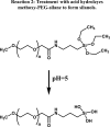
Figure 3. Reaction 2: methoxy–polyethylene glycol (PEG)‐silane was treated with acid to hydrolyze the ester linkages, thus producing PEG‐silanols with exposed OH groups.
Figure 4
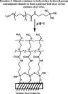
Figure 4. Reaction 3: PEG‐silanols condensed with OH groups both on the wire and on neighboring silanols to form a polymerized primer layer.
Figure 5
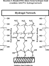
Figure 5. Reaction 4: grafting of the polyvinyl alcohol/polyvinyl pyrrolidone/polyvinyl acetate hydrogel to the primer layer is achieved via hydrogen bonding interactions between the carbonyl, ether and amine groups of polyethylene glycol and the OH groups of polyvinyl alcohol.
Figure 6
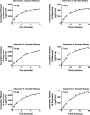
Figure 6. Line graphs demonstrate the elution profile of adenosine from 6 adenosine‐loaded guidewires (Adenowires) in vitro.
The release was curvilinear over ≈60 minutes, a time frame which is ideal for an interventional procedure.
Figure 7
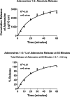
Figure 7. Line graphs depict the average elution profile from the 6 adenosine‐loaded guidewires shown in Figure 6.
The results are plotted either as cumulative release of absolute amounts of adenosine (top panel) or as % release relative to the 60‐minute time point (bottom panel). The average release at 60 minutes was 2.7±0.2 mg per 10 cm of length. Values are means±SEM.
Figure 8
Figure 8. Line graphs summarize the effects on coronary flow velocity (CFV) of adenosine‐releasing guidewires (n=8) inserted into the LAD or circumflex coronary artery of Yucatan mini‐pigs.
Coronary flow velocity (A) was markedly increased immediately upon wire insertion and remained significantly elevated for 33 minutes. B, The fold increase in CFV, which equates to coronary flow reserve. The coronary flow reserve was >2.0 for the first 15 minutes. The small spike in CFV when the guidewire was removed was likely attributable to an incremental surge in adenosine release caused by wire manipulation. The adenosine‐releasing wire did not affect mean blood pressure (MABP) or heart rate (C and D, respectively). Values are means and SEMs. RM 1‐Factor ANOVA indicates repeated measures 1‐factor analysis of variance. Bonferroni indicates Bonferroni test for comparisons to baseline (control) of subsequent time points after insertion of the Adenowire.
Figure 9
Figure 9. Coronary angiogram of circumflex in left anterior oblique projection.
A, Baseline; (B) adenosine guidewire positioned in distal vessel with radio‐opaque tip (solid red arrow) clearly visible; (C) post wire removal after 30 minutes showing normal flow. Also shown (dashed yellow arrow) is the doppler Combowire used to measure coronary flow velocity.
Figure 10
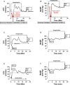
Figure 10. Line graphs summarize 3 experiments in which acetylcholine was infused into the coronary artery before the adenosine‐loaded guidewire was inserted into the artery.
Experiment 1 is summarized in (A and B); experiment 2 in (C and D); and experiment 3 in (E and F). Coronary flow velocities (CFV) are illustrated in (A, C, and E) and associated mean arterial blood pressures (MABPs) are shown in (B, D, and F). In experiment 1, the animal developed severe ischemia that induced ventricular fibrillation requiring cardioversion. In all 3 experiments, acetylcholine caused coronary vasoconstriction that was reversed by insertion of the adenosine‐releasing guidewire.
Figure 11
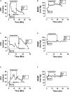
Figure 11. Line graphs summarize 3 experiments in which acetylcholine was infused into the coronary artery after the adenosine‐loaded guidewire was inserted into the artery.
Experiment 1 is summarized in (A and B); experiment 2 in (C and D); and experiment 3 in (E and F). Coronary flow velocities (CFVs) are illustrated in (A, C, and E) and associated mean arterial blood pressures (MABPs) are shown in (B, D, and F). In all 3 experiments, insertion of the adenosine‐releasing guidewire immediately increased CFV and prevented acetylcholine from reducing CFV below baseline.
Figure 12
Figure 12. Line graphs summarize 2 additional experiments in which acetylcholine was infused into the coronary artery after the adenosine‐loaded guidewire was inserted into the artery.
Experiment 1 is summarized in (A and B); and experiment 2 in (C and D). Coronary flow velocities (CFVs) are illustrated in (A and C) and associated mean arterial blood pressures (MABPs) are shown in (B and D). In both experiments, insertion of the adenosine‐releasing guidewire immediately increased CFV and prevented acetylcholine from reducing CFV below baseline. Note that in (C) removal of the adenosine‐releasing guidewire while maintaining the intracoronary infusion of acetylcholine resulted in a rapid and profound decline in CFV.
Figure 13
Figure 13. Coronary angiography showing an example of the no‐reflow phenomenon (NRF) reversed by an adenosine‐releasing guidewire.
A, A baseline angiogram was obtained with the Combowire (dashed yellow arrow) in the proximal LAD. B, Soon after administration of acetylcholine, severe NRF occurred with total cessation of blood flow in the proximal LAD (solid red arrow). C, Upon placement of an adenosine‐releasing guidewire (solid purple arrow) in the LAD, coronary flow was rapidly restored and maintained even while infusing acetylcholine for an additional 20 minutes. D, After stopping acetylcholine and removing the guidewire, a final angiogram showed that normal coronary flow was maintained.
Figure 14
Figure 14. Track forces (g) of 4 Adenowires and 3 commercial wires with a proprietary hydrophilic coating is illustrated.
Note that the insertion and withdrawal forces were similar, which was indicative of comparable lubricity properties.
Figure 15
Figure 15. Effects of Adenowires and Control wires (wires coated with the novel coating formulation but without adenosine) on area under the aggregation curve (AUC), maximum aggregation, rate of aggregation (slope) and lag time between addition of agonist and the onset of aggregation.
A, Representative platelet aggregation tracings showing aggregation responses to ADP (2 µmol/L), collagen (1 µg/mL), ADP+Adenowire, collagen+Adenowire, ADP+Control wire and collagen+Control wire. Adenowires were coated with our novel hydrophilic coating that contained adenosine; Control wires were similarly coated but did not contain adenosine. Bar graphs summarize the effects of treatments on (B) area under the aggregation curve, (C) maximum aggregation, (D) slope of the aggregation curve and (E) the lag time between adding the agonist and onset of aggregation. Note that Adenowires markedly inhibited platelet aggregation. The inert coating also manifested antiplatelet properties that was similar to, but not quite as efficacious as, Adenowires. Data were analyzed by 1‐factor ANOVA after Box‐Cox transformation. *Significantly different vs agonist without wire; Significantly different vs corresponding Control wire.

References

    1. Go AS, Mozaffarian D, Roger VL, Benjamin EJ, Berry JD, Blaha MJ, Dai S, Ford ES, Fox CS, Franco S, et al. Heart disease and stroke statistics ‐2014 update: a report from the American Heart Association. Circulation. 2014;129:e28–e292. - PMC - PubMed
    1. Keely EC, Boura JA, Grines CL. Primary angioplasty versus intravenous thrombolytic therapy for acute infarction: a quantitative review of 23 randomized trials. Lancet. 2013;361:13–20. - PubMed
    1. Jaffe R, Dick A, Strauss BH. Prevention and treatment of microvascular obstruction–related myocardial injury and coronary no‐reflow following percutaneous intervention: a systematic approach. JACC Cardiovasc Interv. 2010;3:695–704. - PubMed
    1. Forman MB, Jackson EK. Importance of tissue perfusion in ST segment elevation myocardial infarction undergoing reperfusion strategies: role of adenosine. Clin Cardiol. 2007;30:583–585. - PMC - PubMed
    1. Forman MB, Stone GW, Jackson EK. Role of adenosine as adjunctive therapy in acute myocardial infarction. Cardiovasc Drug Rev. 2006;24:116–147. DOI: 10.1111/j.1527-3466.2006.00116.x . - DOI - PubMed

Publication types

LinkOut - more resources