

Determinants of left atrial reservoir and pump strain and use of atrial strain for evaluation of left ventricular filling pressure
- PMID: 33496314
- PMCID: PMC8685600
- DOI: 10.1093/ehjci/jeaa415
Determinants of left atrial reservoir and pump strain and use of atrial strain for evaluation of left ventricular filling pressure
Erratum in
-
Corrigendum to: Determinants of left atrial reservoir and pump strain and use of atrial strain for evaluation of left ventricular filling pressure.Eur Heart J Cardiovasc Imaging. 2021 Dec 18;23(1):136. doi: 10.1093/ehjci/jeab194. Eur Heart J Cardiovasc Imaging. 2021. PMID: 34608485 Free PMC article. No abstract available.
Abstract
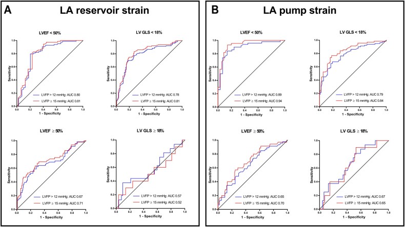
Aims: The aim of this study is to investigate determinants of left atrial (LA) reservoir and pump strain and if these parameters may serve as non-invasive markers of left ventricular (LV) filling pressure.
Methods and results: In a multicentre study of 322 patients with cardiovascular disease of different aetiologies, LA strain and other echocardiographic parameters were compared with invasively measured LV filling pressure. The strongest determinants of LA reservoir and pump strain were LV global longitudinal strain (GLS) (r-values 0.64 and 0.51, respectively) and LV filling pressure (r-values -0.52 and -0.57, respectively). Left atrial volume was another independent, but weaker determinant of both LA strains. For both LA strains, association with LV filling pressure was strongest in patients with reduced LV ejection fraction. Left atrial reservoir strain <18% and LA pump strain <8% predicted elevated LV filling pressure better (P < 0.05) than LA volume and conventional Doppler parameters. Accuracy to identify elevated LV filling pressure was 75% for LA reservoir strain alone and 72% for pump strain alone. When combined with conventional parameters, accuracy was 82% for both LA strains. In patients with normal LV systolic function by GLS, LA pump strain >14% identified normal LV filling pressure with 92% accuracy.
Conclusion: Left atrial reservoir and pump strain are determined predominantly by LV GLS and filling pressure. Accuracy of LA strains to identify elevated LV filling pressure was best in patients with reduced LV systolic function. High values of LA pump strain, however, identified normal LV filling pressure with good accuracy in patients with normal systolic function.
Keywords: catheterization; diastolic dysfunction; heart failure; left atrial strain; left ventricular filling pressure.
© The Author(s) 2021. Published by Oxford University Press on behalf of the European Society of Cardiology.
Figures


Comment in
-
Left atrial strain: evaluating left ventricular filling pressure from an upstream vantage point.Eur Heart J Cardiovasc Imaging. 2021 Dec 18;23(1):71-73. doi: 10.1093/ehjci/jeab015. Eur Heart J Cardiovasc Imaging. 2021. PMID: 33822927 No abstract available.
References
-
- Nagueh SF, Smiseth OA, Appleton CP, Byrd BF 3rd, Dokainish H, Edvardsen T et al. Recommendations for the evaluation of left ventricular diastolic function by echocardiography: an update from the American Society of Echocardiography and the European Association of Cardiovascular Imaging. Eur Heart J Cardiovasc Imaging 2016;17:1321–60. - PubMed
-
- Andersen OS, Smiseth OA, Dokainish H, Abudiab MM, Schutt RC, Kumar A et al. Estimating left ventricular filling pressure by echocardiography. J Am Coll Cardiol 2017;69:1937–48. - PubMed
-
- Lancellotti P, Galderisi M, Edvardsen T, Donal E, Goliasch G, Cardim N et al. Echo-Doppler estimation of left ventricular filling pressure: results of the multicentre EACVI Euro-Filling study. Eur Heart J Cardiovasc Imaging 2017;18:961–8. - PubMed
-
- Badano LP, Kolias TJ, Muraru D, Abraham TP, Aurigemma G, Edvardsen T, Industry representatives et al. Standardization of left atrial, right ventricular, and right atrial deformation imaging using two-dimensional speckle tracking echocardiography: a consensus document of the EACVI/ASE/Industry Task Force to standardize deformation imaging. Eur Heart J Cardiovasc Imaging 2018;19:591–600. - PubMed
-
- Rahimtoola SH, Loeb HS, Ehsani A, Sinno MZ, Chuquimia R, Lal R et al. Relationship of pulmonary artery to left ventricular diastolic pressures in acute myocardial infarction. Circulation 1972;46:283–90. - PubMed
Publication types
MeSH terms
LinkOut - more resources
Full Text Sources
Other Literature Sources
Miscellaneous
