. 2021 Nov;40(11):2339-2351.
doi: 10.1002/jum.15617. Epub 2021 Jan 26.

Lung Ultrasound for Identification of Patients Requiring Invasive Mechanical Ventilation in COVID-19

Affiliations

Lung Ultrasound for Identification of Patients Requiring Invasive Mechanical Ventilation in COVID-19

Claudia Seiler et al. J Ultrasound Med. 2021 Nov.

Abstract

Objectives: Indication for invasive mechanical ventilation in COVID-19 pneumonia has been a major challenge. This study aimed to evaluate if lung ultrasound (LUS) can assist identification of requirement of invasive mechanical ventilation in moderate to severe COVID-19 pneumonia.

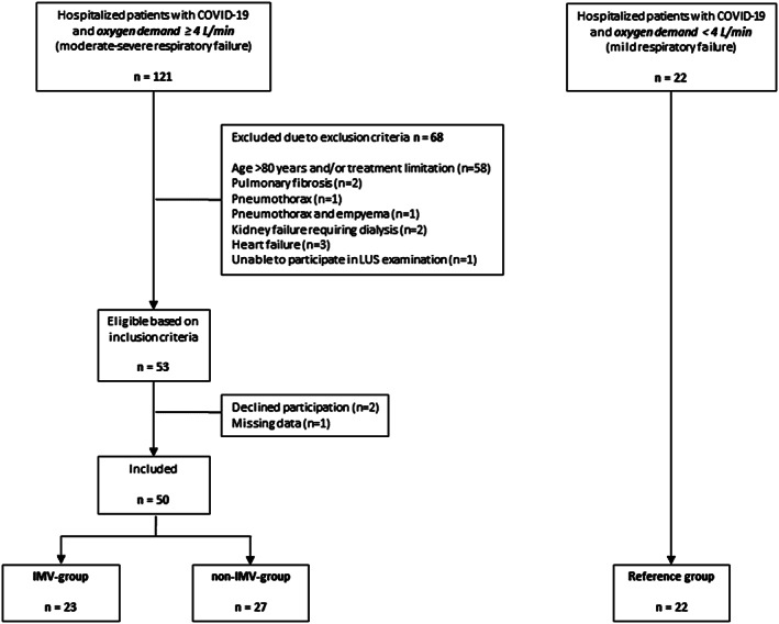
Materials and methods: Between April 23 and November 12, 2020, hospitalized patients with moderate to severe COVID-19 (oxygen demand ≥4 L/min) were included consecutively. Lung ultrasound was performed daily until invasive mechanical ventilation (IMV-group) or spontaneous recovery (non-IMV-group). Clinical parameters and lung ultrasound findings were compared between groups, at intubation (IMV-group) and highest oxygen demand (non-IMV-group). A reference group with oxygen demand <4 L/min was examined at hospital admission.

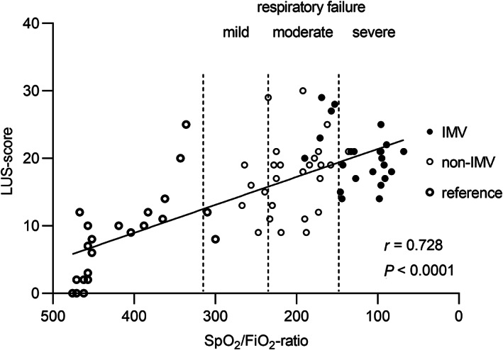
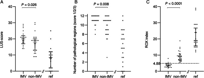
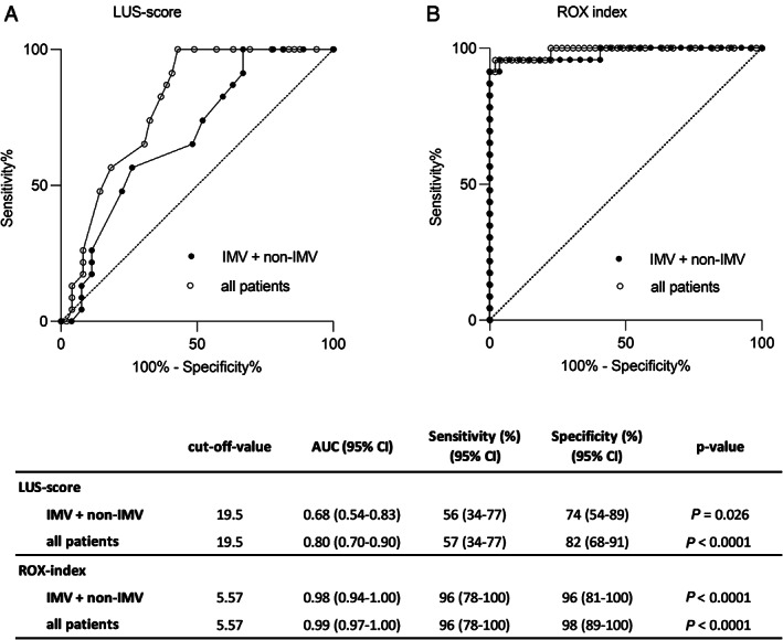
Results: Altogether 72 patients were included: 50 study patients (IMV-group, n = 23; non-IMV-group, n = 27) and 22 reference patients. LUS-score correlated to oxygen demand (SpO2 /FiO2 -ratio) (r = 0.728; p < .0001) and was higher in the IMV-group compared to the non-IMV-group (20.0 versus 18.0; p = .026). Based on receiver operating characteristic analysis, a LUS-score of 19.5 was identified as cut-off for requirement of invasive mechanical ventilation (area under the curve 0.68; sensitivity 56%, specificity 74%). In 6 patients, LUS identified critical coexisting conditions. Respiratory rate and oxygenation index ((SpO2 /FiO2 )/respiratory rate) ≥4.88 identified no requirement of invasive mechanical ventilation with a positive predictive value of 87% and negative predictive value of 100%.

Conclusions: LUS-score had only a moderate diagnostic value for requirement of invasive mechanical ventilation in moderate to severe COVID-19. However, LUS proved valuable as complement to respiratory parameters in guidance of disease severity and identifying critical coexisting conditions.

Keywords: COVID-19; Coronavirus; mechanical; ultrasonography; ventilators.


Figures

Figure 1
Figure 1
Study flowchart. Patients positive for SARS‐CoV‐2 by reverse transcription polymerase chain reaction and oxygen demand ≥4 L/min were eligible for the study. Patients were followed daily until either initiation of IMV (IMV‐group) or spontaneous improvement without requirement of IMV (non‐IMV‐group). Patients in the reference group were included based on the same inclusion or exclusion criteria but with oxygen demand <4 L/min. IMV = invasive mechanical ventilation.
Figure 2
Figure 2
Correlation between LUS‐score and oxygen demand defined as SpO2/FiO2‐ratio. Cut‐off values (dotted lines) for mild, moderate and severe respiratory failure are defined as SpO2/FiO2‐ratio 315, 235 and 148, respectively. Data presented prior to intubation (IMV‐group) and on the day with highest oxygen demand (non‐IMV‐group). For the reference group data were collected within 48 hours after hospital admission. Correlation coefficient and p‐value given for Pearson correlation. See Figure 1 legend for definition of groups. LUS = lung ultrasound; IMV = invasive mechanical ventilation; SpO2 = peripheral oxygen saturation; FiO2 = inspired fraction of oxygen.
Figure 3
Figure 3
LUS findings and ROX index presented for IMV‐, non‐IMV‐ and reference groups. See Figure 1 legend for definition of groups. (A) LUS‐score (0–36) for each patient based on 12 lung regions examined. (B) Total number of regions with pathological findings (score 1/2/3) for each patient. (C) ROX index ((SpO2/FiO2)/respiratory rate) based on Roca et al. The dotted line marks suggested cut‐off for low risk for intubation (≥4.88). Comparisons between IMV‐ and non‐IMV‐group using the Mann–Whitney U test. LUS = Lung ultrasound; ROX index = Respiratory rate and oxygenation index; IMV = invasive mechanical ventilation; SpO2 = peripheral oxygen saturation; FiO2 = inspired fraction of oxygen.
Figure 4
Figure 4
Distribution of LUS‐findings over anterior, lateral and posterior lung fields. Each field presents the sum of 4 regions examined by LUS (2 right and 2 left regions ‐ upper and basal) (A) Different qualities of the LUS‐score and (B) categories of consolidations are presented as percentage for 3 examined fields for each patient (anterior, lateral, posterior). Data are presented separately for each study group: IMV‐, non‐IMV‐ and the reference group; see Figure 1 legend for definition of groups. P‐values given for comparison between fields of large consolidations (score 3) using Friedman's test. Post hoc test for comparison of score 3 between anterior and posterior field using Wilcoxon signed rank test (IMV p = .013; non‐IMV p = .015). LUS = Lung ultrasound; IMV = invasive mechanical ventilation.
Figure 6
Figure 6
Reciever operating characteristic (ROC) curve analysis evaluating the diagnostic value of LUS‐score and ROX index for prediction of IMV treatment. ROC curves presented for IMV‐ and non‐IMV‐groups and for all patients (IMV, non‐IMV and reference group). See Figure 1 legend for definition of groups. (A) LUS‐score and (B) ROX index as diagnostic tests for determination of requirement of IMV treatment. The table presents suggested cut‐off values (based on Youden index) together with area under the curve (95% CI) and sensitivity and specificity. LUS = Lung ultrasound; ROX index = Respiratory rate and oxygenation index; IMV = invasive mechanical ventilation.
Figure 5
Figure 5
Photos illustrating the different qualities of LUS findings accounting for the LUS‐score and subpleural consolidations found in patients with COVID‐19. Score 0: normal findings with A‐lines or ≤2 B‐lines; Score 1: 3 or more well separated B‐lines; Score 2: coalescent B‐lines; Score 3: large consolidation with air bronchogram; Small subpleural consolidation <1 cm; Large subpleural consolidation >1 cm. LUS = Lung ultrasound.

References

    1. World Health Organization timeline : WHO's COVID‐19 response. https://www.who.int/emergencies/diseases/novel‐coronavirus‐2019/interact...
    1. Guan W, Ni Z, Hu Y, et al. Clinical characteristics of coronavirus disease 2019 in China. N Engl J Med 2020; 382:1708–1720. - PMC - PubMed
    1. Gattinoni L, Chiumello D, Caironi P, et al. COVID‐19 pneumonia: different respiratory treatments for different phenotypes? Intensive Care Med 2020; 46:1099–1102. - PMC - PubMed
    1. Brochard L, Slutsky A, Pesenti A. Mechanical ventilation to minimize progression of lung injury in acute respiratory failure. Am J Respir Crit Care Med 2017; 195:438–442. - PubMed
    1. Volpicelli G, Elbarbary M, Blaivas M, et al. International evidence‐based recommendations for point‐of‐care lung ultrasound. Intensive Care Med 2012; 38:577–591. - PubMed

Grants and funding