Review
. 2021 Feb;59(1-2):e23410.
doi: 10.1002/dvg.23410. Epub 2021 Jan 26.

Aquatic models of human ciliary diseases

Affiliations
Review

Aquatic models of human ciliary diseases

Mark E Corkins et al. Genesis. 2021 Feb.

Abstract

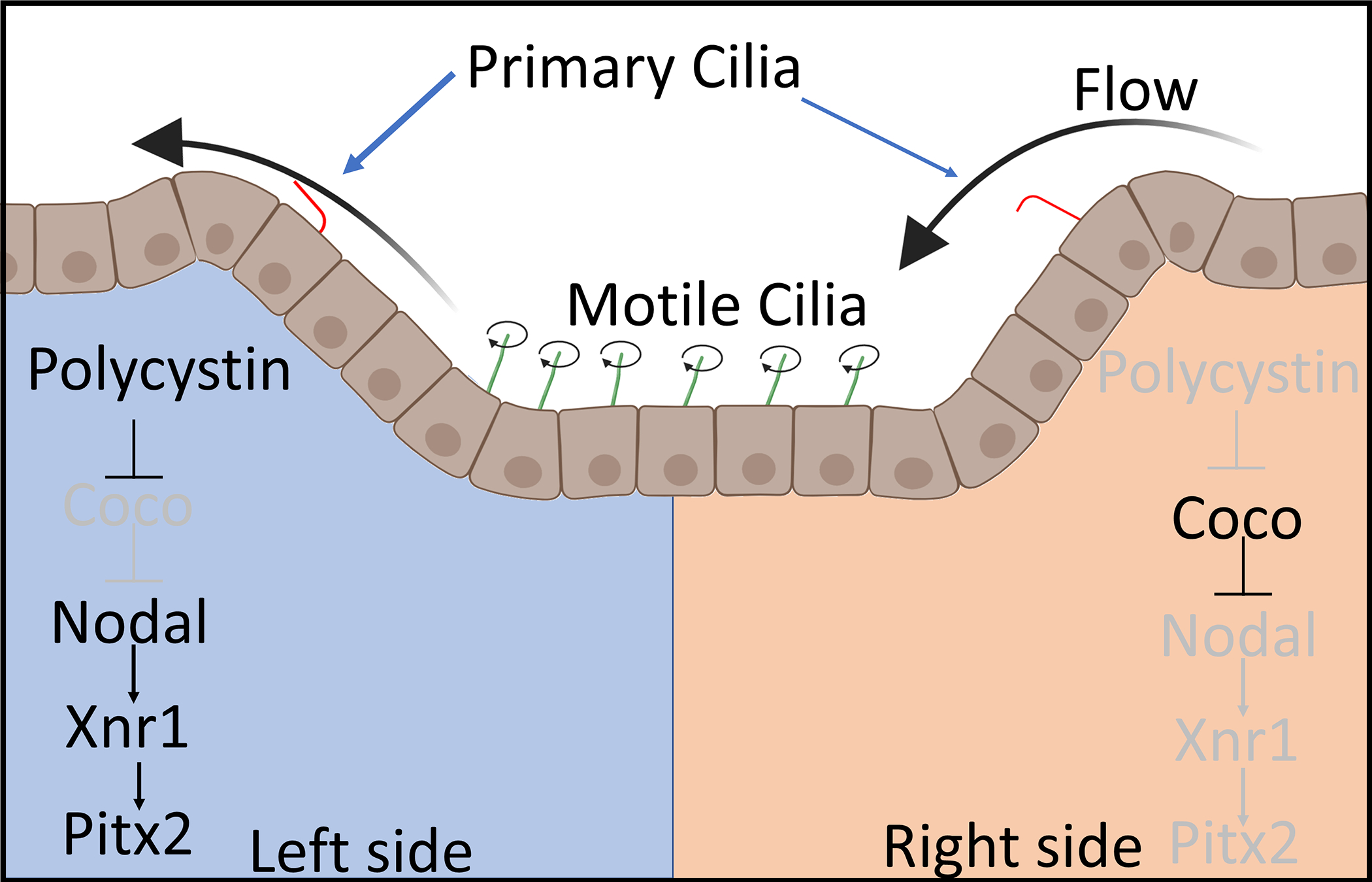
Cilia are microtubule-based structures that either transmit information into the cell or move fluid outside of the cell. There are many human diseases that arise from malfunctioning cilia. Although mammalian models provide vital insights into the underlying pathology of these diseases, aquatic organisms such as Xenopus and zebrafish provide valuable tools to help screen and dissect out the underlying causes of these diseases. In this review we focus on recent studies that identify or describe different types of human ciliopathies and outline how aquatic organisms have aided our understanding of these diseases.

Keywords: Xenopus; cilia; ciliopathy; cystic kidney; kidney; nasal; node; zebrafish.


Figures

Figure 1:
Figure 1:
Structure of cilia. The cilia are composed of three main sections, the axoneme which performs the sensory or movement function, the transition zone which likely contains over 100 proteins which function to anchor the cilia and regulate transport to and from the cilia, and the basal body which is a centriole that functions as a tubulin organizing center to form the cilia. Diagram showing the cross section of the axoneme of common types of motile and primary cilia in vertebrates.
Figure 2:
Figure 2:
Confocal images of wholemount zebrafish (3dpf) and Xenopus laevis (Stage 37) kidney cilia. Cilia were stained using an acetylated alpha-tubulin antibody (Sigma T6793) which labels the neurons and cilia. Kidney cilia are pseudocolored in green while neurons and epithelial cilia are pseudocolored in red. The zebrafish and Xenopus kidney are outlined in white dashed lines, and motile multiciliated cells in the kidney are pseudocolored in magenta. Images were taken on a Zeiss LSM800 confocal microscope.
Figure 3:
Figure 3:
Images of Xenopus laevis motile epidermal cilia. A-C) Confocal imaging of acetylated alpha-tubulin stained whole mount Xenopus embryo. D-E) Scanning electron micrograph of the skin of whole mount Xenopus embryo. E-F) Transmission electron microscopy showing sections through cilia. F) Image showing basal body and axoneme of motile cilia G.) Image showing cross-section and the 9+2 microtubule structure of motile cilia. C,E) Zoomed in image of white dashed box in B and D.
Figure 4:
Figure 4:
Confocal images of the motile cilia lining the zebrafish nasal (olfactory) pit. Dorsal view of 8dpf zebrafish embryos with head towards the top of the image. Embryos were fixed and stained with acetylated alpha-tubulin (Green) (Sigma T6793) and DAPI (Blue). Acetylated tubulin labels both the cilia and neurons. Nasal pits are circled in white, and neural mast cells are circled in red.
Figure 5:
Figure 5:
Diagram of a posterior view of the Left-Right organizer and its functions. Motile cilia (green) create a leftward flow of fluid over the cleft. This leftward flow activated primary cilia (red) on the left half of the cleft resulting in the opening of polycystin calcium channels. Calcium influx inhibits a protein Coco leading to activation of Nodal signaling.

Similar articles

Cited by

References

    1. Ajima Rieko, and Hamada Hiroshi. 2011. “Wnt Signalling Escapes to Cilia.” Nature Cell Biology. - PubMed
    1. Antic Dragana, Stubbs Jennifer L., Suyama Kaye, Kintner Chris, Scott Matthew P., and Axelrod Jeffrey D.. 2010. “Planar Cell Polarity Enables Posterior Localization of Nodal Cilia and Left-Right Axis Determination during Mouse and Xenopus Embryogenesis.” PLoS ONE. - PMC - PubMed
    1. Aslan Yetki, Tadjuidje Emmanuel, Zorn Aaron M., and Cha Sang Wook. 2017. “High-Efficiency Non-Mosaic CRISPR-Mediated Knock-in and Indel Mutation in F0 Xenopus.” Development (Cambridge). - PMC - PubMed
    1. Austin-Tse Christina, Halbritter Jan, Zariwala Maimoona A., Gilberti Renée M., Gee Heon Yung, Hellman Nathan, Pathak Narendra, Liu Yan, Panizzi Jennifer R., Patel-King Ramila S., Tritschler Douglas, Bower Raqual, O’Toole Eileen, Porath Jonathan D., Hurd Toby W., Chaki Moumita, Diaz Katrina A., Kohl Stefan, Lovric Svjetlana, Daw Yang Hwang Daniela A. Braun, Schueler Markus, Airik Rannar, Otto Edgar A., Leigh Margaret W., Noone Peadar G., Carson Johnny L., Davis Stephanie D., Pittman Jessica E., Ferkol Thomas W., Atkinson Jeffry J., Olivier Kenneth N., Sagel Scott D., Dell Sharon D., Rosenfeld Margaret, Milla Carlos E., Loges Niki T., Omran Heymut, Porter Mary E., King Stephen M., Knowles Michael R., Drummond Iain A., and Hildebrandt Friedhelm. 2013. “Zebrafish Ciliopathy Screen plus Human Mutational Analysis Identifies C21orf59 and CCDC65 Defects as Causing Primary Ciliary Dyskinesia.” American Journal of Human Genetics. - PMC - PubMed
    1. Baier Herwig, Klostermann Stefan, Trowe Torsten, Karlstrom Rolf O., Nüsslein-Volhard Christiane, and Bonhoeffer Friedrich. 1996. “Genetic Dissection of the Retinotectal Projection.” Development. - PubMed

Publication types

LinkOut - more resources