

doi: 10.3201/eid2702.203884.
Protective Immunity and Persistent Lung Sequelae in Domestic Cats after SARS-CoV-2 Infection
- PMID: 33496650
- PMCID: PMC7853591
- DOI: 10.3201/eid2702.203884
Item in Clipboard
Protective Immunity and Persistent Lung Sequelae in Domestic Cats after SARS-CoV-2 Infection
Emerg Infect Dis.
2021 Feb.
Abstract
Severe acute respiratory syndrome coronavirus 2 readily transmits between domestic cats. We found that domestic cats that recover from an initial infection might be protected from reinfection. However, we found long-term persistence of inflammation and other lung lesions after infection, despite a lack of clinical symptoms and limited viral replication in the lungs.
Keywords: COVID-19; SARS-CoV-2; cats; coronavirus disease; lung sequelae; protective immunity; reinfection; respiratory infections; severe acute respiratory syndrome coronavirus 2; viruses; zoonoses.
Figures
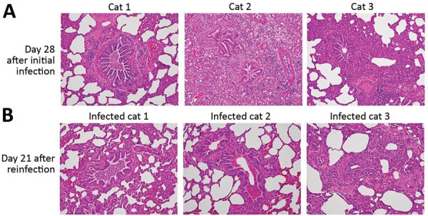
Comparison of histopathology between cats on day 28 after initial infection with severe acute respiratory syndrome coronavirus 2 and on day 21 after reinfection. Bronchioles and alveoli of cats (cats 1–3 in Appendix Figure 6) on day 28 after initial infection (A) and those of cats (infected cats 1–3 in Appendix Figure 6, upper half) on day 21 after reinfection (49 days after the initial infection) (B); original magnification × 20. Cats from both groups showed histiocytic bronchiolitis with occlusive plugs, peribronchiolar fibrosis, and thickening of alveolar septa. Mild acute hemorrhage was detected in affected and less affected regions of the lung on day 21 after reinfection, with a trend toward an increase compared with day 28 (severity score 1.8 + SEM 0.8 on day 21 vs. 0.3 + SEM 0.2 on day 28; p = 0.187 by unpaired t-test).
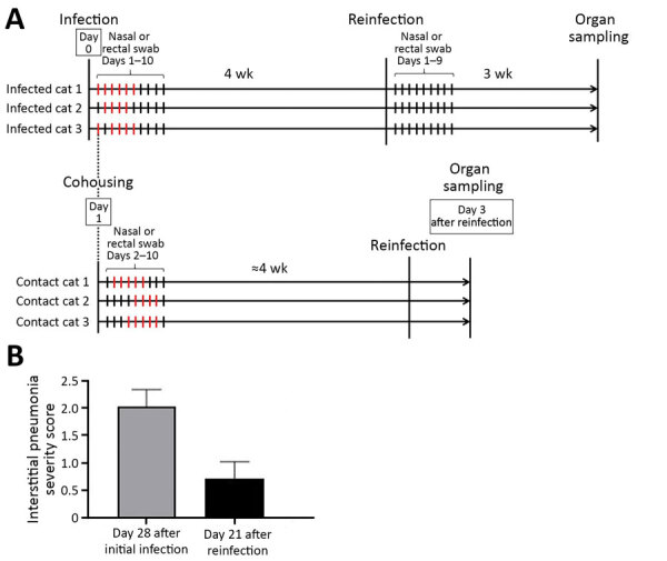
Timeline of severe acute respiratory syndrome coronavirus 2 infection and reinfection of cats and distribution of interstitial thickening. A) Timeline of infection and reinfection. As reported previously (1), a group of cats was inoculated with severe acute respiratory syndrome coronavirus 2 on day 0 (infected cats 1–3, upper half). A virus-naive cat was cohoused with each of the infected cats from day 1 (contact cats 1–3, lower half). The days on which infectious virus was detected in the nasal swabs are shown as red bars for each animal. In this study, we infected the cats with the same severe acute respiratory syndrome coronavirus 2 isolate at ≈4 weeks after initial infection or exposure to infected cats. After reinfection of the group shown in the upper half of the figure, no infectious virus was detected in the nasal swabs. The cats were confirmed to be seronegative before the initial infection or cohousing with infected cats, and seropositive before reinfection, on the basis of neutralization assay results. B) The distribution of interstitial thickening (interstitial pneumonia severity score) was decreased on day 21 after reinfection compared with day 28 (p = 0.041 by unpaired t-test).
Similar articles
-
Household Cases Suggest That Cats Belonging to Owners with COVID-19 Have a Limited Role in Virus Transmission.Viruses. 2021 Apr 14;13(4):673. doi: 10.3390/v13040673. Viruses. 2021. PMID: 33919936 Free PMC article.
-
SARS-CoV-2 Infection and Antibody Response in a Symptomatic Cat from Italy with Intestinal B-Cell Lymphoma.Viruses. 2021 Mar 23;13(3):527. doi: 10.3390/v13030527. Viruses. 2021. PMID: 33806922 Free PMC article.
-
SARS-CoV-2 in Quarantined Domestic Cats from COVID-19 Households or Close Contacts, Hong Kong, China.Emerg Infect Dis. 2020 Dec;26(12):3071-3074. doi: 10.3201/eid2612.202786. Epub 2020 Sep 16. Emerg Infect Dis. 2020. PMID: 32938527 Free PMC article.
-
Anthropogenic Infection of Cats during the 2020 COVID-19 Pandemic.Viruses. 2021 Jan 26;13(2):185. doi: 10.3390/v13020185. Viruses. 2021. PMID: 33530620 Free PMC article. Review.
-
Insights into SARS-CoV-2 Persistence and Its Relevance.Viruses. 2021 May 29;13(6):1025. doi: 10.3390/v13061025. Viruses. 2021. PMID: 34072390 Free PMC article. Review.
Cited by
-
Cats and SARS-CoV-2: A Scoping Review.Animals (Basel). 2022 May 30;12(11):1413. doi: 10.3390/ani12111413. Animals (Basel). 2022. PMID: 35681877 Free PMC article.
-
Higher Frequency of SARS-CoV-2 RNA Shedding by Cats than Dogs in Households with Owners Recently Diagnosed with COVID-19.Viruses. 2024 Oct 11;16(10):1599. doi: 10.3390/v16101599. Viruses. 2024. PMID: 39459932 Free PMC article.
-
Zoonotic and Reverse Zoonotic Transmissibility of SARS-CoV-2.Virus Res. 2021 Sep;302:198473. doi: 10.1016/j.virusres.2021.198473. Epub 2021 Jun 9. Virus Res. 2021. PMID: 34118360 Free PMC article. Review.
-
SARS-CoV-2 antibodies in dogs and cats in a highly infected area of Brazil during the pandemic.Front Vet Sci. 2023 Feb 23;10:1111728. doi: 10.3389/fvets.2023.1111728. eCollection 2023. Front Vet Sci. 2023. PMID: 36908526 Free PMC article.
-
Infection Dynamics, Pathogenesis, and Immunity to SARS-CoV-2 in Naturally Susceptible Animal Species.J Immunol. 2023 Oct 15;211(8):1195-1201. doi: 10.4049/jimmunol.2300378. J Immunol. 2023. PMID: 37782853 Free PMC article. Review.
References
Publication types
MeSH terms
Grants and funding
LinkOut - more resources
Full Text Sources
Other Literature Sources
Medical
Miscellaneous
