

Effects of childhood adversity on the volumes of the amygdala subnuclei and hippocampal subfields in individuals with major depressive disorder
- PMID: 33497169
- PMCID: PMC7955852
- DOI: 10.1503/jpn.200034
Effects of childhood adversity on the volumes of the amygdala subnuclei and hippocampal subfields in individuals with major depressive disorder
Abstract
Background: Reductions in total hippocampus volume have frequently been reported in MRI studies in major depressive disorder (MDD), but reports of differences in total amygdala volume have been inconsistent. Childhood maltreatment is an important risk factor for MDD in adulthood and may affect the volume of the hippocampus and amygdala. In the present study, we examined associations between the volumes of the amygdala subnuclei and hippocampal subfields and history of childhood maltreatment in participants with MDD.
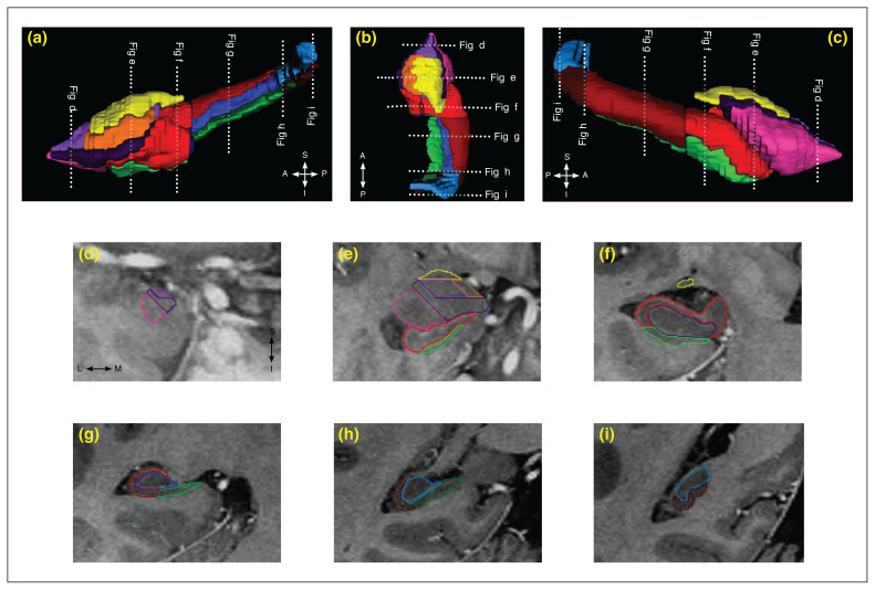
Methods: We recruited 35 patients who met the DSM-IV criteria for MDD and 35 healthy controls. We acquired MRI data sets on a 4.7 T Varian Inova scanner. We manually delineated the amygdala subnuclei (lateral, basal and accessory basal nuclei, and the cortical and centromedial groups) and hippocampal subfields (cornu ammonis, subiculum and dentate gyrus) using reliable volumetric methods. We assessed childhood maltreatment using the Childhood Trauma Questionnaire in participants with MDD.
Results: In participants with MDD, a history of childhood maltreatment had significant negative associations with volume in the right amygdala, anterior hippocampus and total cornu ammonis subfield bilaterally. For volumes of the amygdala subnuclei, such effects were limited to the basal, accessory basal and cortical subnuclei in the right hemisphere, but they did not survive correction for multiple comparisons. We did not find significant effects of MDD or antidepressant treatment on volumes of the amygdala subnuclei.
Limitations: Our study was a cross-sectional study.
Conclusion: Our results provide evidence of negative associations between history of childhood maltreatment and volumes of medial temporal lobe structures in participants with MDD. This may help to identify potential mechanisms by which maltreatment leads to clinical impacts.
© 2021 Joule Inc. or its licensors
Conflict of interest statement
None declared.
Figures
References
-
- Global health estimates 2016: disease burden by cause, age, sex, by country and by region, 2000–2016. Geneva: World Health Organization; 2018.
-
- Lorenzetti V, Allen NB, Fornito A, et al. Structural brain abnormalities in major depressive disorder: a selective review of recent MRI studies. J Affect Disord. 2009;117:1–17. - PubMed
-
- Malykhin NV, Coupland NJ. Hippocampal neuroplasticity in major depressive disorder. Neuroscience. 2015;309:200–13. - PubMed
Publication types
MeSH terms
Grants and funding
LinkOut - more resources
Full Text Sources
Other Literature Sources
Miscellaneous