. 2021 Mar 8;6(5):e145185.
doi: 10.1172/jci.insight.145185.

Particulate matter causes skin barrier dysfunction

Affiliations

Particulate matter causes skin barrier dysfunction

Byung Eui Kim et al. JCI Insight. .

Abstract

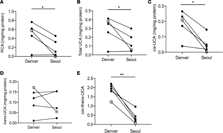
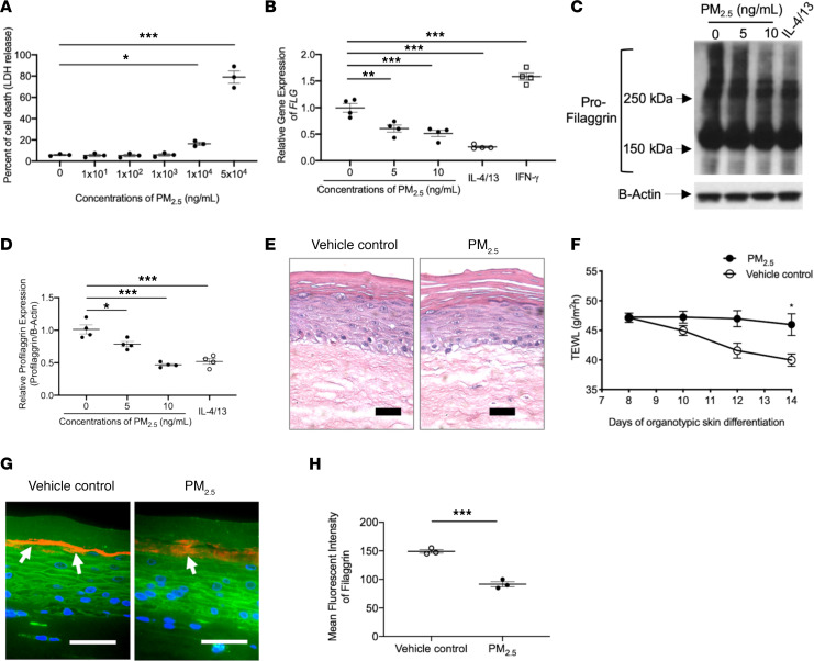
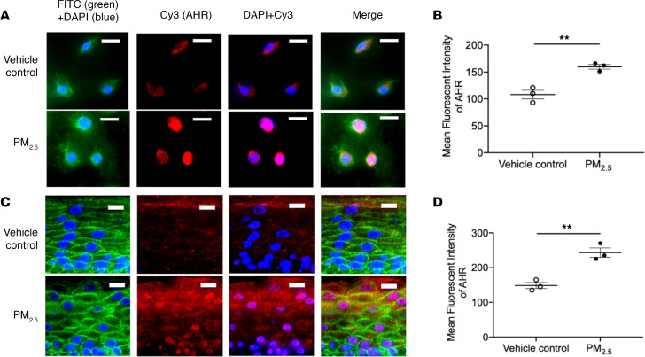
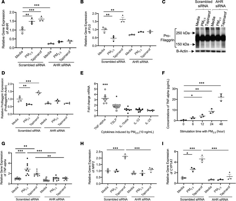
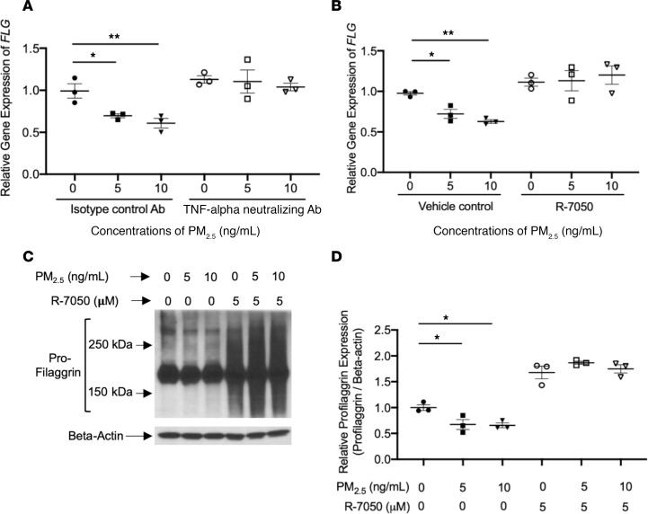
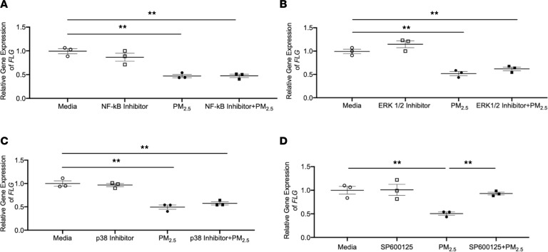
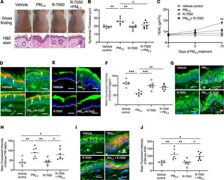
The molecular mechanisms that underlie the detrimental effects of particulate matter (PM) on skin barrier function are poorly understood. In this study, the effects of PM2.5 on filaggrin (FLG) and skin barrier function were investigated in vitro and in vivo. The levels of FLG degradation products, including pyrrolidone carboxylic acid, urocanic acid (UCA), and cis/trans-UCA, were significantly decreased in skin tape stripping samples of study subjects when they moved from Denver, an area with low PM2.5, to Seoul, an area with high PM2.5 count. Experimentally, PM2.5 collected in Seoul inhibited FLG, loricrin, keratin-1, desmocollin-1, and corneodesmosin but did not modulate involucrin or claudin-1 in keratinocyte cultures. Moreover, FLG protein expression was inhibited in human skin equivalents and murine skin treated with PM2.5. We demonstrate that this process was mediated by PM2.5-induced TNF-α and was aryl hydrocarbon receptor dependent. PM2.5 exposure compromised skin barrier function, resulting in increased transepidermal water loss, and enhanced the penetration of FITC-dextran in organotypic and mouse skin. PM2.5-induced TNF-α caused FLG deficiency in the skin and subsequently induced skin barrier dysfunction. Compromised skin barrier due to PM2.5 exposure may contribute to the development and the exacerbation of allergic diseases such as atopic dermatitis.

Keywords: Dermatology; Inflammation; Mouse models; Skin.


Conflict of interest statement

Conflict of interest: The authors have declared that no conflict of interest exists.

Figures

Figure 1
Figure 1. FDP levels decrease in the same subjects after moving from Denver, USA, to Seoul, South Korea, during the same winter season.
STS samples were collected from 4 normal, healthy subjects and 1 convalescent AD subject. A total of 10 consecutive STS samples were collected from the same subjects several months apart while they lived in Denver and Seoul. STS samples were processed, and FDP levels were evaluated using liquid chromatography electrospray ionization tandem mass spectrometry (LC-ESI-MS/MS). (A) PCA. (B) Total UCA. (C) Cis-UCA. (D) Trans-UCA. (E) Cis/trans-UCA. Circles, normal healthy subjects; squares, convalescent AD subjects. n = 5 per group. *P < 0.05, **P < 0.01 by paired 2-tailed Student’s t test.
Figure 2
Figure 2. Effects of PM2.5 on FLG and skin barrier function in cultured keratinocytes and organotypic skin.
(A) The percentage of cell death (lactate dehydrogenase release into cell culture media) is increased after exposure to PM2.5. Gene (B) and protein (C and D) expressions of FLG in cultured HEKs were evaluated using reverse transcriptase PCR (RT-PCR) and Western blotting, respectively, and demonstrated reduced FLG mRNA and protein expression in PM2.5-treated cultures. H&E staining (E) and TEWL (F) in organotypic skin. FLG protein expression (G and H) was evaluated in organotypic skin using immunofluorescence staining. Arrows point to FLG staining (shown in red). Wheat germ agglutinin–conjugated FITC (green) was used to stain the cytoskeleton. Nuclei were visualized with DAPI (blue). Data are representative of 3 independent experimental repetitions using 3 different lots of HEKs. The data are shown as the mean ± SEM. n = 3–4 per group. Scale bar: 50 μm. *P < 0.05, **P < 0.01, ***P < 0.001 by 1-way ANOVA with Tukey-Kramer test (A, B, and D) and 2-tailed Student’s t test (F and H).
Figure 3
Figure 3. Effect of PM2.5 on AHR in both human primary keratinocytes and organotypic skin.
Expressions of AHR (red) in both cultured HEKs (A and B) and organotypic skin (C and D) were evaluated using immunofluorescence staining and demonstrated a reduction in FLG expression after PM2.5 exposure. Wheat germ agglutinin–conjugated FITC (green) was used to stain the cytoskeleton. Nuclei were visualized with DAPI (blue). Data are representative of 3 independent experimental repetitions. The data are shown as the mean ± SEM. n = 3 per group. Scale bar: 50 μm. **P < 0.01 by 2-tailed Student’s t test.
Figure 4
Figure 4. PM2.5 modulates expressions of FLG and TNF-α in human primary keratinocytes.
Gene expressions of AHR (A), FLG (B), TNFA (G), NRF2 (H), and CYP1A1 (I) were examined in cultured HEKs using real-time RT-PCR. n = 3–9 per group. (C and D) Protein expression of FLG was evaluated in cultured HEKs using Western blotting. n = 4 per group. (E) Gene expressions of keratinocyte-derived cytokines were examined in cultured HEKs treated with PM2.5 (10 ng/mL) using real-time RT-PCR. n = 8 per group. (F) Protein levels of TNF-α were evaluated in culture media using ELISA. Data are representative of 3 independent experimental repetitions. The data are shown as the mean ± SEM. *P < 0.05, **P < 0.01, ***P < 0.001 by 1-way ANOVA with Tukey-Kramer post hoc test.
Figure 5
Figure 5. PM2.5-induced TNF-α inhibits FLG expression in human primary keratinocytes.
Gene expression of FLG (A and B) was examined in cultured HEKs using real-time RT-PCR. Protein expression of FLG (C and D) was evaluated using Western blotting. Data are representative of 3 independent experimental repetitions. The data are shown as the mean ± SEM. n = 3 per group. *P < 0.05, **P < 0.01 by 1-way ANOVA with Tukey-Kramer post hoc test.
Figure 6
Figure 6. PM2.5 inhibits FLG through the MAPK/c-JNK pathway.
Gene expression of FLG was examined using real-time RT-PCR in cultured HEKs stimulated with PM2.5 or combinations of each inhibitor and PM2.5. (A) NF-κB inhibitor. (B) ERK1/2 inhibitor. (C) P38 inhibitor. (D) SP600125 inhibitor. Data are representative of 3 independent experimental repetitions. The data are shown as the mean ± SEM. n = 3 per group. **P < 0.01 by 1-way ANOVA with Tukey-Kramer post hoc test.
Figure 7
Figure 7. PM2.5 inhibits FLG and causes skin barrier dysfunction in murine skin.
Hairless mice were treated with a vehicle, PM2.5, R-7050, or a combination of PM2.5 and R-7050 on the back of each mouse twice daily for 10 days. FITC-dextran was applied to the left side of the back of each mouse for 60 minutes on day 10 and demonstrated enhanced barrier penetration of the PM2.5-treated skin. (A) Skin appearance and H&E staining (original magnification, ×100) of the skin biopsy samples in the study groups. Epidermal thickness (B) and TEWL (C) were evaluated and illustrated increased epidermal thickness and TEWL in PM2.5-exposed skin. (D) The penetration of FITC-dextran is enhanced in PM2.5-treated skin and is attenuated by TNF-α inhibitors. Protein expressions of FLG (E and F), TNF-α (G and H), and AHR (I and J) were evaluated using immunofluorescence staining. Arrows point to FLG staining (red). The dotted line represents a border between epidermis and dermis. Wheat germ agglutinin–conjugated FITC (green) was used to stain the cytoskeleton. Nuclei were visualized with DAPI (blue). Data are representative of 2 independent experiments. The data are shown as the mean ± SEM. Each point indicates individual mice, n = 7 mice per group. Scale bar: 25 μm. *P < 0.05, **P < 0.01, ***P < 0.001 by 1-way ANOVA with Tukey-Kramer post hoc test.

References

    1. Guarnieri M, Balmes JR. Outdoor air pollution and asthma. Lancet. 2014;383(9928):1581–1592. doi: 10.1016/S0140-6736(14)60617-6. - DOI - PMC - PubMed
    1. Cooper DM, Loxham M. Particulate matter and the airway epithelium: the special case of the underground? Eur Respir Rev. 2019;28(153):190066. doi: 10.1183/16000617.0066-2019. - DOI - PMC - PubMed
    1. Lelieveld J, et al. Cardiovascular disease burden from ambient air pollution in Europe reassessed using novel hazard ratio functions. Eur Heart J. 2019;40(20):1590–1596. doi: 10.1093/eurheartj/ehz135. - DOI - PMC - PubMed
    1. Zhang P, Zhou X. Health and economic impacts of particulate matter pollution on hospital admissions for mental disorders in Chengdu, Southwestern China. Sci Total Environ. 2020;733:139114. - PubMed
    1. Burnett R, et al. Global estimates of mortality associated with long-term exposure to outdoor fine particulate matter. Proc Natl Acad Sci U S A. 2018;115(38):9592–9597. doi: 10.1073/pnas.1803222115. - DOI - PMC - PubMed

Publication types