. 2021 Mar 15;131(6):e145427.
doi: 10.1172/JCI145427.

Severe SARS-CoV-2 placenta infection can impact neonatal outcome in the absence of vertical transmission

Affiliations

Severe SARS-CoV-2 placenta infection can impact neonatal outcome in the absence of vertical transmission

Fulvia Milena Cribiù et al. J Clin Invest. .

Abstract

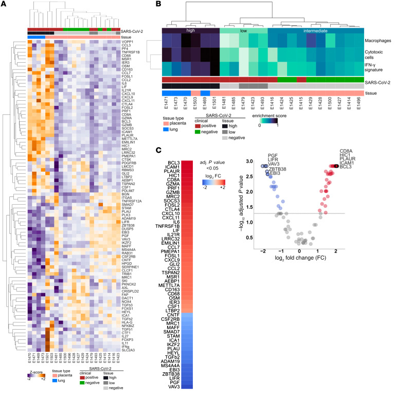
The effect of severe acute respiratory syndrome coronavirus 2 (SARS-CoV-2) infection on the pathophysiology of the placenta and its impact on pregnancy outcome has not yet been fully elucidated. Here, we present a comprehensive clinical, morphological, and molecular analysis of placental tissues from pregnant women with and without SARS-CoV-2 infection. SARS-CoV-2 could be detected in half of placental tissues from SARS-CoV-2-positive women. The presence of the virus was not associated with any distinctive pathological, maternal, or neonatal outcome features. SARS-CoV-2 tissue load was low in all but one patient who exhibited severe placental damage leading to neonatal neurological manifestations. The placental transcriptional response induced by high viral load of SARS-CoV-2 showed an immunopathology phenotype similar to autopsy lung tissues from patients with severe coronavirus disease 2019. This finding contrasted with the lack of inflammatory response in placental tissues from SARS-CoV-2-positive women with low viral tissue load and from SARS-CoV-2-negative women. Importantly, no evidence of vertical transmission of SARS-CoV-2 was found in any newborns, suggesting that the placenta may be an effective maternal-neonatal barrier against the virus even in the presence of severe infection. Our observations suggest that severe placental damage induced by the virus may be detrimental for the neonate independently of vertical transmission.

Keywords: COVID-19; Embryonic development; Molecular pathology; Obstetrics/gynecology; Reproductive Biology.


Conflict of interest statement

Conflict of interest: The authors have declared that no conflict of interest exists.

Figures

Figure 1
Figure 1. Examination of SARS-CoV-2 in Patient 1.
(A) Case timeline. (B) Histopathological and molecular features of the placenta with severe injury. Massive fibrin deposition associated with syncytiotrophoblast layer necrosis and ghost villi (left, H&E staining). Original magnification, ×10. See also Supplemental Figure 1. Placental SARS-CoV-2 in situ hybridization reveals an intense positivity (red staining) of perivillous trophoblastic cells (middle, RNA-ISH). Marked peri-villous trophoblastic cells apoptosis outlined by cleaved caspase-3 brown staining (right, immunohistochemistry). Scale bar: 100 μm.
Figure 2
Figure 2. Gene expression analyses.
(A) Heatmap representing the expression values (z-scaled) of all genes in the Nanostring panel, excluding housekeeping genes (n = 90; rows), across lung and placenta samples (n = 21; columns). (B) Heatmap representing the enrichment scores of cytotoxic cells, macrophages, and IFN-γ signature (rows) in the placenta and lung cohorts (n = 21). E1501 and E1503 are 2 independent samples of severe injured placenta from Patient 1. On top, the resulting dendrogram of sample aggregation (columns) according to the expression values of all genes (A) and enrichment of the 3 signatures (B) are shown. On the left, the dendrogram of gene/signature aggregation (rows) is shown. Sample tissue and SARS-CoV-2 status are highlighted as colored heatmap annotations. (C) Differential genes expression analysis comparing severe injured placenta from Patient 1 (n = 2) and lung samples (n = 6) with the rest of the placental samples (n = 15). In the volcano plot (right), each dot represents a gene and is colored according to Mann Whitney U test results and the log2 fold change (FC). Significantly overexpressed genes (adjusted P < 0.05 and FC > 0) are colored red and significantly underexpressed genes (adjusted P < 0.05 and FC < 0) are colored blue; nonsignificant differentially expressed genes are colored gray. A dashed line represents the significance threshold, adjusted P = 0.05. The top 5 significantly over- and underexpressed genes are highlighted. Column heatmap representing all significant results (adjusted P < 0.05) of the differential expression analysis in (B) (left). The column represents the log2 FC.

References

    1. Liu W, et al. Clinical characteristics of 19 neonates born to mothers with COVID-19. Front Med. 2020;14(2):193–198. doi: 10.1007/s11684-020-0772-y. - DOI - PMC - PubMed
    1. Zhu H, et al. Clinical analysis of 10 neonates born to mothers with 2019-nCoV pneumonia. Transl Pediatr. 2020;9(1):51–60. doi: 10.21037/tp.2020.02.06. - DOI - PMC - PubMed
    1. Chen H, et al. Clinical characteristics and intrauterine vertical transmission potential of COVID-19 infection in nine pregnant women: a retrospective review of medical records. Lancet. 2020;395(10226):809–815. doi: 10.1016/S0140-6736(20)30360-3. - DOI - PMC - PubMed
    1. Yan J, et al. Coronavirus disease 2019 in pregnant women: a report based on 116 cases. Am J Obstet Gynecol. 2020;223(1):111.e1–111.e14. doi: 10.1016/j.ajog.2020.04.014. - DOI - PMC - PubMed
    1. Schwartz DA. An analysis of 38 pregnant women with COVID-19, their newborn infants, and maternal-fetal transmission of SARS-CoV-2: maternal Coronavirus infections and pregnancy outcomes. Arch Pathol Lab Med. 2020;144(7):799–805. doi: 10.5858/arpa.2020-0901-SA. - DOI - PubMed

MeSH terms