Review
. 2021 Jan-Jun:296:100338.
doi: 10.1016/j.jbc.2021.100338. Epub 2021 Jan 23.

Structure, function, and substrates of Clp AAA+ protease systems in cyanobacteria, plastids, and apicoplasts: A comparative analysis

Affiliations
Review

Structure, function, and substrates of Clp AAA+ protease systems in cyanobacteria, plastids, and apicoplasts: A comparative analysis

Imen Bouchnak et al. J Biol Chem. 2021 Jan-Jun.

Erratum in

Abstract

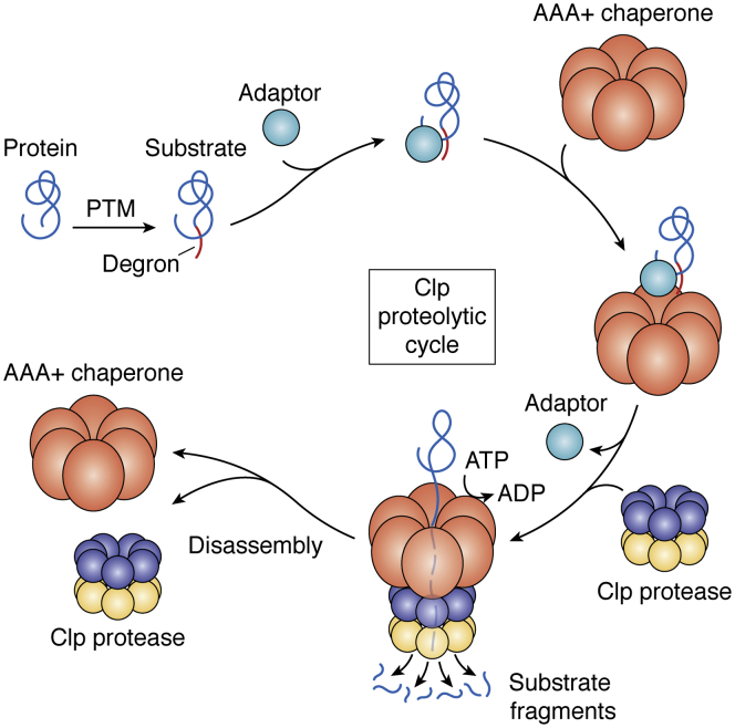
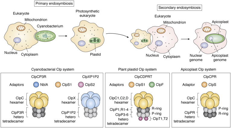
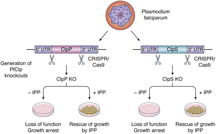
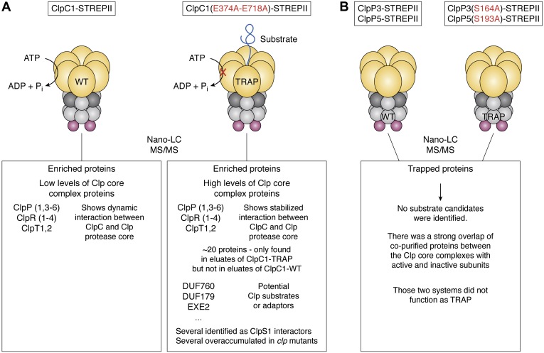
ATPases Associated with diverse cellular Activities (AAA+) are a superfamily of proteins that typically assemble into hexameric rings. These proteins contain AAA+ domains with two canonical motifs (Walker A and B) that bind and hydrolyze ATP, allowing them to perform a wide variety of different functions. For example, AAA+ proteins play a prominent role in cellular proteostasis by controlling biogenesis, folding, trafficking, and degradation of proteins present within the cell. Several central proteolytic systems (e.g., Clp, Deg, FtsH, Lon, 26S proteasome) use AAA+ domains or AAA+ proteins to unfold protein substrates (using energy from ATP hydrolysis) to make them accessible for degradation. This allows AAA+ protease systems to degrade aggregates and large proteins, as well as smaller proteins, and feed them as linearized molecules into a protease chamber. This review provides an up-to-date and a comparative overview of the essential Clp AAA+ protease systems in Cyanobacteria (e.g., Synechocystis spp), plastids of photosynthetic eukaryotes (e.g., Arabidopsis, Chlamydomonas), and apicoplasts in the nonphotosynthetic apicomplexan pathogen Plasmodium falciparum. Recent progress and breakthroughs in identifying Clp protease structures, substrates, substrate adaptors (e.g., NblA/B, ClpS, ClpF), and degrons are highlighted. We comment on the physiological importance of Clp activity, including plastid biogenesis, proteostasis, the chloroplast Protein Unfolding Response, and metabolism, across these diverse lineages. Outstanding questions as well as research opportunities and priorities to better understand the essential role of Clp systems in cellular proteostasis are discussed.

Keywords: AAA+ proteins; Clp protease; N-degrons; Proteases; apicoplasts; chloroplasts; plastids; protease adaptors; proteostasis.


Conflict of interest statement

Conflict of interest The authors declare that they have no conflicts of interest with the contents of this article.

Figures

Figure 1
Figure 1
Evolution of the Clp protease system from cyanobacteria to chloroplasts in higher plants to apicoplasts in apicomplexan pathogens. Primary plastids evolved from cyanobacteria that were engulfed by a eukaryotic cell (primary endosymbiosis). Apicoplasts evolved from a plastid containing eukaryote that was engulfed by another nonphotosynthetic eukaryotic cell (secondary endosymbiosis). The so far studied cyanobacterial Clp systems consist of (1) ClpS1 and ClpS2 and NblA as the adaptor proteins and (2) ClpC and ClpX (yellow and blue) as the chaperone subunits for ClpP3R and ClpP1P2 heterotetradecameric protease core complexes, respectively. The plant plastid Clp system consists of (1) ClpS1 and ClpF as the adaptor proteins, (2) ClpC1, C2, and D (yellow) as the chaperone subunits, (3) heterotetradecameric protease core complexes consisting of the so-called R-ring with ClpP1, R1-4, and the P-ring with ClpP3-6 subunits, (4) ClpT1 and ClpT2 (pink) as the accessory proteins associated with the P-ring. The apicoplast Clp system consists of (1) ClpS as the adaptor protein, (2) ClpC (yellow) as the chaperone, and tetradecameric ClpP3 (gray) and ClpR (black) likely assembled as a single heterotetradecameric core complex. A list of Clp subunits and their functional role for the different biological systems is provided in Table 1. Clp, caseinolytic protease.
Figure 2
Figure 2
Working model of the Clp proteolytic cycle and the Clp components. Proteins become substrates after undergoing a post-translational modification (PTM) that must result in the generation of a substrate recognition signal, a degron. The substrate is recruited to the Clp chaperone complex, possibly in dependence of an adaptor (or an adaptor complex). ATP-dependent unfolding and threading of the substrate through the central pore of the Clp hexamer follows, and the Clp protease core complex docks onto the hexameric Clp chaperone. Small substrate fragments (∼6–9 aa) are released from the Clp protease core through dynamic lateral pores, and once the substrate degradation is completed, the Clp chaperone–protease complex disassembles. Clp chaperones may accumulate as dimers when not engaged in the degradation cycle and the formation of the chaperone hexamer requires priming of the chaperone by adaptors and/or ATP, leading to the formation of the activate hexamer in ATP-bound state. Clp, caseinolytic protease.
Figure 3
Figure 3
Schematic representation summarizing the essential role of Clp components for apicoplast biogenesis and survival of Plasmodium falciparum. ClpPKO and ClpSKO lines were generated by CRISPR-cas9. The absence of either ClpP or ClpS proteins led to the loss of function and growth arrest, suggesting that ClpP and ClpS are essential for apicoplast biogenesis and survival of the parasite. This growth inhibition was completely rescued by addition of the isoprenoid precursor IPP (isopentenyl pyrophosphate) (53). Clp, caseinolytic protease.
Figure 4
Figure 4
Schema summarizing in vivo trapping in Arabidopsis.A, the in vivo trap was generated by expressing ClpC1 mutated in two critical glutamate residues in the two Walker B domains of ClpC1 required for the hydrolysis of ATP and with a C-terminal STREPII affinity tag for purification (ClpC1(E374A-E718A)-STREPII). ClpP (1, 3, 4, 5, 6), ClpR (1, 2, 3, 4) and ClpT1,2 subunits were strongly enriched in the ClpC1–TRAP, providing the first robust support for ClpC and Clp protease physical and functional interaction. A dozen potential substrates were only detected in eluates of ClpC1–TRAP but not in eluates of ClpC1-WT. Several of these trapped proteins also overaccumulated in Clp mutants and were previously identified as ClpS1 interactors, supporting the role of Clp proteases in the degradation of these targets (29). B, in vivo traps were generated by expressing ClpP3 or ClpP5 mutated in one critical serine residue and with a C-terminal STREPII affinity tag for purification (ClpP3(S164A)-STREPII and ClpP5(S193A)-STREPII). Those two systems did not function well as a TRAP compared with the ClpC1 TRAP. No substrate candidates were identified, suggesting that the bottle neck for degradation is likely substrate recognition and unfolding by Clp adaptors and chaperones, upstream of the Clp core (45).
Figure 5
Figure 5
Schematic representation of the dual degron of UmuD substrate of ClpCPR and ClpXP in cyanobacteria.A, amino acid sequence of the first 19 residues of UmuD in Synechocystis sp. PCC6803 (42). N-terminal region ((MPANVLPEIERPSRRTVYE) of UmuD protein (1–19 AA) fused to GFP (UmuD1-19AA-GFP). B, the N-terminus leucine residue (L6) is mutated to aspartate (D) (UmuD1-19AA-GFP(L6D)). The N-terminus leucine residue (L6) and the arginine residues (R11, R14, and R15) are mutated to aspartate (UmuD1-19AA-GFP (L6DR11DR14–15D)). L6 is essential for ClpS triggered degradation by ClpCPR and arginine residues in the N-terminal region contributed to degradation by ClpXP1P2 (42).
Figure 6
Figure 6
Schema illustrating how GluTR N-degrons can be masked and unmasked by the GluTR-binding protein (GBP). In the absence of heme, GBP binds to the N-degron of GluTR protein and prevents its degradation by the Clp system (masked N-degron). When heme levels increase, the release of GBP from GluTR1 enables the binding of ClpS1 and ClpF adaptors to the latter and its concomitant proteolytic degradation (unmasked N-degron) (106).

Similar articles

Cited by

References

    1. Dohmen R.J., Wu P., Varshavsky A. Heat-inducible degron: A method for constructing temperature-sensitive mutants. Science. 1994;263:1273–1276. - PubMed
    1. Bachmair A., Finley D., Varshavsky A. In vivo half-life of a protein is a function of its amino-terminal residue. Science. 1986;234:179–186. - PubMed
    1. Varshavsky A. N-degron and C-degron pathways of protein degradation. Proc. Natl. Acad. Sci. U. S. A. 2019;116:358–366. - PMC - PubMed
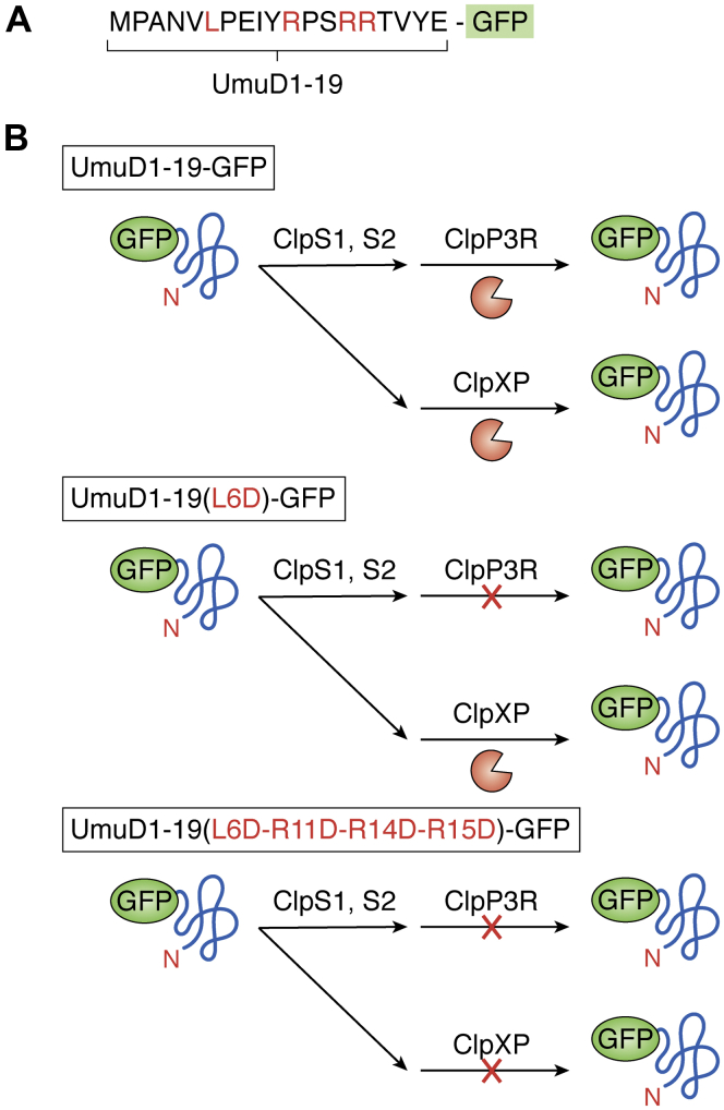
    1. Bouchnak I., van Wijk K.J. N-degron pathways in plastids. Trends Plant Sci. 2019;24:917–926. - PubMed
    1. Rawlings N.D., Barrett A.J., Finn R. Twenty years of the MEROPS database of proteolytic enzymes, their substrates and inhibitors. Nucleic Acids Res. 2016;44:D343–D350. - PMC - PubMed

Publication types

Substances