

Labeling lateral prefrontal sulci using spherical data augmentation and context-aware training
- PMID: 33497773
- PMCID: PMC8366030
- DOI: 10.1016/j.neuroimage.2021.117758
Labeling lateral prefrontal sulci using spherical data augmentation and context-aware training
Abstract
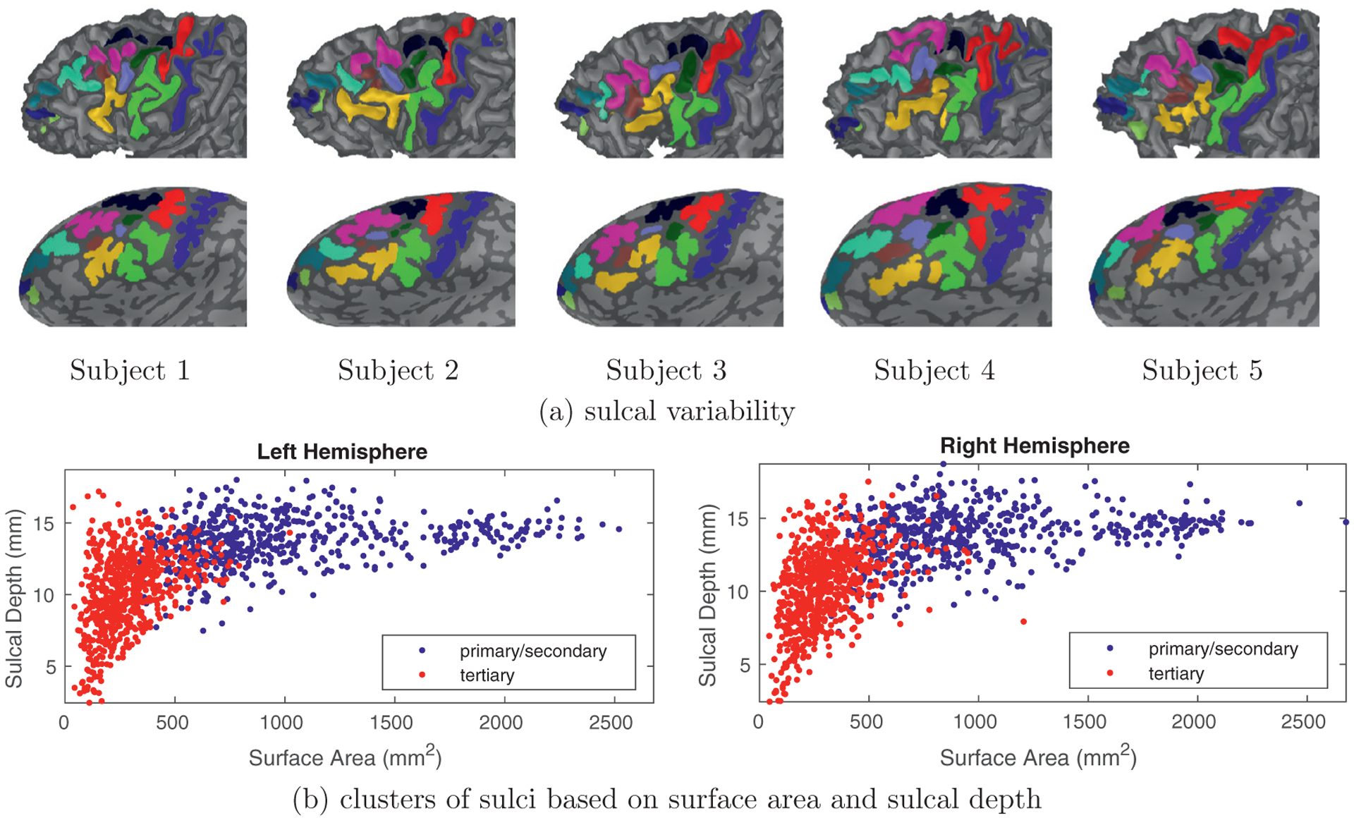
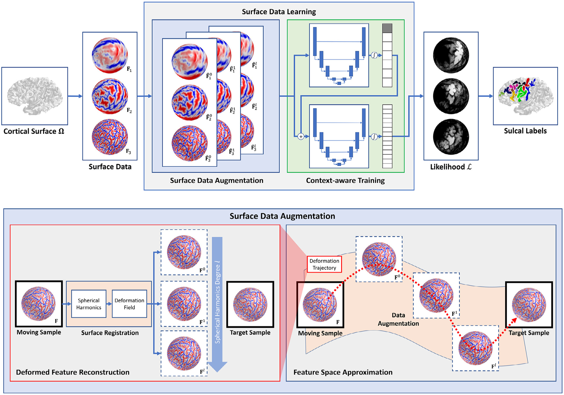
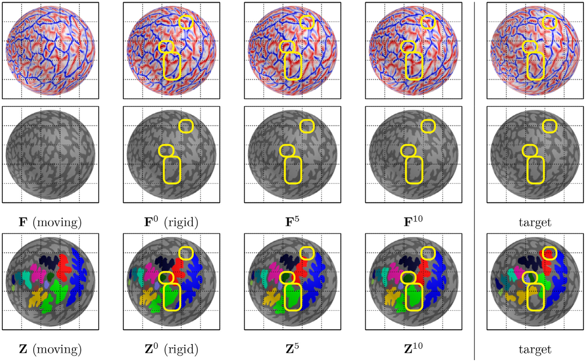
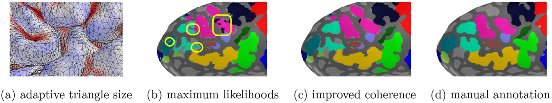
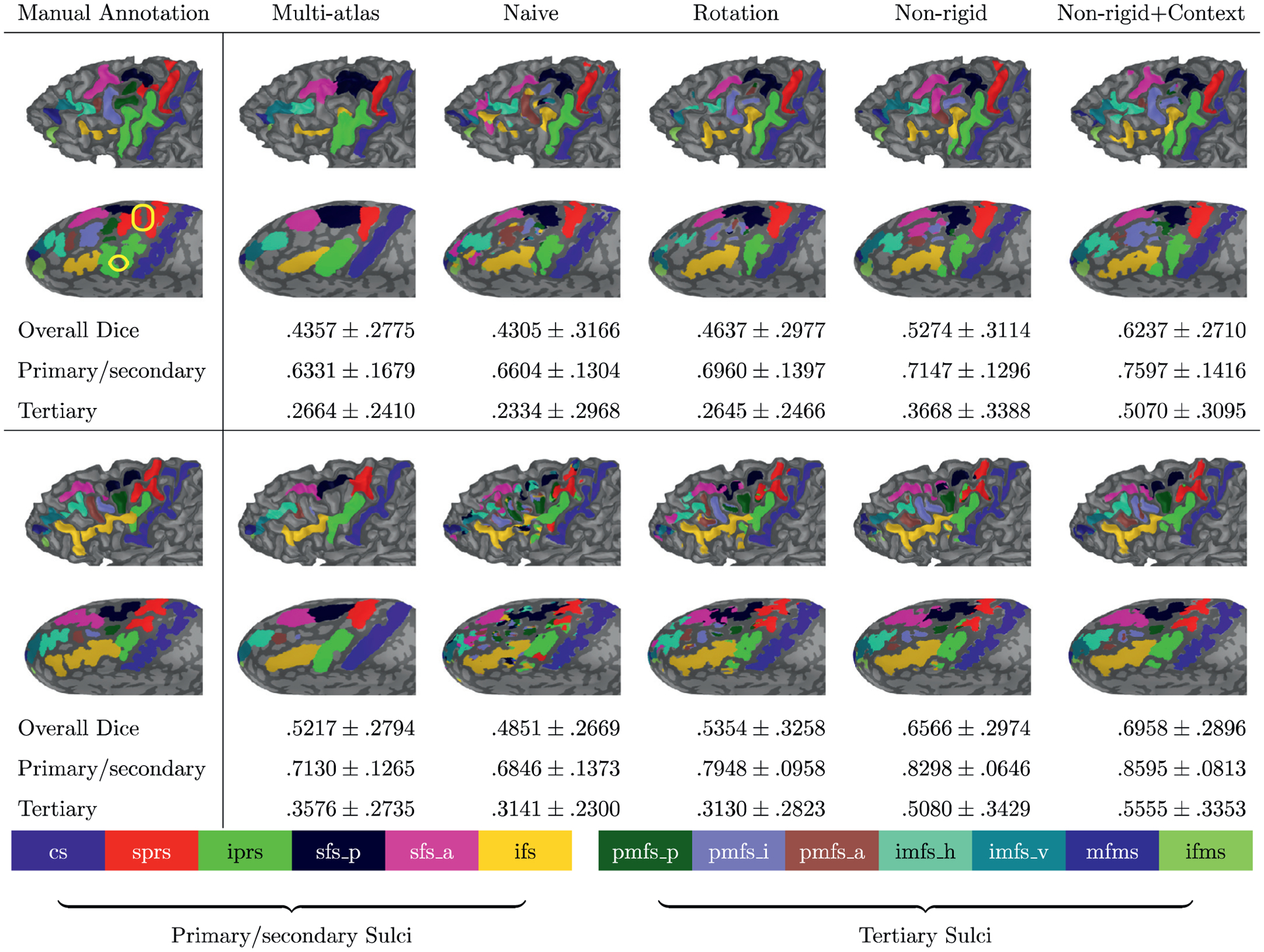
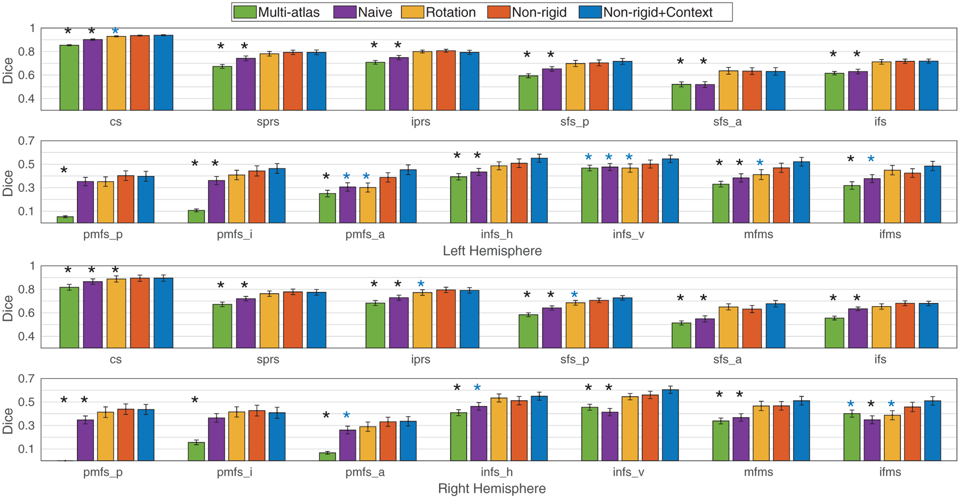
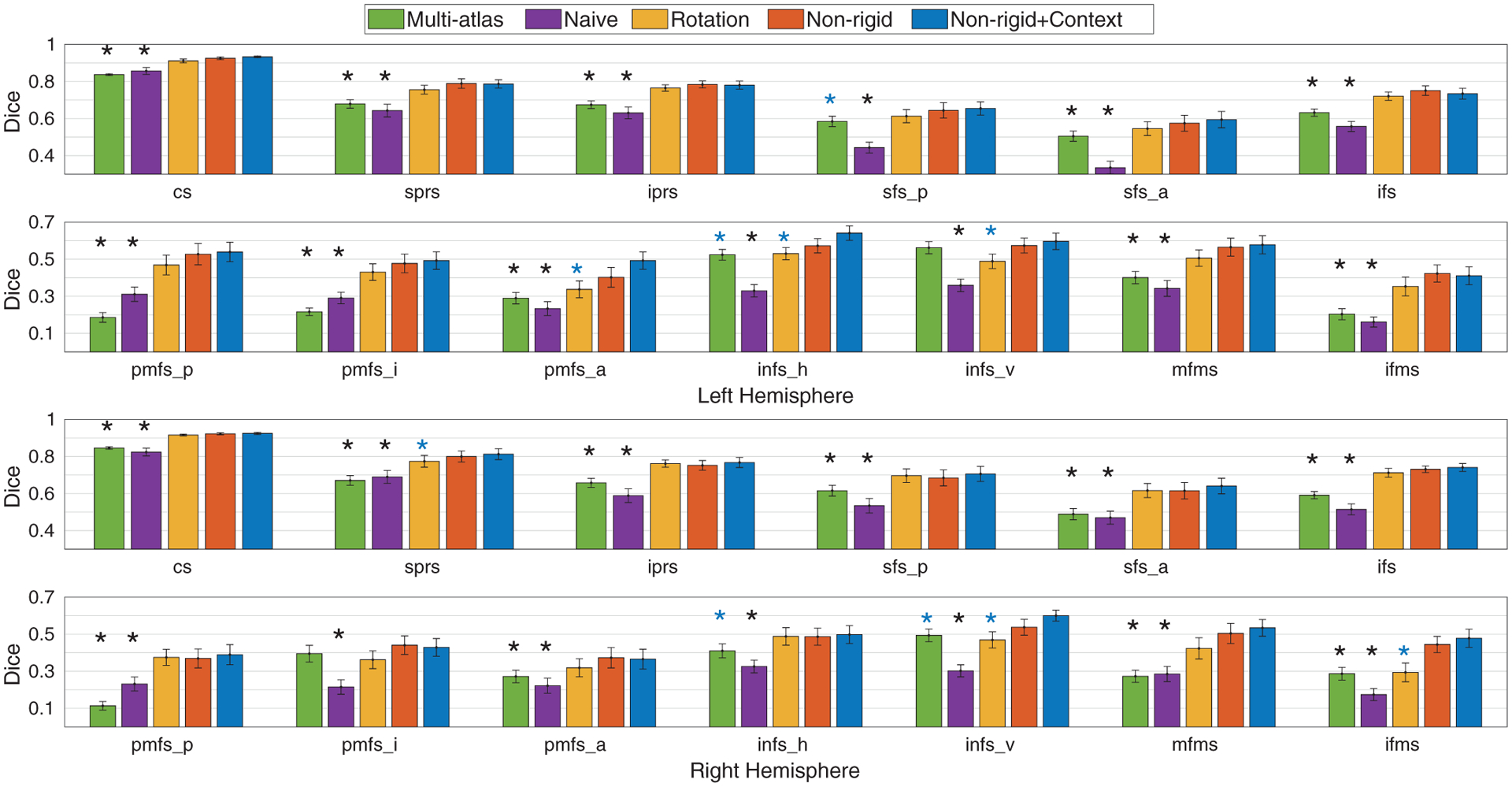
The inference of cortical sulcal labels often focuses on deep (primary and secondary) sulcal regions, whereas shallow (tertiary) sulcal regions are largely overlooked in the literature due to the scarcity of manual/well-defined annotations and their large neuroanatomical variability. In this paper, we present an automated framework for regional labeling of both primary/secondary and tertiary sulci of the dorsal portion of lateral prefrontal cortex (LPFC) using spherical convolutional neural networks. We propose two core components that enhance the inference of sulcal labels to overcome such large neuroanatomical variability: (1) surface data augmentation and (2) context-aware training. (1) To take into account neuroanatomical variability, we synthesize training data from the proposed feature space that embeds intermediate deformation trajectories of spherical data in a rigid to non-rigid fashion, which bridges an augmentation gap in conventional rotation data augmentation. (2) Moreover, we design a two-stage training process to improve labeling accuracy of tertiary sulci by informing the biological associations in neuroanatomy: inference of primary/secondary sulci and then their spatial likelihood to guide the definition of tertiary sulci. In the experiments, we evaluate our method on 13 deep and shallow sulci of human LPFC in two independent data sets with different age ranges: pediatric (N=60) and adult (N=36) cohorts. We compare the proposed method with a conventional multi-atlas approach and spherical convolutional neural networks without/with rotation data augmentation. In both cohorts, the proposed data augmentation improves labeling accuracy of deep and shallow sulci over the baselines, and the proposed context-aware training offers further improvement in the labeling of shallow sulci over the proposed data augmentation. We share our tools with the field and discuss applications of our results for understanding neuroanatomical-functional organization of LPFC and the rest of cortex (https://github.com/ilwoolyu/SphericalLabeling).
Keywords: Context encoder; Cortical surface; Frontal cortex; Spherical data augmentation; Sulcal labeling.
Copyright © 2021 The Author(s). Published by Elsevier Inc. All rights reserved.
Conflict of interest statement
Declaration of competing interest The authors declare no conflict of interest.
Figures
Similar articles
-
Overlooked Tertiary Sulci Serve as a Meso-Scale Link between Microstructural and Functional Properties of Human Lateral Prefrontal Cortex.J Neurosci. 2021 Mar 10;41(10):2229-2244. doi: 10.1523/JNEUROSCI.2362-20.2021. Epub 2021 Jan 21. J Neurosci. 2021. PMID: 33478989 Free PMC article.
-
Defining putative tertiary sulci in lateral prefrontal cortex in chimpanzees using human predictions.Brain Struct Funct. 2024 Nov;229(8):2059-2068. doi: 10.1007/s00429-023-02638-7. Epub 2023 May 17. Brain Struct Funct. 2024. PMID: 37195311
-
Leveraging Input-Level Feature Deformation With Guided-Attention for Sulcal Labeling.IEEE Trans Med Imaging. 2025 Feb;44(2):915-926. doi: 10.1109/TMI.2024.3468727. Epub 2025 Feb 4. IEEE Trans Med Imaging. 2025. PMID: 39325613
-
Automated extraction and variability analysis of sulcal neuroanatomy.IEEE Trans Med Imaging. 1999 Mar;18(3):206-17. doi: 10.1109/42.764891. IEEE Trans Med Imaging. 1999. PMID: 10363699 Review.
-
Organization of cognitive control within the lateral prefrontal cortex in schizophrenia.Arch Gen Psychiatry. 2009 Apr;66(4):377-86. doi: 10.1001/archgenpsychiatry.2009.10. Arch Gen Psychiatry. 2009. PMID: 19349307 Review.
Cited by
-
Graph-Based Deep Learning for Medical Diagnosis and Analysis: Past, Present and Future.Sensors (Basel). 2021 Jul 12;21(14):4758. doi: 10.3390/s21144758. Sensors (Basel). 2021. PMID: 34300498 Free PMC article. Review.
-
Functionally and structurally distinct fusiform face area(s) in over 1000 participants.Neuroimage. 2023 Jan;265:119765. doi: 10.1016/j.neuroimage.2022.119765. Epub 2022 Nov 23. Neuroimage. 2023. PMID: 36427753 Free PMC article.
-
Neuroanatomical and functional dissociations between variably present anterior lateral prefrontal sulci.bioRxiv [Preprint]. 2023 May 25:2023.05.25.542301. doi: 10.1101/2023.05.25.542301. bioRxiv. 2023. Update in: J Cogn Neurosci. 2023 Nov 1;35(11):1846-1867. doi: 10.1162/jocn_a_02049. PMID: 37292839 Free PMC article. Updated. Preprint.
-
Updating the sulcal landscape of the human lateral parieto-occipital junction provides anatomical, functional, and cognitive insights.bioRxiv [Preprint]. 2024 May 14:2023.06.08.544284. doi: 10.1101/2023.06.08.544284. bioRxiv. 2024. PMID: 38798426 Free PMC article. Preprint.
-
TopoFit: Rapid Reconstruction of Topologically-Correct Cortical Surfaces.Proc Mach Learn Res. 2022 Jul;172:508-520. Proc Mach Learn Res. 2022. PMID: 37220495 Free PMC article.
References
-
- Armstrong E, Schleicher A, Omran H, Curtis M, Zilles K, 1995. The ontogeny of human gyrification. Cereb. Cortex 5 (1), 56–63. - PubMed
-
- Auzias G, Lefevre J, Le Troter A, Fischer C, Perrot M, Régis J, Coulon O, 2013. Model-driven harmonic parameterization of the cortical surface: Hip-hop. IEEE Trans. Med. Imaging 32 (5), 873–887. - PubMed
Publication types
MeSH terms
Grants and funding
LinkOut - more resources
Full Text Sources
Other Literature Sources
Medical
Miscellaneous
