Review
. 2021 Apr 15:178:113004.
doi: 10.1016/j.bios.2021.113004. Epub 2021 Jan 16.

Optical technologies for the detection of viruses like COVID-19: Progress and prospects

Affiliations
Review

Optical technologies for the detection of viruses like COVID-19: Progress and prospects

Jijo Lukose et al. Biosens Bioelectron. .

Abstract

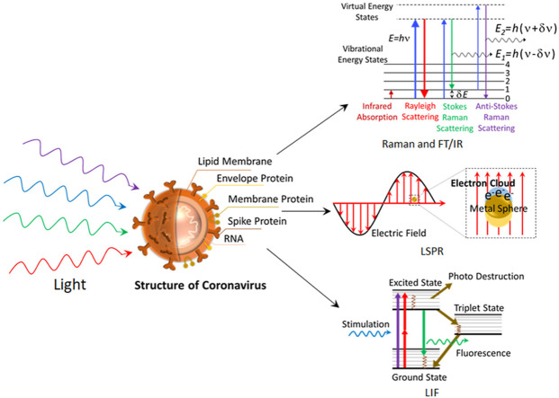
The outbreak of life-threatening pandemic like COVID-19 necessitated the development of novel, rapid and cost-effective techniques that facilitate detection of viruses like SARS-CoV-2. The presently popular approach of a collection of samples using the nasopharyngeal swab method and subsequent detection of RNA using the real-time polymerase chain reaction suffers from false-positive results and a longer diagnostic time scale. Alternatively, various optical techniques namely optical sensing, spectroscopy, and imaging shows a great promise in virus detection. Herein, a comprehensive review of the various photonics technologies employed for virus detection, particularly the SARS-CoV family, is discussed. The state-of-art research activities in utilizing the photonics tools such as near-infrared spectroscopy, Fourier transform infrared spectroscopy, Raman spectroscopy, fluorescence-based techniques, super-resolution microscopy, surface plasmon resonance-based detection, for virus detection accounted extensively with an emphasis on coronavirus detection. Further, an account of emerging photonics technologies of SARS-CoV-2 detection and future possibilities is also explained. The progress in the field of optical techniques for virus detection unambiguously show a great promise in the development of rapid photonics-based devices for COVID-19 detection.

Keywords: COVID-19; Optical imaging; Optical spectroscopy; Photonics; Surface plasmon resonance; Virus.


Conflict of interest statement

The authors declare that they have no known competing financial interests or personal relationships that could have appeared to influence the work reported in this paper.

Figures

Image 1
Graphical abstract
Fig. 1
Fig. 1
Nano-FTIR spectra of influenza virus particles at neutral pH (green) and pH 5 (red). Reprinted from (Gamage et al., 2018). (https://creativecommons.org/licenses/by/4.0/). (For interpretation of the references to color in this figure legend, the reader is referred to the Web version of this article.)
Fig. 2
Fig. 2
VIRRION testing of respiratory viruses. (A) Raman spectra. (B) PCA plot of Raman fingerprint of the different viruses. Each dot represents a collected spectrum. Reproduced from (Yeh et al., 2020) (https://creativecommons.org/licenses/by/4.0/).
Fig. 3
Fig. 3
Visualization of interactions of pVN3a-pVC3a (top panel) and pVN3a-pVC7a (bottom panel) using a split-EYFP based bimolecular fluorescence complementation assay. The rest of the 31 interactions among accessory proteins were also visualized under the same conditions (data not shown). BiFC, bimolecular fluorescence complementation assay images. DIC, differential interference contrast images. Merged, merged images of DIC and BiFC Reprinted from (Kong et al., 2015), copyright (2015), with permission from Elsevier.
Fig. 4
Fig. 4
SPR sensorgrams for (a) sensitive and (b) selective detection of anti-SCVme using the GBP-E-SCVme immobilized gold sensor chip at various concentrations (0.1, 1, 10, 50, and 100 μg mL−1) of anti-SCVme and (1 and 10 μg mL−1) of mouse IgG as negative controls. Reprinted from (Park et al., 2009), copyright (2009), with permission from Elsevier. (For interpretation of the references to color in this figure legend, the reader is referred to the Web version of this article.)
Fig. 5
Fig. 5
Specific Interactions between SARS-CoV-2-S1 and SARS-CoV-2-CTD with hACE2, Characterized by SPR. The indicated mFc-tagged proteins in the supernatant were captured by anti-mIgG antibodies that were immobilized on the chip and subsequently tested for binding with gradient concentrations of hACE2 or hCD26, with the following binding profiles shown. (A) SARS-RBD binding to hACE2. (B) SARS-RBD binding to hCD26. (C) MERS-RBD binding to hACE2. (D) MERS-RBD binding to hCD26. (E) SARS-CoV-2-S1 binding to hACE2. (F) SARS-CoV-2-S1 binding to hCD26. (G) SARS-CoV-2-NTD binding to hACE2. (H) SARS-CoV-2-NTD binding to hCD26. (I) SARS-CoV-2-CTD binding to hACE2. (J) SARS-CoV-2-CTD binding to hCD26. (K) Culture supernatant of HEK293T cells without transfection (NC) binding to hACE2. (L) Culture supernatant of HEK293T cells without transfection (NC) binding to hCD26. The values shown are the mean ± SD of three independent experiments. This article was published in Wang et al. (2020), copyright Elsevier (2020)

References

    1. Ambartsumyan O., Gribanyov D., Kukushkin V., Kopylov A., Zavyalova E. Int. J. Mol. Sci. 2020;21(9):3373. - PMC - PubMed
    1. Bächi T. J. Cell Biol. 1988;107(5):1689–1695. - PMC - PubMed
    1. Baulcombe D.C., Chapman S., Santa Cruz S. Plant J. 1995;7(6):1045–1053. - PubMed
    1. Broughton J.P., Deng X., Yu G., Fasching C.L., Servellita V., Singh J., Miao X., Streithorst J.A., Granados A., Sotomayor-Gonzalez A., Zorn K., Gopez A., Hsu E., Gu W., Miller S., Pan C.-Y., Guevara H., Wadford D.A., Chen J.S., Chiu C.Y. Nat. Biotechnol. 2020;38:870–874. - PMC - PubMed
    1. Chang Y.-F., Wang W.-H., Hong Y.-W., Yuan R.-Y., Chen K.-H., Huang Y.-W., Lu P.-L., Chen Y.-H., Chen Y.-M.A., Su L.-C. Anal. Chem. 2018;90(3):1861–1869. - PubMed

Supplementary concepts