. 2021 Jan 20;10(2):199.
doi: 10.3390/cells10020199.

Genetic Approach to Elucidate the Role of Cyclophilin D in Traumatic Brain Injury Pathology

Affiliations

Genetic Approach to Elucidate the Role of Cyclophilin D in Traumatic Brain Injury Pathology

Ryan D Readnower et al. Cells. .

Abstract

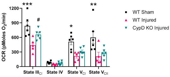
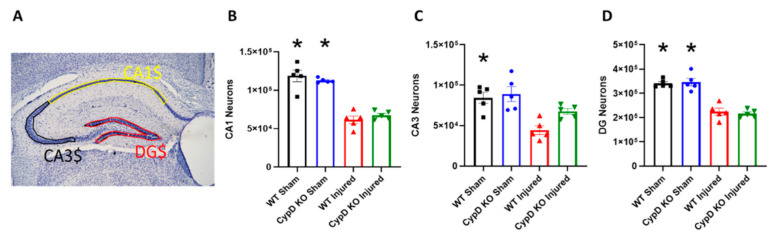
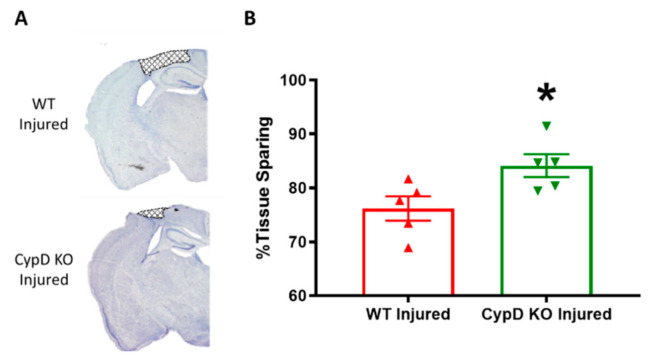
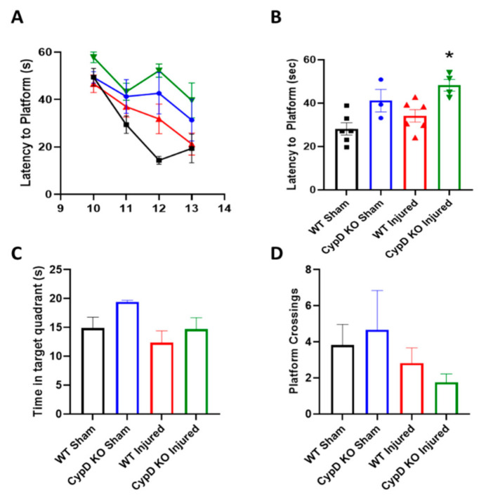
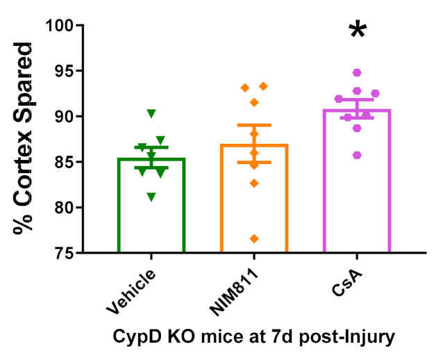
Cyclophilin D (CypD) has been shown to play a critical role in mitochondrial permeability transition pore (mPTP) opening and the subsequent cell death cascade. Studies consistently demonstrate that mitochondrial dysfunction, including mitochondrial calcium overload and mPTP opening, is essential to the pathobiology of cell death after a traumatic brain injury (TBI). CypD inhibitors, such as cyclosporin A (CsA) or NIM811, administered following TBI, are neuroprotective and quell neurological deficits. However, some pharmacological inhibitors of CypD have multiple biological targets and, as such, do not directly implicate a role for CypD in arbitrating cell death after TBI. Here, we reviewed the current understanding of the role CypD plays in TBI pathobiology. Further, we directly assessed the role of CypD in mediating cell death following TBI by utilizing mice lacking the CypD encoding gene Ppif. Following controlled cortical impact (CCI), the genetic knockout of CypD protected acute mitochondrial bioenergetics at 6 h post-injury and reduced subacute cortical tissue and hippocampal cell loss at 18 d post-injury. The administration of CsA following experimental TBI in Ppif-/- mice improved cortical tissue sparing, highlighting the multiple cellular targets of CsA in the mitigation of TBI pathology. The loss of CypD appeared to desensitize the mitochondrial response to calcium burden induced by TBI; this maintenance of mitochondrial function underlies the observed neuroprotective effect of the CypD knockout. These studies highlight the importance of maintaining mitochondrial homeostasis after injury and validate CypD as a therapeutic target for TBI. Further, these results solidify the beneficial effects of CsA treatment following TBI.

Keywords: NIM811; Ppif; controlled cortical impact; cyclosporin a; mitochondria; mitochondrial bioenergetics; mitochondrial permeability transition; neuroprotection.


Conflict of interest statement

The authors declare no conflict of interest. The funders had no role in the design of the study; in the collection, analyses, or interpretation of data; in the writing of the manuscript, or in the decision to publish the results.

Figures

Figure 1
Figure 1
Cyclophilin D (CypD) knockout attenuates mitochondrial dysfunction following traumatic brain injury (TBI). At 6 h post-injury, complex I driven State III respiration was significantly reduced compared to sham in wild-type (WT) animals. However, CypD knockout attenuated State III mitochondrial dysfunction after TBI. For State V respiration, there was no significant difference between WT Injured and CypD KO Injured. * p = 0.0046 vs. WT Injured, ** p < 0.002 vs. WT Injured and knockout (KO) Injured, *** p = 0.0001 vs. WT Injured, # p = 0.047 vs. WT Injured. Five µg mitochondrial protein were added to each well. Data points represent group mean ± SEM. N = 5–6/group.
Figure 2
Figure 2
CypD knockout mitigates TBI-related CA3 neuronal loss. (A) Outline of hippocampal sub-regions, DG, CA3, and CA1. (B) Injury significantly decreased the number of CA1 neurons in both WT and CypD KO mice. (C) There was a significant decrease in CA3 neurons after TBI in WT mice. However, CypD knockout protected CA3 neurons from injury-induced cell loss. (D) TBI resulted in a significant decrease of DG neurons in both WT and CypD KO mice. * p < 0.003 compared to injured counterpart in each genotype. Data points represent group mean ± SEM. N = 5/group.
Figure 3
Figure 3
CypD knockout increases tissue sparing following TBI. (A) Representative coronal brain sections displaying lesion area (bregma level −1.4 mm). (B) Quantitative assessment of tissue sparing revealed that CypD knockout animals had significantly higher tissue sparing percentage compared to WT animals. * p = 0.033. Data points represent group mean ± SEM. N = 5/group.
Figure 4
Figure 4
CypD knockout does not improve memory function following TBI. (A) Average learning progression, characterized by latency to platform, over days 10 to 13 after injury. (B) Morris water maze (MWM) latency to platform data collapsed across all training days. CypD knockout in injured animals resulted in an increase in latency to platform compared to WT Injured mice. * p = 0.033 vs. WT Injured. (C) At 14 d after TBI, mice performed the probe trial. Time in the target quadrant was calculated. (D) During the probe trial, the number of times mice crossed the platform area was also quantified. Data points represent group mean ± SEM. N = 3–6/group.
Figure 5
Figure 5
Cyclosporin A (CsA) and NIM811 treatment in CypD KO animals following TBI. While NIM811 did not alter tissue sparing in CypD KO animals compared to vehicle, CsA administration improved tissue sparing following experimental TBI mice. * p = 0.039 vs. Vehicle. Data points represent group mean ± SEM. N = 7–8/group.

References

    1. Centers for Disease Control and Prevention TBI Data and Statistics. [(accessed on 26 October 2020)]; Available online: https://www.cdc.gov/traumaticbraininjury/data/
    1. Yonutas H., Vekaria H., Sullivan P.G. Mitochondrial specific therapeutic targets following brain injury. Brain Res. 2016;1640:77–93. doi: 10.1016/j.brainres.2016.02.007. - DOI - PubMed
    1. Kulbe J.R., Hill R.L., Singh I.N., Wang J.A., Hall E.D. Synaptic Mitochondria Sustain More Damage than Non-Synaptic Mitochondria after Traumatic Brain Injury and Are Protected by Cyclosporine A. J. Neurotrauma. 2017;34:1291–1301. doi: 10.1089/neu.2016.4628. - DOI - PMC - PubMed
    1. Springer J.E., Azbill D., Knapp P.E. Activation of the caspase-3 apoptotic cascade in traumatic spinal cord injury. Nat. Med. 1999;5:943–946. doi: 10.1038/11387. - DOI - PubMed
    1. Sullivan P.G., Keller J.N., Bussen W.L., Scheff S.W. Cytochrome c release and caspase activation after traumatic brain injury. Brain Res. 2002;949:88–96. doi: 10.1016/S0006-8993(02)02968-2. - DOI - PubMed

Publication types

MeSH terms

LinkOut - more resources