Visualization of Chromatin in the Yeast Nucleus and Nucleolus Using Hyperosmotic Shock
- PMID: 33498839
- PMCID: PMC7866036
- DOI: 10.3390/ijms22031132
Visualization of Chromatin in the Yeast Nucleus and Nucleolus Using Hyperosmotic Shock
Abstract
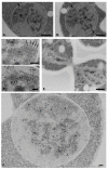
Unlike in most eukaryotic cells, the genetic information of budding yeast in the exponential growth phase is only present in the form of decondensed chromatin, a configuration that does not allow its visualization in cell nuclei conventionally prepared for transmission electron microscopy. In this work, we studied the distribution of chromatin and its relationships to the nucleolus using different cytochemical and immunocytological approaches applied to yeast cells subjected to hyperosmotic shock. Our results show that osmotic shock induces the formation of heterochromatin patches in the nucleoplasm and intranucleolar regions of the yeast nucleus. In the nucleolus, we further revealed the presence of osmotic shock-resistant DNA in the fibrillar cords which, in places, take on a pinnate appearance reminiscent of ribosomal genes in active transcription as observed after molecular spreading ("Christmas trees"). We also identified chromatin-associated granules whose size, composition and behaviour after osmotic shock are reminiscent of that of mammalian perichromatin granules. Altogether, these data reveal that it is possible to visualize heterochromatin in yeast and suggest that the yeast nucleus displays a less-effective compartmentalized organization than that of mammals.
Keywords: chromatin; electron cytochemistry; immunoelectron microscopy; nucleolus; nucleus; perichromatin granules; yeast.
Conflict of interest statement
The authors declare no conflict of interest.
Figures


Similar articles
-
Ultrastructural detection of nucleic acids within heat shock-induced perichromatin granules of HeLa cells by cytochemical and immunocytological methods.J Struct Biol. 2009 Jun;166(3):329-36. doi: 10.1016/j.jsb.2009.03.002. Epub 2009 Mar 12. J Struct Biol. 2009. PMID: 19285557
-
Nucleolar structure across evolution: the transition between bi- and tri-compartmentalized nucleoli lies within the class Reptilia.J Struct Biol. 2011 May;174(2):352-9. doi: 10.1016/j.jsb.2011.02.003. Epub 2011 Feb 16. J Struct Biol. 2011. PMID: 21335089
-
Ultrastructural cytochemistry of the nucleus in Zea mays embryos during germination.J Cell Sci. 1979 Dec;40:43-62. doi: 10.1242/jcs.40.1.43. J Cell Sci. 1979. PMID: 536392
-
[The perichromatin compartment of the cell nucleus].Tsitologiia. 2014;56(6):399-409. Tsitologiia. 2014. PMID: 25696976 Review. Russian.
-
Localization of nucleolar chromatin by immunocytochemistry and in situ hybridization at the electron microscopic level.Electron Microsc Rev. 1991;4(1):85-110. doi: 10.1016/0892-0354(91)90017-7. Electron Microsc Rev. 1991. PMID: 1873488 Review.
Cited by
-
When Phased without Water: Biophysics of Cellular Desiccation, from Biomolecules to Condensates.Chem Rev. 2023 Jul 26;123(14):9010-9035. doi: 10.1021/acs.chemrev.2c00659. Epub 2023 May 3. Chem Rev. 2023. PMID: 37132487 Free PMC article. Review.
-
Detecting material state changes in the nucleolus by label-free digital holographic microscopy.EMBO Rep. 2024 Jun;25(6):2786-2811. doi: 10.1038/s44319-024-00134-5. Epub 2024 Apr 23. EMBO Rep. 2024. PMID: 38654122 Free PMC article.
-
Large organellar changes occur during mild heat shock in yeast.J Cell Sci. 2022 Mar 1;135(5):jcs258325. doi: 10.1242/jcs.258325. Epub 2021 Aug 11. J Cell Sci. 2022. PMID: 34378783 Free PMC article.
-
Maltose accumulation-induced cell death in Saccharomyces cerevisiae.FEMS Yeast Res. 2024 Jan 9;24:foae012. doi: 10.1093/femsyr/foae012. FEMS Yeast Res. 2024. PMID: 38565313 Free PMC article.
-
Increasing Complexity of Ribosomes and Their Biogenesis.Int J Mol Sci. 2022 Jul 27;23(15):8264. doi: 10.3390/ijms23158264. Int J Mol Sci. 2022. PMID: 35897840 Free PMC article.
References
MeSH terms
Substances
LinkOut - more resources
Full Text Sources
Other Literature Sources
Molecular Biology Databases

