. 2021 Jan 24;13(2):173.
doi: 10.3390/v13020173.

Development of a Cell-Based Luciferase Complementation Assay for Identification of SARS-CoV-2 3CLpro Inhibitors

Affiliations

Development of a Cell-Based Luciferase Complementation Assay for Identification of SARS-CoV-2 3CLpro Inhibitors

Jonathan M O Rawson et al. Viruses. .

Abstract

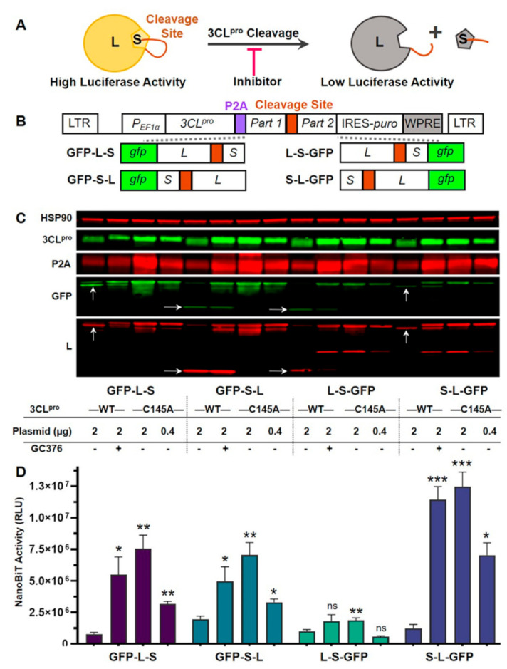
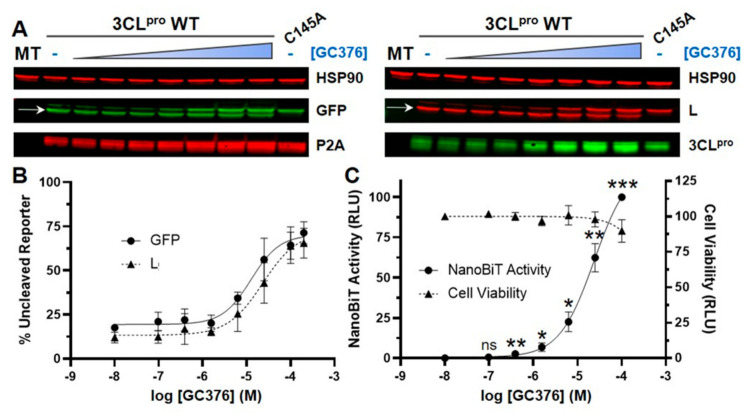
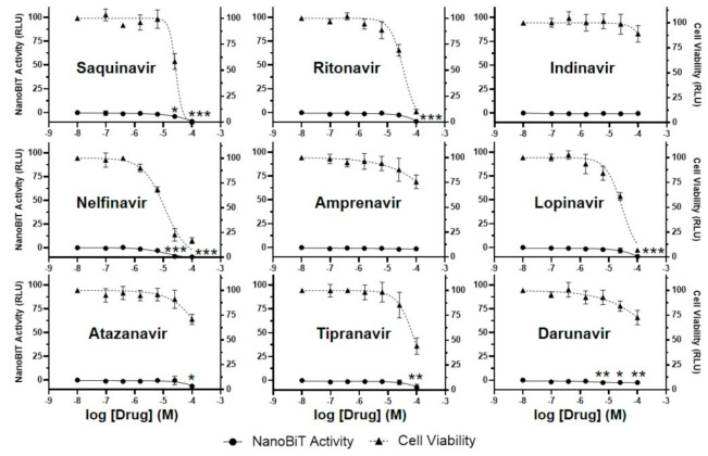
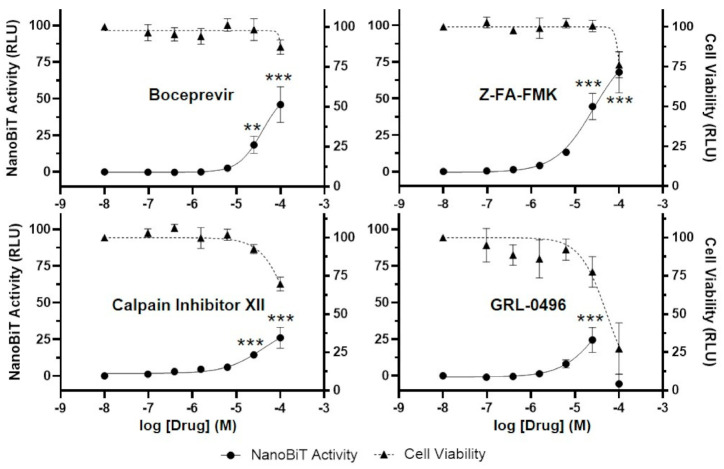
The 3C-like protease (3CLpro) of SARS-CoV-2 is considered an excellent target for COVID-19 antiviral drug development because it is essential for viral replication and has a cleavage specificity distinct from human proteases. However, drug development for 3CLpro has been hindered by a lack of cell-based reporter assays that can be performed in a BSL-2 setting. Current efforts to identify 3CLpro inhibitors largely rely upon in vitro screening, which fails to account for cell permeability and cytotoxicity of compounds, or assays involving replication-competent virus, which must be performed in a BSL-3 facility. To address these limitations, we have developed a novel cell-based luciferase complementation reporter assay to identify inhibitors of SARS-CoV-2 3CLpro in a BSL-2 setting. The assay is based on a lentiviral vector that co-expresses 3CLpro and two luciferase fragments linked together by a 3CLpro cleavage site. 3CLpro-mediated cleavage results in a loss of complementation and low luciferase activity, whereas inhibition of 3CLpro results in 10-fold higher levels of luciferase activity. The luciferase reporter assay can easily distinguish true 3CLpro inhibition from cytotoxicity, a powerful feature that should reduce false positives during screening. Using the assay, we screened 32 small molecules for activity against SARS-CoV-2 3CLpro, including HIV protease inhibitors, HCV protease inhibitors, and various other compounds that have been reported to inhibit SARS-CoV-2 3CLpro. Of these, only five exhibited significant inhibition of 3CLpro in cells: GC376, boceprevir, Z-FA-FMK, calpain inhibitor XII, and GRL-0496. This assay should greatly facilitate efforts to identify more potent inhibitors of SARS-CoV-2 3CLpro.

Keywords: 3CLpro; COVID-19; SARS-CoV-2; antiviral; inhibitor; luciferase; protease.


Conflict of interest statement

The authors declare no conflict of interest. The funders had no role in the design of the study; in the collection, analyses, or interpretation of data; in the writing of the manuscript, or in the decision to publish the results.

Figures

Figure 1
Figure 1
Development of cell-based luciferase complementation reporters to detect inhibition of SARS-CoV-2 3CLpro activity. (A) Strategy for detection of 3CLpro inhibition using NanoBiT, a luciferase complementation reporter comprised of Large BiT (L) and Small BiT (S). When connected by a linker containing a 3CLpro cleavage site, 3CLpro cleavage should result in a loss of complementation and low luciferase activity. Conversely, 3CLpro inhibitors should prevent cleavage, resulting in high luciferase activity. (B) Lentiviral vectors that express 3CLpro and reporters. P2A, a self-cleaving peptide, was inserted between 3CLpro and reporters. Reporters consisted of GFP linked to L and S in various orders. L and S were separated by the SARS-CoV-2 nsp4-nsp5 cleavage site. LTR, long terminal repeat; PEF1α, EF1α promoter; IRES, internal ribosome entry site; puro, puromycin resistance gene; WPRE, woodchuck hepatitis virus post-transcriptional regulatory element. (C) Western blotting to examine reporter expression and cleavage. Lentiviral vectors expressing 3CLpro WT or C145A (a catalytically inactive mutant) were transfected into 293T cells, and DMSO (control) or the 3CLpro inhibitor GC376 (100 µM) was added. Cell lysates were collected 30 h post-transfection. 3CLpro was detected using anti-SARS-CoV 3CLpro or anti-P2A antibodies, whereas reporters were detected using anti-GFP or anti-L antibodies. Arrows denote cleavage products of expected sizes. For 3CLpro C145A, two different plasmid amounts were transfected: 2 or 0.4 µg. HSP90 was included as a loading control. (D) NanoBiT luciferase activity. 293T cells were transfected (same sample order as in C), and NanoBiT activity (expressed as relative luciferase units, or RLU) was measured 30 h post-transfection. The data represent the mean ± standard deviation of four independent experiments; ns: not significant; * p < 0.05, ** p < 0.01, *** p < 0.001 (relative to 3CLpro WT without drug; mixed effects analysis with Dunnett’s post-test).
Figure 2
Figure 2
The luciferase complementation assay accurately reflects concentration-dependent inhibition of SARS-CoV-2 3CLpro-mediated cleavage. (A) Western blotting to examine reporter cleavage. 293T cells were transfected with the lentiviral vector containing 3CLpro WT and the S-L-GFP reporter cassette, and DMSO (control) or GC376 (0.1, 0.4, 1.6, 6.3, 25, 100, or 200 µM) was added. Mock transfection (MT) and 3CLpro C145A S-L-GFP were included as controls. On the GFP and LgBiT (L) panels, the bottom band corresponds to cleaved reporter (L-GFP, 46 kDa, indicated by arrows), while the top band corresponds to uncleaved reporter (S-L-GFP, 49 kDa). (B) Quantification of the percentage of uncleaved reporter from three independent western blots based on GFP (circle) or L (triangle) detection. (C) NanoBiT luciferase activity and cell viability. 293T cells were transfected with the S-L-GFP lentiviral vector, and DMSO (control) or GC376 (0.1, 0.4, 1.6, 6.3, 25, or 100 µM) was added. NanoBiT activity (circle, left axis) and cell viability (measured with the CellTiter-Glo 2.0 assay; triangle, right axis) were analyzed 30 h post-transfection and are expressed as relative luciferase units (RLU). Cell viability data were normalized to the DMSO sample, which was set to 100%. NanoBiT data were normalized to the DMSO and 100 µM GC376 samples, which were set to 0 and 100%, respectively. The data represent the mean ± standard deviation of four independent experiments; ns: not significant; * p < 0.05, ** p < 0.01, *** p < 0.001 (relative to no-drug; one-way ANOVA with Dunnett’s post-test).
Figure 3
Figure 3
The luciferase complementation assay can easily distinguish 3CLpro inhibition from cytotoxicity. To determine if the luciferase complementation reporter assay can differentiate 3CLpro inhibition from cytotoxicity, 293T cells were transfected with the S-L-GFP lentiviral vector, and DMSO (control) or the anti-cancer drug doxorubicin (0.1–100 µM) was added. NanoBiT activity (circle, left axis) and cell viability (measured with the CellTiter-Glo 2.0 assay; triangle, right axis) were analyzed 30 h post-transfection and are expressed as relative luciferase units (RLU). Cell viability data were normalized to the DMSO sample, which was set to 100%. NanoBiT data were normalized to the DMSO and 100 µM GC376 samples, which were set to 0 and 100%, respectively. The data represent the mean ± standard deviation of three independent experiments.
Figure 4
Figure 4
HIV protease inhibitors do not block SARS-CoV-2 3CLpro activity in a cell-based luciferase complementation assay. To determine if HIV protease inhibitors are active against SARS-CoV-2 3CLpro in cells, 293T cells were transfected with the S-L-GFP lentiviral vector, and DMSO (control) or inhibitors (0.1, 0.4, 1.6, 6.3, 25, or 100 µM) were added. NanoBiT activity (circle, left axis) and cell viability (measured with the CellTiter-Glo 2.0 assay; triangle, right axis) were analyzed 30 h post-transfection and are expressed as relative luciferase units (RLU). Cell viability data were normalized to the DMSO sample, which was set to 100%. NanoBiT data were normalized to the DMSO and 100 µM GC376 samples, which were set to 0 and 100%, respectively. The data represent the mean ± standard deviation of three independent experiments; * p < 0.05, ** p < 0.01, *** p < 0.001 (relative to no-drug; one-way ANOVA with Dunnett’s post-test).
Figure 5
Figure 5
Boceprevir, Z-FA-FMK, calpain inhibitor XII, and GRL-0496, inhibit SARS-CoV-2 3CLpro in a cell-based luciferase complementation assay. To determine if other compounds are active against SARS-CoV-2 3CLpro in cells, 293T cells were transfected with the S-L-GFP lentiviral vector, and DMSO (control) or compounds (0.1, 0.4, 1.6, 6.3, 25, or 100 µM) were added. NanoBiT activity (circle, left axis) and cell viability (measured with the CellTiter-Glo 2.0 assay; triangle, right axis) were analyzed 30 h post-transfection and are expressed as relative luciferase units (RLU). Cell viability data were normalized to the DMSO sample, which was set to 100%. NanoBiT data were normalized to the DMSO and 100 µM GC376 samples, which were set to 0 and 100%, respectively. The data represent the mean ± standard deviation of three independent experiments; ** p < 0.01, *** p < 0.001 (relative to no-drug; one-way ANOVA with Dunnett’s post-test).

Similar articles

Cited by

References

    1. Wu F., Zhao S., Yu B., Chen Y.-M., Wang W., Song Z.-G., Hu Y., Tao Z.-W., Tian J.-H., Pei Y.-Y., et al. A new coronavirus associated with human respiratory disease in China. Nature. 2020;579:265–269. - PMC - PubMed
    1. Zhou P., Yang X.-L., Wang X.-G., Hu B., Zhang L., Zhang W., Si H.-R., Zhu Y., Li B., Huang C.-L., et al. A pneumonia outbreak associated with a new coronavirus of probable bat origin. Nature. 2020;579:270–273. - PMC - PubMed
    1. Jackson L.A., Anderson E.J., Rouphael N.G., Roberts P.C., Makhene M., Coler R.N., McCullough M.P., Chappell J.D., Denison M.R., Stevens L.J., et al. An mRNA Vaccine against SARS-CoV-2—Preliminary Report. N. Engl. J. Med. 2020;383:1920–1931. - PMC - PubMed
    1. Mulligan M.J., Lyke K.E., Kitchin N., Absalon J., Gurtman A., Lockhart S., Neuzil K., Raabe V., Bailey R., Swanson K.A., et al. Phase I/II study of COVID-19 RNA vaccine BNT162b1 in adults. Nature. 2020;586:589–593. - PubMed
    1. Widge A.T., Rouphael N.G., Jackson L.A., Anderson E.J., Roberts P.C., Makhene M., Chappell J.D., Denison M.R., Stevens L.J., Pruijssers A.J., et al. Durability of Responses after SARS-CoV-2 mRNA-1273 Vaccination. N. Engl. J. Med. 2020;384:80–82. - PMC - PubMed

Publication types

MeSH terms