Review
. 2021 Jan 22;13(3):415.
doi: 10.3390/cancers13030415.

Primary Pulmonary B-Cell Lymphoma: A Review and Update

Affiliations
Review

Primary Pulmonary B-Cell Lymphoma: A Review and Update

Francesca Sanguedolce et al. Cancers (Basel). .

Abstract

Primary pulmonary B-cell lymphomas (PP-BCLs) comprise a group of extranodal non-Hodgkin lymphomas of B-cell origin, which primarily affect the lung without evidence of extrapulmonary disease at the time of diagnosis and up to 3 months afterwards. Primary lymphoid proliferations of the lung are most often of B-cell lineage, and include three major entities with different clinical, morphological, and molecular features: primary pulmonary marginal zone lymphoma of mucosa-associated lymphoid tissue (PP-MZL, or MALT lymphoma), primary pulmonary diffuse large B cell lymphoma (PP-DLBCL), and lymphomatoid granulomatosis (LYG). Less common entities include primary effusion B-cell lymphoma (PEL) and intravascular large B cell lymphoma (IVLBCL). A proper workup requires a multidisciplinary approach, including radiologists, pneumologists, thoracic surgeons, pathologists, hemato-oncologists, and radiation oncologists, in order to achieve a correct diagnosis and risk assessment. Aim of this review is to analyze and outline the clinical and pathological features of the most frequent PP-BCLs, and to critically analyze the major issues in their diagnosis and management.

Keywords: BALT; MALT lymphoma; diffuse large B-cell lymphoma; intravascular large B-cell lymphoma; lymphomatoid granulomatosis; primary effusion lymphoma; pulmonary B-cell lymphoma.


Conflict of interest statement

The authors declare no conflict of interest.

Figures
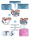
Figure 1
Figure 1
The stepwise progression from BALT to MALT lymphoma.
Figure 2
Figure 2
Primary pulmonary MALT lymphoma. (A,B). A dense infiltrate of mostly centrocyte-like lymphocytes effaces the lung parenchyma (A H&E stain, 40×; B H&E stain, 400×). (C,D). Neoplastic cells are CD20+ with a minor population of reactive CD3+ T-cells; both antibodies feature a membranous staining ((C) CD20 immunostain, 200×; (D) CD3 immunostain, 100×). (E). Lymphoepitelial lesions due to neoplastic cells infiltrating the epithelium are highlighted by cytokeratins (CK-AE1/AE3 immunostain, 200×). (F). The proliferative index, i.e., the percentage of Ki-67 stained nuclei, is low in the neoplastic cells, but higher in residual germinal centers (Ki-67 immunostain, 100×).
Figure 3
Figure 3
Primary pulmonary diffuse large B-cell lymphoma. (A,B). At low and high power, respectively, a heavy infiltrate of large tumor cell, the majority with centroblastic morphology (A H&E stain, 40×; B H&E stain, 400×). (C,D). Neoplastic cells show diffuse and intense expression of CD20, with fewer reactive CD3+ T-cells ((C) CD20 immunostain, 200×; (D) CD3 immunostain, 100×). (E). In this GCB-type PP-DLBCL there is diffuse nuclear expression of BCL6 (BCL6 immunostain, 200×). (F). Tumor cells are intensely positive for CD30 in another case with immunoblastic morphology (CD30 immunostain, 100×).
Figure 4
Figure 4
Lymphomatoid granulomatosis. (A). A number of atypical medium- to large- lymphoid cells within a polymorphous infiltrate (H&E stain, 200×). (B,C). Neoplastic cells show diffuse and intense expression of CD20 in this case of grade 3 LYG, with fewer reactive CD3+ T-cells ((B) CD20 immunostain, 200×; (C) CD3 immunostain, 200×). (D,E). Few cells show positivity for CD30 in another case of grade 2 LYG (CD30 immunostain, 400×); the same cells are intensely positive for EBER (EBER ISH, 400×). (F). Grade 3 LYG has a high proliferation index, i.e., >50% tumor cells showing nuclear immunoreactivity (Ki-67 immunostain, 400×).
Figure 5
Figure 5
Primary effusion lymphoma. (A). Large tumor cells are abundant in this pleural fluid sample (Papanicolau stain, 400×). (B). There is diffuse and intensemembranous and cytoplasmic staining for CD45 (Leukocyte Common Antigen) (CD45 immunostain, 400×).
Figure 6
Figure 6
Intravascular large B-cell lymphoma. (A). Large neoplastic cells fill the vessels within the interstitial spaces (H&E stain, 200×). (BD). Tumor cells are positive for CD45 (B), CD20 (C), and show a high proliferative index, i.e., >90% tumor cells showing nuclear immunoreactivity (D) ((B) CD45 immunostain, 200×; (C) CD20 immunostain, 200×; (D) Ki-67 immunostain, 400×).

References

    1. Shinoda K., Taki H., Tsuda T., Hayashi R., Nomoto K., Tobe K. Primary pulmonary lymphoma presenting with multiple lung nodules. Am. J. Respir. Crit. Care Med. 2014;190:e30–e31. doi: 10.1164/rccm.201401-0086IM. - DOI - PubMed
    1. Poletti V., Ravaglia C., Tomassetti S., Gurioli C., Casoni G., Asioli S., Dubini A., Piciucchi S., Chilosi M. Lymphoproliferative lung disorders: Clinicopathological aspects. Eur. Respir. Rev. 2013;22:427–436. doi: 10.1183/09059180.00004313. - DOI - PMC - PubMed
    1. Swerdlow S., Campo E., Harris N.L., Jaffe E.S., Pileri S.A., Stein H., Thiele J., Arber D.A., Hasserjian R.P., Le Beau M.M., et al. WHO Classification of Tumours of Haematopoietic and Lymphoid Tissues. 4th ed. IARC; Lyon, France: 2017.
    1. Piña-Oviedo S., Weissferdt A., Kalhor N., Moran C.A. Primary pulmonary lymphomas. Adv. Anat. Pathol. 2015;22:355–375. doi: 10.1097/PAP.0000000000000090. - DOI - PubMed
    1. Hare S.S., Souza C.A., Bain G., Seely J.M., Gomes M.M., Quigley M. The radiological spectrum of pulmonary lymphoproliferative disease. Br. J. Radiol. 2012;85:848–864. doi: 10.1259/bjr/16420165. - DOI - PMC - PubMed

LinkOut - more resources