Review
. 2021 Jan 22;10(3):427.
doi: 10.3390/jcm10030427.

Linking Labile Heme with Thrombosis

Affiliations
Review

Linking Labile Heme with Thrombosis

Marie-Thérèse Hopp et al. J Clin Med. .

Abstract

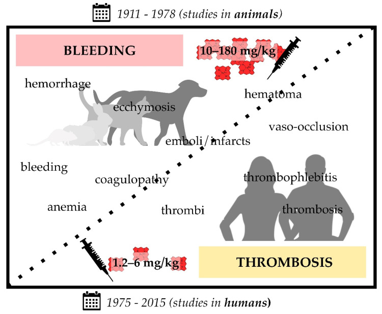
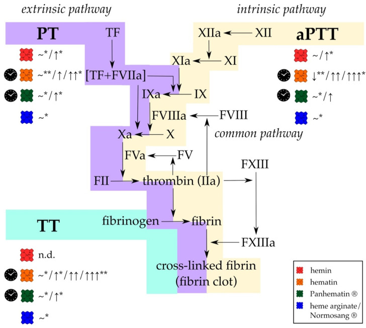
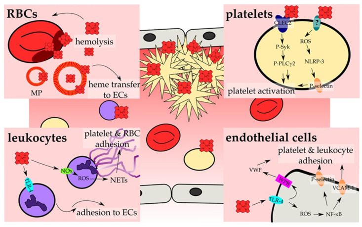
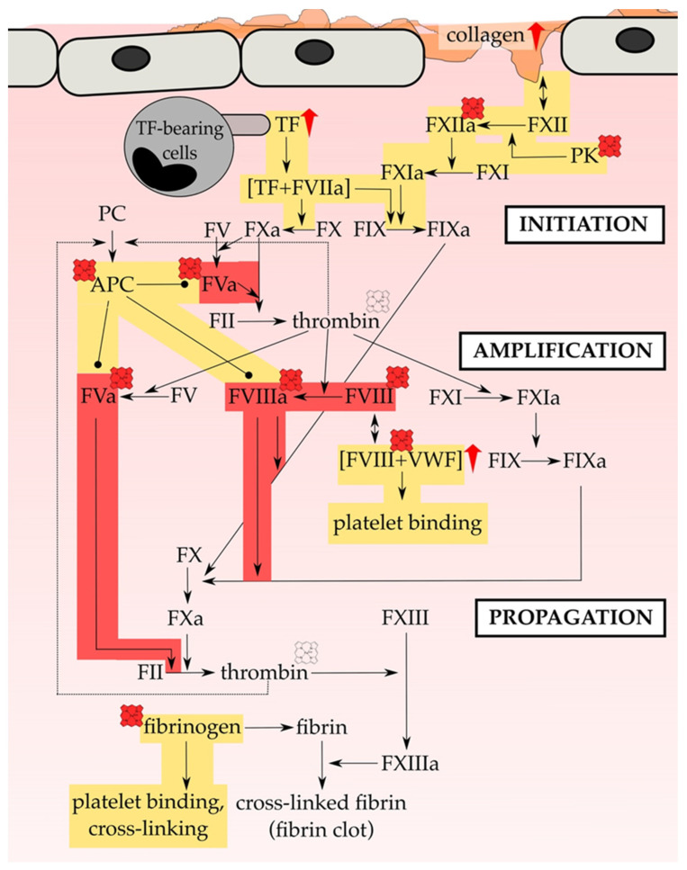
Thrombosis is one of the leading causes of death worldwide. As such, it also occurs as one of the major complications in hemolytic diseases, like hemolytic uremic syndrome, hemorrhage and sickle cell disease. Under these conditions, red blood cell lysis finally leads to the release of large amounts of labile heme into the vascular compartment. This, in turn, can trigger oxidative stress and proinflammatory reactions. Moreover, the heme-induced activation of the blood coagulation system was suggested as a mechanism for the initiation of thrombotic events under hemolytic conditions. Studies of heme infusion and subsequent thrombotic reactions support this assumption. Furthermore, several direct effects of heme on different cellular and protein components of the blood coagulation system were reported. However, these effects are controversially discussed or not yet fully understood. This review summarizes the existing reports on heme and its interference in coagulation processes, emphasizing the relevance of considering heme in the context of the treatment of thrombosis in patients with hemolytic disorders.

Keywords: blood coagulation; coagulation factors; heme binding; hemolysis; hemolytic diseases; hemorrhage; labile heme; platelets; thrombosis.


Conflict of interest statement

The authors declare no conflict of interest.

Figures

Figure 1
Figure 1
Infusion of heme in different formulations (i.e., hematin, hemin, heme arginate (Normosang®), Panhematin®) leads to coagulation disorders in animals (1911–1978; rats, guinea pigs, rabbits, cats, and dogs) and humans (1975–2015). Heme infusions of rather high concentrations (10–180 mg/kg) were shown to cause bleeding symptoms in animals, such as hemorrhage and coagulopathy, whereas administration of comparatively low concentrations (1.2–6 mg/kg) resulted in prothrombotic symptoms, such as vaso-occlusion and thrombophlebitis. The transition between bleeding and thrombotic events seems to be smooth, since there are also studies that report both effects in the same study (10–25 mg/kg hematin) [141,143]. Moreover, there are single exceptions (three studies) that also report bleeding upon administration of lower hematin (3.5–9 mg/kg) [140,162] or heme arginate (2–3 mg/kg) [168] concentrations.
Figure 2
Figure 2
Different formulations of heme affect the results of standard coagulation diagnostic tests, (PT, aPTT, and TT) in vitro and in vivo. While PT is used to evaluate effects on the extrinsic and common pathway (violet), changes in the intrinsic and common pathways (yellow) can be determined using aPTT. With TT only the last step of fibrin generation and fibrin clot formation (turquoise) can be analyzed. Hemin (red symbol; concentration ranges: 50 mg/kg, 50 µmol/kg, 0.01–100 µM) either did not affect PT and aPTT or a slight prolongation was observed. TT was not determined (n.d.). In contrast, hematin (orange symbol; concentration ranges: 4–12 mg/kg, 3 nmol, 10–100 µg/mL) has been reported to induce significant prolongation of all clotting times. For Panhematin® (green symbol; concentration ranges: 4 mg/kg, 70–78 µg/mL), longer clotting times were also recorded, but to a lesser extent than with hematin. However, from detailed investigations of different researcher it can be assumed that just aged hematin and Panhematin® solutions can have such strong effects, whereas fresh hematin and Panhematin® solutions are ineffective (clock symbol) [156,157,175]. Heme arginate/Normosang® (blue symbol; concentration: 3 mg/kg) did not influence these diagnostic tests at all. FIIa = thrombin, FII = prothrombin, FV = factor V, FVa = activated factor V, FVIIa = activated factor VII, FVIII = factor VIII, FVIIIa = activated factor VIII, FIX = factor IX, FIXa = activated FIX, FX = factor X, FXa = activated FX, FXI = factor XI, FXIa = activated FXI, FXII = factor FXII, FXIIa = activated factor XII, FXIII = factor XIII, FXIIIa = activated factor XIII, n.d. = not determined, ~ = no effect, VWF = von Willebrand factor, ↓ = 30% decrease, ↑ = < 2-fold increase, ↑↑ > 2-fold increase, ↑↑↑ > 3-fold increase. * only observed in in vivo experiments; ** only observed in in vitro experiments.
Figure 3
Figure 3
Heme activates cellular components of hemostasis. The main investigated pathways and consequences that result in the activation of cellular components of hemostasis and, thus, prothrombotic reactions by heme are depicted. On cellular level, hemostasis results from an interplay of RBCs (red, Section 4.3), platelets (yellow; Section 4.1), leukocytes (violet; Section 4.4) and endothelial cells (ECs, grey; Section 4.2). RBCs contribute to heme-induced hemostasis through the release of heme upon hemolysis, which can be further strengthened by heme itself (Section 4.3). Moreover, erythrocyte membrane particles (MP) incorporate and accumulate heme within the membrane, and allow for the transfer of heme to ECs (Section 4.2). These, in turn, become activated by heme in a TLR-4 -dependent manner (turquoise), which can lead either to the secretion of the contents of Weibel Palade bodies (WPBs; pink) (Section 4.2), among them VWF, or to ROS generation that triggers the increase of surface expression of adhesion proteins, such as P-selectin and VCAM-1 (orange) (Section 4.2). The exposure of those adhesion molecules as well as the secretion of VWF leads to the adhesion of platelets and leukocytes onto the endothelium. In contrast, activation of TLR-4 in leukocytes promotes the rolling and adhesion to ECs (Section 4.4). In addition, heme-induced NADPH oxidase (NOx; green)-dependent ROS generation in neutrophils can lead to NET formation, forming the scaffold for the adhesion of platelets and RBCs (Section 4.4). Finally, heme can also directly activate platelets. Two main mechanisms have been proposed. On the one hand, heme binding to CLEC2 was shown, leading to the phosphorylation of Syk (P-Syk) and PLCγ2 (P-PLCγ2) and eventually to the activation of platelets (Section 4.1). On the other hand, a ROS-dependent activation of the inflammasome via NLRP-3 has been demonstrated, which culminates in the expression of for example P-selectin, which again allows for the adhesion and activation of platelets (Section 4.1). Furthermore, the induction of ferroptosis (platelets) as well as apoptosis and necroptosis (endothelial cells) by heme has been demonstrated, which further might support the activation of endothelial cells and platelets (not shown; Section 4.1 and Section 4.2).
Figure 4
Figure 4
Heme promotes plasmatic hemostasis. Heme can directly affect various proteins of the blood coagulation cascade. While usually activated through the exposure of TF (e.g., by monocytes or the subendothelium), in pathologic thrombotic situations hemostasis activation through the exposure of negatively charged surfaces (e.g., of collagen) plays a supportive role. Indeed, initiation, amplification and propagation of hemostasis on the surface of cells (TF-bearing cells and platelets) is targeted by heme, either through upregulation of proteins’ expression level (red arrow) or regulation of proteins’ function (heme symbol). Direct heme-binding with functional consequences was only demonstrated for APC, FVIII(a) and fibrinogen. Contradictory results were obtained in case of the impact of heme on the activity of thrombin (pale heme symbol). The investigations of more than 35 years research allow for the assumption that heme is able to initiate hemostasis via both the upregulation of TF expression on leukocytes and endothelial cells as well as of collagen in the subendothelium. Most of the analyzed interactions tend to a procoagulant/prothrombotic (yellow) impact of heme. In contrast, heme-induced FVIIIa and FVa inactivation is exclusively described leading to anticoagulant (red) consequences. FVIII and FV are central cofactors of the coagulation cascade. Thus, the inactivation of FVIIIa and FVa by heme could constitute kind of a control center of heme-mediated initiation, amplification and propagation of the coagulation process. Plasma level changes of clotting factors that were recorded in humans upon heme infusion are not included. APC = activated protein C, FIIa = thrombin, FII = prothrombin, FV = factor V, FVa = activated factor V, FVIIa = activated factor VII, FVIII = factor VIII, FVIIIa = activated factor VIII, FIX = factor IX, FIXa = activated FIX, FX = factor X, FXa = activated FX, FXI = factor XI, FXIa = activated FXI, FXII = factor FXII, FXIIa = activated factor XII, FXIII = factor XIII, FXIIIa = activated factor XIII, PC = protein C, PK = plasma kallikrein, VWF = von Willebrand factor.

References

    1. Raskob G.E., Angchaisuksiri P., Blanco A.N., Buller H., Gallus A., Hunt B.J., Hylek E.M., Kakkar A., Konstantinides S.V., McCumber M., et al. Thrombosis: A major contributor to global disease burden. Thromb. Res. 2014;134:931–938. doi: 10.1016/j.thromres.2014.08.014. - DOI - PubMed
    1. Furie B., Furie B.C. Mechanisms of thrombus formation. N. Engl. J. Med. 2008;359:938–949. doi: 10.1056/NEJMra0801082. - DOI - PubMed
    1. Oklu R. Thrombosis. Cardiovasc. Diagn. Ther. 2017;7:S131–S133. doi: 10.21037/cdt.2017.11.08. - DOI - PMC - PubMed
    1. Koupenova M., Kehrel B.E., Corkrey H.A., Freedman J.E. Thrombosis and platelets: An update. Eur. Heart J. 2016;38:785–791. doi: 10.1093/eurheartj/ehw550. - DOI - PMC - PubMed
    1. Prandoni P. Venous and arterial thrombosis: Two aspects of the same disease? Clin. Epidemiol. 2009;1:1–6. doi: 10.2147/CLEP.S4780. - DOI - PMC - PubMed

LinkOut - more resources