. 2021 Mar 19;128(6):738-751.
doi: 10.1161/CIRCRESAHA.120.317715. Epub 2021 Jan 27.

Myocardial Blood Flow Control by Oxygen Sensing Vascular Kvβ Proteins

Affiliations

Myocardial Blood Flow Control by Oxygen Sensing Vascular Kvβ Proteins

Vahagn Ohanyan et al. Circ Res. .

Abstract

[Figure: see text].

Keywords: aldo-keto reductases; coronary circulation; ion channels; physiology; potassium channels.


Figures

Figure 1:
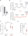
Figure 1:. Loss of Kvβ2 impairs cardiac pump function during stress.
(A) Representative M-mode echocardiographic images obtained from wild type (WT; 129SvEv), and Kvβ2−/− mice during infusion of 5 μg/kg∙min−1 norepinephrine. (B) Box-and-whisker plot of ejection fraction data for WT and Kvβ2−/− mice at baseline, after administration of hexamethonium (HX; 5 mg∙kg−1, i.v.), and during norepinephrine infusions (0.5 – 5 μg/kg∙min−1; 2–3 min duration). n = 8 each, **P<0.01, ***P<0.001 (two-way RM ANOVA). (C) Arterial blood pressure recordings obtained via femoral artery catheter in WT and Kvβ2−/− mice, before and after norepinephrine treatment (NE, 5 μg/kg∙min−1, indicated by arrows).
Figure 2:
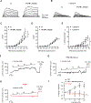
Figure 2:. Relationship between myocardial blood flow and cardiac workload in Kvβ-null mice.
(A) Long axis MCE images showing signal intensity from myocardial tissue and cavity before destruction frame and during replenishment phase (~10 sec). The left ventricular wall is outlined with a yellow dashed line in the destruction frame. (B) Signal intensity versus time (following destruction frame) in region of interest in the anterior left ventricular myocardial wall of WT (129SvEv), Kvβ1.1−/−, and Kvβ2−/− mice. Data were fit with exponential function (see inset). (C,D) Summary of MBF as a function of cardiac workload (double product; heart rate x mean arterial pressure) in Kvβ1.1−/− (C) and Kvβ2−/− (D) versus strain-matched wild type (WT) control mice. Data were fit with a simple linear regression model with slopes: WT (0.00192 ± 0.00031), Kvβ1.1−/− (0.00279 ± 0.00016); n = 6–8 mice; WT (0.00241 ± 0.00014), Kvβ2−/− (0.00162 ± 0.00022); n = 4–8 mice, *P<0.05, slope of Kvβ2−/− vs. WT.
Figure 3:
Figure 3:. Ablation of Kvβ2 attenuates hypoxia-induced coronary vasodilation.
(A) Summarized bath O2 (%) measured in normoxic and hypoxic conditions (perfusate aerated with 5% CO2, balance N2, + 1 mM Na2S2O4); data are pooled from measurements obtained with wild type (129SvEv) and Kvβ2−/− coronary arteries. n = 7–9, ***P<0.001 (Mann Whitney U). (B) Representative arterial diameter recordings in isolated preconstricted (100 nM U46619) coronary arteries from wild type (WT; 129SvEv) and Kvβ2−/− mice in normoxic and hypoxic conditions. Ca2+-free perfusate containing nifedipine (nifed; 1 μM) and forskolin (fsk; 0.5 μM) was introduced at the end of the experiment to induce maximum dilation. (C) Scatter-plot and mean ± SEM showing percent decrease in diameter recorded under normoxic (- hypoxia) and hypoxic (+ hypoxia) conditions for arteries from WT and Kvβ2−/− mice. Normoxic and hypoxic conditions were both applied in continuous presence of U46619, see above (B). n = 5 arteries, 3–4 mice *P<0.05, ns: P≥0.05 (one-way ANOVA, Tukey). (D) Scatter-plot and mean ± SEM showing hypoxia-induced dilation (%) for arteries from WT and Kvβ2−/− mice. **P<0.01 (Mann-Whitney U test).
Figure 4:
Figure 4:. L-lactate enhances IKv in coronary arterial myocytes and promotes coronary vasodilation via Kvβ2.
(A, B) Representative outward K+ current recordings normalized to cell capacitance (pA/pF) in response to step-wise (10 mV) depolarization to +50 mV from a holding potential of −70 mV in isolated coronary arterial myocytes. Currents were recorded before and after application of 10 mM L-lactate in bath solution lacking (A) or containing (B) 500 nM psora-4. (C, D) Summary current-voltage relationships obtained in coronary arterial myocytes before and after application of 10 mM L-lactate in bath solution lacking (C) or containing (D) 500 nM psora-4. n = 5–7 cells from 4–7 mice. *P <0.05, ns: P≥0.05 (two-way RM ANOVA). (E) Summary of L-lactate-induced currents recorded in the absence and presence of 500 nM psora-4. n = 5–7 cells from 4–7 mice. *P <0.05 (mixed-effects). (F-H) Arterial diameter traces obtained from pressurized (80 mmHg) coronary arteries isolated from wild type (WT; 129SvEv; F,G) and Kvβ2−/− (H) mice in the absence and presence of L-lactate (5–20 mM, as indicated). Arteries were preconstricted with 100 nM U46619; for WT arteries, L-lactate was applied in the absence (top) and presence (bottom) of psora-4 (500 nM). Maximum passive diameter was recorded at the end of each experiment in Ca2+-free saline solution with nifedipine (nifed; 1 μM) and forskolin (fsk; 0.5 μM). (I) Summary plot showing L-lactate-induced dilation, expressed as a percent change from baseline diameter relative to maximum passive diameter, for arteries isolated from WT (129SvEv; ± 500 nM psora-4) and Kvβ2−/− mice. n = 4 arteries from 4 mice for each. *P<0.001; ns: P≥0.05, lactate vs. baseline (Friedman).
Figure 5:
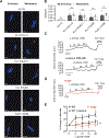
Figure 5:. Kvβ2 controls redox-dependent vasoreactivity in resistance mesenteric arteries.
(A) Representative fluorescence images showing PLA-associated fluorescent punctae (red) in wild type coronary and mesenteric arterial myocytes. Cells were labelled for Kv1.5 alone, or co-labelled for Kv1.5 and Kv1.2, Kv1.5 and Kvβ1.1, Kv1.5 and Kvβ2, or Kvβ1.1 and Kvβ2 proteins. DAPI nuclear stain is shown for each condition (blue). Scale bars represent 5 μm. (B) Summary of PLA-associated punctate sites normalized to total cell footprint area for conditions and groups as in D. P values are shown for coronary versus mesenteric arteries (Mann Whitney U). (C,D) Arterial diameter traces obtained from pressurized (80 mmHg) mesenteric arteries isolated from wild type (C; 129SvEv) and Kvβ2−/− (D) mice in the absence and presence of L-lactate (5–20 mM, as indicated). Arteries were preconstricted with 100 nM U46619 and L-lactate was applied in the absence (top) and presence (bottom) of the selective Kv1 channel inhibitor psora-4 (500 nM). Maximum passive diameters were recorded at the end of each experiment in Ca2+-free saline solution with nifedipine (nifed; 1 μM) and forskolin (fsk; 0.5 μM). (E) Summary plot of L-lactate-induced dilation, expressed as the percent change from baseline diameter relative to maximum passive diameter, for arteries isolated from WT (129SvEv; ± psora-4) and Kvβ2−/− mice. n = 5 arteries from 4–5 mice for each. *P<0.05; ns: P≥0.05, lactate vs. baseline (Friedman).
Figure 6:
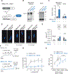
Figure 6:. Increasing the ratio of Kvβ1.1:Kvβ2 subunits in smooth muscle inhibits L-lactate-induced vasodilation and suppresses myocardial blood flow.
(A) Schematic diagram describing the SM22α-rtTA:TRE-β1 model. Double transgenic animals (+dox) results in activation of the reverse tetracycline trans-activator (rtTA) in smooth muscle cells, and drives expression of Kvβ1.1. (B) Western blots showing immunoreactive bands for Kvβ1 in whole mesenteric artery and brain lysates from SM22α-rtTA (single transgenic control) and SM22α-rtTA:TRE-β1 (double transgenic) mice after doxycycline treatment. Ponceau-stained membrane (mol. Wt.: ~30–55 kDa) is shown as an internal control for total loaded protein. (C) Summarized relative densities of Kvβ1.1-associated immunoreactive bands in mesenteric arteries and brains of SM22α-rtTA:TRE-β1 relative to SM22α-rtTA. n = 3 each. *P<0.05, ns: P≥0.05 (one sample t test). (D) Representative fluorescence images showing PLA-associated fluorescent punctae (red) in coronary arterial myocytes isolated from SM22α-rtTA and SM22α-rtTA:TRE-β1mice. Cells were labelled for Kv1.5 alone, or co-labelled for Kv1.5 and Kvβ1, or Kv1.5 and Kvβ2 proteins. DAPI nuclear stain is shown for each condition (blue). Scale bars represent 5 μm. (E) Summary of PLA-associated punctate sites normalized to total cell footprint area for conditions and groups as in D. n = 6–19 cells from 2–3 mice for each; *P<0.05, **P<0.001 (Mann Whitney U). (F) Representative arterial diameter recordings from 100 nM U46619-preconstricted mesenteric arteries isolated from SM22α-rtTA and SM22α-rtTA:TRE-β1 mice in the absence and presence of L-lactate (5–20 mM), as in Figure 5C,D. Passive dilation in the presence of Ca2+-free solution + nifedipine (1 μM) and forskolin (fsk; 0.5 μM) is shown for each recording. (G) Summary plot of L-lactate-induced dilation for arteries isolated from SM22α-rtTA and SM22α-rtTA:TRE-β1 mice. n = 6–10 arteries from 5–6 mice; *P<0.05; ns: P≥0.05, lactate vs. baseline (Friedman). (H) Summary relationships between myocardial blood flow (MBF) and cardiac workload (double product; heart rate x mean arterial pressure) in SM22α-rtTA:TRE-β1 vs. SM22α-rtTA control mice. n = 5 each; ***P<0.001 (linear regression).

Comment in

References

    1. Virani SS, Alonso A, Benjamin EJ, Bittencourt MS, Callaway CW, Carson AP, Chamberlain AM, Chang AR, Cheng S, Delling FN, Djousse L, Elkind MSV, Ferguson JF, Fornage M, Khan SS, Kissela BM, Knutson KL, Kwan TW, Lackland DT, Lewis TT, Lichtman JH, Longenecker CT, Loop MS, Lutsey PL, Martin SS, Matsushita K, Moran AE, Mussolino ME, Perak AM, Rosamond WD, Roth GA, Sampson UKA, Satou GM, Schroeder EB, Shah SH, Shay CM, Spartano NL, Stokes A, Tirschwell DL, VanWagner LB, Tsao CW, American Heart Association Council on E, Prevention Statistics C, Stroke Statistics S. Heart disease and stroke statistics-2020 update: A report from the american heart association. Circulation. 2020:CIR0000000000000757 - PubMed
    1. Tsagalou EP, Anastasiou-Nana M, Agapitos E, Gika A, Drakos SG, Terrovitis JV, Ntalianis A, Nanas JN. Depressed coronary flow reserve is associated with decreased myocardial capillary density in patients with heart failure due to idiopathic dilated cardiomyopathy. Journal of the American College of Cardiology. 2008;52:1391–1398 - PubMed
    1. Brush JE Jr., Cannon RO 3rd, Schenke WH, Bonow RO, Leon MB, Maron BJ, Epstein SE. Angina due to coronary microvascular disease in hypertensive patients without left ventricular hypertrophy. The New England journal of medicine. 1988;319:1302–1307 - PubMed
    1. Di Carli MF, Charytan D, McMahon GT, Ganz P, Dorbala S, Schelbert HR. Coronary circulatory function in patients with the metabolic syndrome. J Nucl Med. 2011;52:1369–1377 - PubMed
    1. Fukuda D, Yoshiyama M, Shimada K, Yamashita H, Ehara S, Nakamura Y, Kamimori K, Tanaka A, Kawarabayashi T, Yoshikawa J. Relation between aortic stiffness and coronary flow reserve in patients with coronary artery disease. Heart. 2006;92:759–762 - PMC - PubMed

Publication types

MeSH terms