. 2021 May:113:132-147.
doi: 10.1016/j.semcdb.2020.12.007. Epub 2021 Jan 23.

Genome diversity and instability in human germ cells and preimplantation embryos

Affiliations

Genome diversity and instability in human germ cells and preimplantation embryos

Vallari Shukla et al. Semin Cell Dev Biol. 2021 May.

Abstract

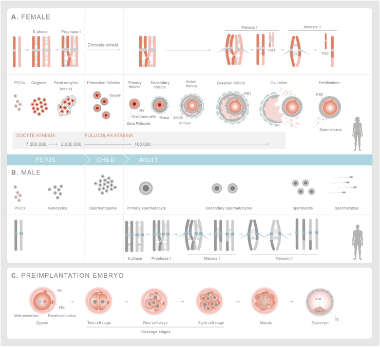
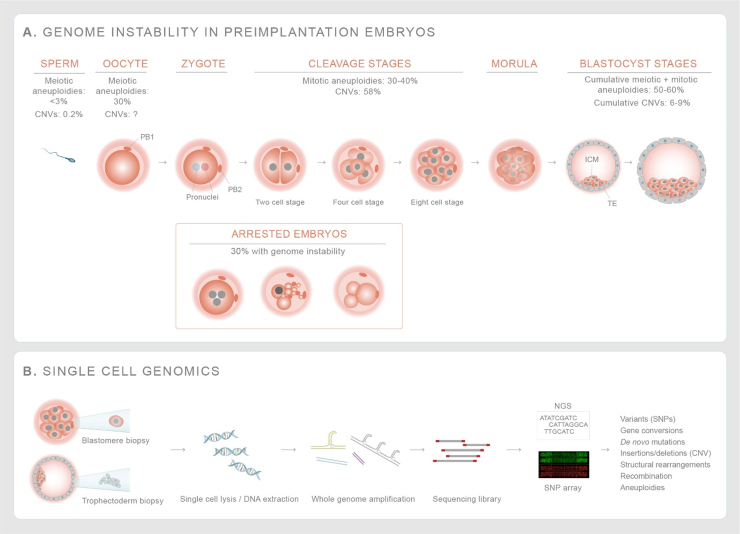
Genome diversity is essential for evolution and is of fundamental importance to human health. Generating genome diversity requires phases of DNA damage and repair that can cause genome instability. Humans have a high incidence of de novo congenital disorders compared to other organisms. Recent access to eggs, sperm and preimplantation embryos is revealing unprecedented rates of genome instability that may result in infertility and de novo mutations that cause genomic imbalance in at least 70% of conceptions. The error type and incidence of de novo mutations differ during developmental stages and are influenced by differences in male and female meiosis. In females, DNA repair is a critical factor that determines fertility and reproductive lifespan. In males, aberrant meiotic recombination causes infertility, embryonic failure and pregnancy loss. Evidence suggest germ cells are remarkably diverse in the type of genome instability that they display and the DNA damage responses they deploy. Additionally, the initial embryonic cell cycles are characterized by a high degree of genome instability that cause congenital disorders and may limit the use of CRISPR-Cas9 for heritable genome editing.

Keywords: Aneuploidy; CNVs; DNA damage response; Genome instability; Genomic disorders; Human oocytes and embryos; Rearrangements.


Figures

Fig. 1
Fig. 1
Germline and preimplantation embryo development in humans. (A) Female-Primordial germ cells expand and are specified as oogonia, which form nests before initiating meiosis. Fetal oocytes replicate their DNA and homologous chromosomes undergo meiotic recombination before entering dictyate (G2/M) arrest. Prior to birth, individual oocytes are surrounded by layer of follicular cells forming primordial follicles. Follicles are recruited throughout life but only after onset of puberty do they mature over 290 days. Once per month, the luteinizing hormone surge causes the ovulation of a single mature egg that has resumed and completed the first meiosis division. At this stage, the cohesion between sister chromatid arms are released and homologous chromosomes segregate forming the mature, secondary oocyte and first polar body (PB1). Upon fertilization the sister chromatids separate giving rise to zygote and second polar body (PB2). (B) Male-The primordial germ cells are specified as gonocytes at fetal stage, which differentiate into spermatogonia upon birth. Meiosis is initiated at the onset of puberty in primary spermatocytes and completed in round spermatids. Round spermatids undergo morphological differentiation to give rise to haploid sperm. (C) Preimplantation embryos-Zygote consists of female and male pronuclei that fuse together and undergo first mitotic division. After initial 2–3 divisions, the cells undergo compaction forming a morula. Subsequent divisions lead to the formation of blastocyst which comprises of a cavity (blastocoel) and inner cell mass with a layer of trophectoderm cells around it. PB1 and PB2 chromosomes are shown in smaller size to indicate that their extrusion after MI and MII division respectively stops their transmission into germline. List of abbreviations - PGCs-Primordial germ cells; GV-Germinal Vesicle; GVBD-Germinal Vesicle Breakdown; TE-Trophectoderm; ICM-Inner Cell Mass.
Fig. 2
Fig. 2
DNA damage repair pathways. (A) Fanconi Anemia pathway described from various studies in somatic cells and Xenopus extracts. Stalled replication forks activate ATR mediated checkpoint response, which recruits the FA core complex, which monoubiquitinates the FANCI/FANCD2 complex. The FANCI/D2 complex with SLX4 promotes the nucleolytic initiation, interstand unhooking and double strand break formation. DNA replication is resumed by translesion synthesis polymerases. The double strand break end initiates resection and repair via homologous recombination . (B) Homologous recombination-meiotic recombination is initiated by SPO11-induced double strand breaks. Invasion structures are based on findings in budding yeast (see text). ZMM-complex of meiosis-specific proteins that are conserved and promotes double Holliday junction formation and their biased resolution into crossovers , . Invasion structures are unwound/disassembled by BLM and SMC5/6 and can re-populate the meiotic recombination pathway (crossover or synthesis-dependent single strand annealing for noncrossovers-not shown). Invasion structures that are not disassembled can be resolved by structure-specific endonucleases, yielding both crossovers and noncrossovers . (C) The Base excision pathway is required by murine primordial germ cells and the pronucleus of embryos to undergo demethylation, which is essential for gametogenesis. TET enzymes oxidize 5mC to 5HmC, which is oxidized. TDP glycosylase removes the oxidized base creating a single strand break recognized by XRCC1 and PARP1 that facilitate DNA demethylation and chromatin remodeling. (D) Non-homologous end joining pathway (NHEJ) - Double strand break ends are recognized and processed by Ku70/80 heterodimer, DNA-PKCs and other enzymes. The ends are ligated by DNA ligase IV/XRCC4/XLF . (E) Microhomology mediated end joining pathway (MMEJ) uses short homologous sequences (shown in yellow) to align the broken ends. FEN1 mediates flap trimming and the ends are ligated by XRCC1/DNA Ligase III . End joining pathways are preferred double strand repair mechanisms in preimplantation embryos , , . List of abbreviations - BER-base excision repair; NHEJ-nonhomologous end joining; MMEJ-microhomology mediated end joining; HR-homologous recombination. (For interpretation of the references to color in this figure legend, the reader is referred to the web version of this article.)
Fig. 3
Fig. 3
Segregation error patterns in human oocytes follows a U-curve with female age. (A) Normal MI division the homologous chromosomes segregate forming the MII oocyte and the first polar body. Normal MII division, where sister chromatids disjoin is shown below as is a MII nondisjunction event leading to an aneuploid conception. (B) During premature separation of sister chromatid (PSSC) one sister chromatid separates from its homolog and ends up either in oocyte or PB1 leading to aneuploidy after MI division. The segregation of the single chromatid in meiosis II results either in a euploid or an aneuploid conception. Meiosis II errors occur independently and would result in an aneuploid conception. (C) Non-disjunction (MI NDJ) of homologous chromosmes during MI division leads to aneuploidy with either the oocyte or the PB1 ending up with both chromosomes. A normal meiosis II division results in an aneuploid conception, whereas a meiosis II error can restore, by chance, the euploid maternal genome. (D) Reverse segregation where sister chromatids of both homologous chromosmes separate during MI division. The segregation of the two nonsister chromatids results in 70% euploid eggs after MII division , . List of abbreviations – MI - Meiosis I division; MII - Meiosis II division; PB1- Polar Body 1; PB2 - Polar Body 2.
Fig. 4
Fig. 4
Phases of high genome instability during human embryogenesis – (A) The incidence of aneuploidy is higher in eggs (30%) than in sperm (< 3%). The incidence of mitotic aneuploidies and gross chromosomal rearrangements increase during cleavage and blastocyst stages. About 30% of human cleavage stage embryos arrest, with evidence of genome instability such as gross chromosomal rearrangements and complex aneuploidies that affect multiple chromosomes , , , , , , . The cumulative aneuploidies from germ cells and mitotic divisions result in a high incidence of blastocyst embryos that are genomically mutated. (B) Use of single cell genomics technologies including whole genome amplification and sequencing of biopsied of cell(s) from cleavage stage embryos or blastocysts helps to map out gross chromosomal rearrangements and aneuploidies giving a better overview of genome instability in human preimplantation embryos. List of abbreviations- CNV - Copy number variants; NGS - next generation sequencing; SNP - single nucleotide polymorphisms.

References

    1. Olivieri M., Cho T., Álvarez-Quilón A., Li K., Schellenberg M.J., Zimmermann M., Hustedt N., Rossi S.E., Adam S., Melo H., Heijink A.M., Sastre-Moreno G., Moatti N., Szilard R.K., McEwan A., Ling A.K., Serrano-Benitez A., Ubhi T., Feng S., Pawling J., Delgado-Sainz I., Ferguson M.W., Dennis J.W., Brown G.W., Cortés-Ledesma F., Williams R.S., Martin A., Xu D., Durocher D. A genetic map of the response to DNA damage in human cells. Cell. 2020;182(2):481–496. e21. - PMC - PubMed
    1. Ford C.E., Jones K.W., Miller O.J., Mittwoch U., Penrose L.S., Ridler M., Shapiro A. The chromosomes in a patient showing both mongolism and the Klinefelter syndrome. Lancet. 1959;1(7075):709–710. - PubMed
    1. Ford C.E., Polani P.E., Briggs J.H., Bishop P.M. A presumptive human XXY/XX mosaic. Nature. 1959;183(4667):1030–1032. - PubMed
    1. Lejeune J., Turpin R., Gautier M. Mongolism; a chromosomal disease (trisomy) Bull. Acad. Natl. Med. 1959;143(11–12):256–265. - PubMed
    1. Jacobs P.A., Baikie A.G., Court Brown W.M., Strong J.A. The somatic chromosomes in mongolism. Lancet. 1959;1(7075):710. - PubMed

Publication types

LinkOut - more resources