. 2021 Jun;11(6):1542-1561.
doi: 10.1158/2159-8290.CD-20-1375. Epub 2021 Jan 26.

Chemotherapy Induces Senescence-Like Resilient Cells Capable of Initiating AML Recurrence

Affiliations

Chemotherapy Induces Senescence-Like Resilient Cells Capable of Initiating AML Recurrence

Cihangir Duy et al. Cancer Discov. 2021 Jun.

Abstract

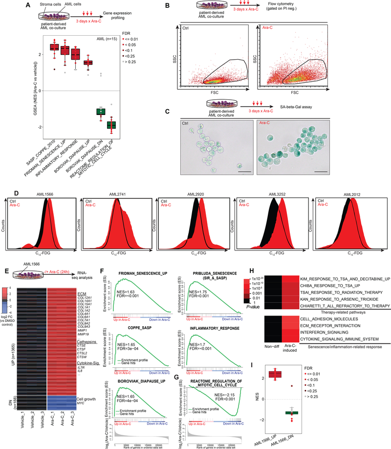
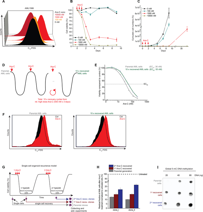
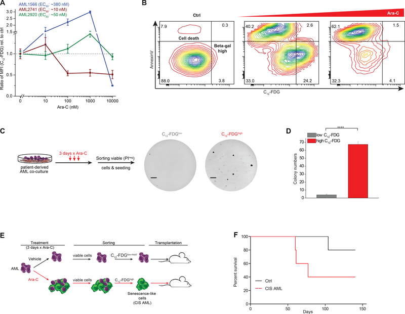
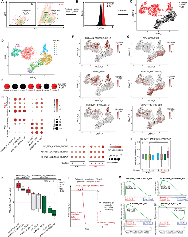
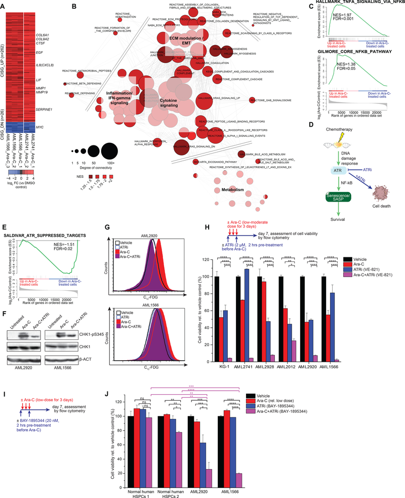
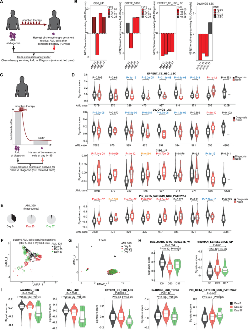
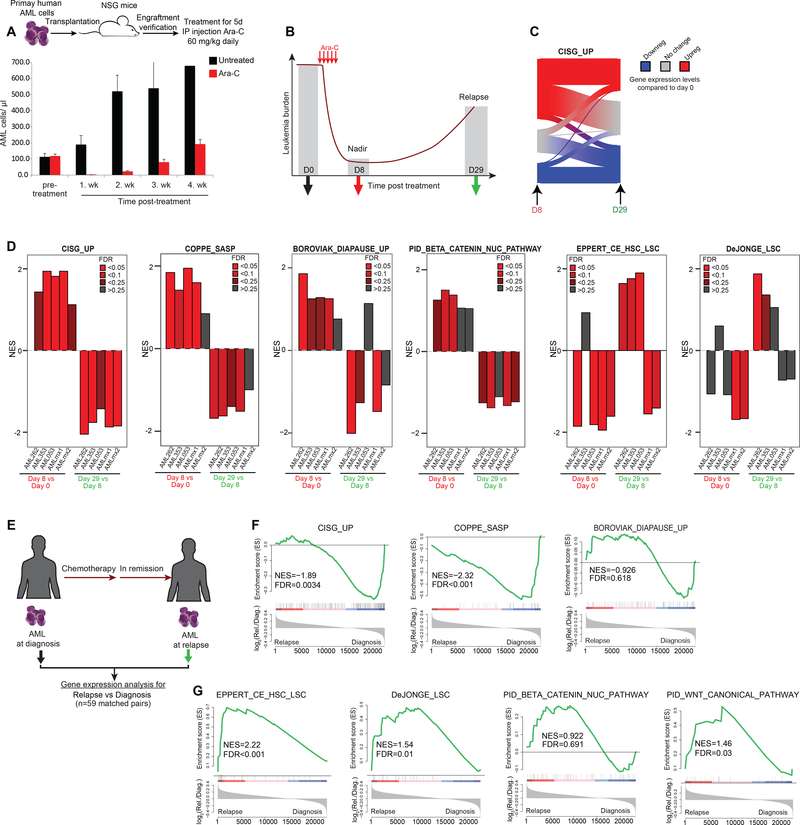
Patients with acute myeloid leukemia (AML) frequently relapse after chemotherapy, yet the mechanism by which AML reemerges is not fully understood. Herein, we show that primary AML cells enter a senescence-like phenotype following chemotherapy in vitro and in vivo. This is accompanied by induction of senescence/inflammatory and embryonic diapause transcriptional programs, with downregulation of MYC and leukemia stem cell genes. Single-cell RNA sequencing suggested depletion of leukemia stem cells in vitro and in vivo, and enrichment for subpopulations with distinct senescence-like cells. This senescence effect was transient and conferred superior colony-forming and engraftment potential. Entry into this senescence-like phenotype was dependent on ATR, and persistence of AML cells was severely impaired by ATR inhibitors. Altogether, we propose that AML relapse is facilitated by a senescence-like resilience phenotype that occurs regardless of their stem cell status. Upon recovery, these post-senescence AML cells give rise to relapsed AMLs with increased stem cell potential. SIGNIFICANCE: Despite entering complete remission after chemotherapy, relapse occurs in many patients with AML. Thus, there is an urgent need to understand the relapse mechanism in AML and the development of targeted treatments to improve outcome. Here, we identified a senescence-like resilience phenotype through which AML cells can survive and repopulate leukemia.This article is highlighted in the In This Issue feature, p. 1307.


Figures

Figure 1.
Figure 1.. Chemotherapy-induced senescence in human patient-derived AML.
A, Box-and-whisker plot showing NES scores derived from GSEA using senescence and relevant gene signatures in 15 primary AMLs (33) after ex vivo Ara-C treatment (3 days x 100 nM) compared to their corresponding vehicle-treated conditions. Data is presented with median (bisecting line in the box) and whiskers representing the 1.5 x of the interquartile range (IQR; 25th – 75th percentile, length of the box). Red box indicates that the majority of samples are positively enriched, while green reflects negative enrichment. Each dot represents a primary case, color of dots reflect FDRs calculated by GSEA. NES, Normalized enrichment score; FDR, False discovery rate. B, Upper panel illustrates Ara-C regimen of patient-derived AML cells in an ex vivo co-culture model (36) before subjecting the cells to flow cytometry. FSC vs SSC plots show increased cell size and granularity of patient-derived AML cells after Ara-C exposure for three days. C, Representative images of SA-β-Gal activity in patient-derived AML1566 exposed to Ara-C for three days and stained with X-gal. Scale bar corresponds to 25 μm. D, Flow cytometry histogram depicting SA-β-Gal activity using the fluorogenic β-galactosidase substrate C12-FDG in five patient-derived AML cases, gated on viable cells (PI- exclusion), after 3 days of using a case-dependent moderate-to-high dose of Ara-C exposure (1566: 1000 nM, 2741: 50 nM, 2920: 250 nM, 3252 & 2012: 100 nM). E, RNA-seq analysis of differentially expressed genes (at least 2-FC rel. to mean of vehicle-treated cells and q<0.01) induced by Ara-C treatment (1000 nM) in patient-derived AML1566 cells within 24 hours (n=3 per group). Genes of interest are exemplarily highlighted. F, G, GSEA showing positive enrichment for senescence, diapause and SASP/inflammatory-associated gene signatures unlike a signature of mitotic cell cycle (G) showing negative enrichment after Ara-C exposure. H, Heat map of overrepresented pathways induced by Ara-C treatment in patient-derived AML1566 cells. I, Box-and-whisker plot showing NES scores derived from GSEA using up and down DEG signatures from panel E in the 15 primary patient-derived AMLs (33) from panel A, relative to their corresponding vehicle-treated condition.
Figure 2.
Figure 2.. Chemotherapy-induced senescence as a cellular resilience mechanism.
A, Dose-dependent induction of chemotherapy-induced senescence (CIS) after three days of treatment at the indicated doses measured by C12-FDG. B, C, Ex vivo AML recurrence model with patient-derived AML1566 cells measured by flow cytometry for viability (B; by PI exclusion) and cell numbers (C; total viable cells) at indicated Ara-C doses. High dose Ara-C-treated conditions (≥ 1,000 nM) were measured after one month of recovery (C). D, Scheme demonstrating that patient-derived AML cells (AML2928) treated with a case-dependent high dose of Ara-C (3 days x 500 nM) and retreated after recovery from nadir. E, Dose-response curves for Ara-C (3 days treatment) in parental vs 10 x recovered AML cells including their EC50 values. F, Flow histogram showing C12-FDG signal in parental and 10 x recovered AML cells following Ara-C treatment for three days. G, Patient-derived AML cells (AML2741) were treated with a case-dependent high dose of chemotherapy (3d x 100 nM Ara-C). Single cells were picked at indicated time points before and after chemotherapy. Single cells from untreated leukemia were defined as “parental clones”, while cells derived from Ara-C-treated leukemia at nadir were defined as 1st or 2nd Ara-C recovered clones depending on the treatment cycle. To exclude time-dependent epigenetic variations, all recovered monoclonal populations were processed together. H, Bar diagram shows the viability of parental and Ara-C recovered AML monoclonal populations following re-exposure to the same Ara-C regimen (3d x 100 nM Ara-C) that was used to enrich these cells (G). I, Dot plot for 5-mC methylation in parental and Ara-C recovered clones.
Figure 3.
Figure 3.. Chemotherapy-induced senescence is dose-dependent and capable of repopulating AML.
A, Three different patient-derived AML cases (including their EC50 values for Ara-C) were exposed from relatively low (10 nM) to highly toxic (10,000 nM) doses of Ara-C. SA-β-Gal activity was assessed by the ratio of median fluorescence intensity (MFI) of C12-FDG relative to untreated cells set as 1. B, Exemplary of AML cells measured by flow cytometry for SA-β-Gal activity with C12-FDG and Annexin V co-staining following moderate or high dose Ara-C exposure. Shown is the separation of apoptotic cells with reduced C12-FDG levels and the C12-FDGhigh viable fraction following Ara-C. C, D, Ara-C-treated patient-derived AML cells (AML1566) were sorted for low and high SA-β-gal signal and plated in methylcellulose. Colonies were counted after 3 weeks (F, line = 4 mm). Bar graph indicates mean (n=3) of colony numbers (G). (****P<0.0001, Student’s t-test, means of triplicate measurements ± s.d.). E, Schematic showing the treatment of patient-derived AML cells (AML1566) with Ara-C (1000 nM) for 3 days with subsequent sorting of non-senescent cells (untreated condition) and chemotherapy-induced senescence-like cells (CIS AML cells). F, Overall survival of mice after sorting leukemia cells that were untreated or exposed to Ara-C. Each group consisted of 5 NSG mice.
Figure 4.
Figure 4.. Chemotherapy-surviving AML cells are not enriched for LSCs.
A, Flow cytometry plot displaying primary patient-derived AML1566 cells with and without daily treatment with 1000 nM Ara-C for 3 days. B, Sorted viable C12-FDGhigh senescence-like AML cells collected following three days exposure to 1000 nM Ara-C as well as untreated viable control cells. Sorted viable untreated control cells and Ara-C-treated C12-FDGhigh AML cells were subjected to single-cell RNA-seq. C, UMAP plot of untreated (black; n=1952) and Ara-C-induced senescent AML cells (red, n=2675). D, Same as (C), colored according to the identified 7 clusters. E, Pie chart illustrating the relative distribution within each cluster by untreated control and Ara-C-induced senescent AML cells across the clusters. F, G, UMAP plot displaying the signature scores for senescence and diapause-associated gene signatures (F) as well as LSC/HSC-associated gene signatures (G). H, Dot plot showing the average expression levels (color scale) and the proportion of expressing cells (circle size) per cluster for the indicated gene signatures. Clusters were selected by at least containing >5% of Ara-C-induced senescent cells or untreated control cells, respectively. I, J, Dot plot (I) and box plot (J) showing average expression levels for WNT/β-catenin-associated gene signatures in the AML1566 clusters. P values calculated by Wilcoxon signed-rank test using cluster 6 (Ara-C-treated cells) with any of the cluster in the untreated cells (**** P<0.0001). K, Box-and-whisker plot showing NES scores derived from GSEA using hematopoietic stem cell (LSC/HSC)-relevant gene signatures in 15 primary AMLs (33) after Ara-C treatment (3 days x 100 nM) compared to their corresponding vehicle-treated condition. Data is presented with median (bisecting line in the box) and whiskers representing the 1.5 x of the interquartile range (IQR; 25th – 75th percentile, length of the box). Red box indicates that the majority of samples are positively enriched, while green reflects negative enrichment. Each dot represents a primary case, color of dots reflect FDRs calculated by GSEA. NES, Normalized enrichment score; FDR, False discovery rate. L, Schematic showing the selection of the residual Ara-C-persistent AML cells and depletion of apoptotic cells before RNA-seq analysis. A regimen consisting of 3 days with a case-dependent high dose of Ara-C (100 nM) left ~10% viable cells in AML2741 after chemotherapy that was used to analyze the chemotherapy-persistent leukemia cells. M, GSEA results for senescence and diapause signatures (upper panel) as well as stem cell-related ones (lower panel) in the residual fraction of chemotherapy-persistent leukemia cells vs untreated control cells of AML2741.
Figure 5.
Figure 5.. Chemotherapy-induced senescence program enables survival of AML.
A, Shown are differentially expressed genes in chemotherapy-persistent AML2741 that were also differentially expressed in AML1566 cells after Ara-C treatment compared to their respective untreated controls. Fold change (FC) scores were calculated for each AML case separately. Genes consistently regulated after chemotherapy were defined as chemotherapy-induced stress genes (CISG). Selected up- and downregulated genes are exemplarily shown for genes of interest. B, Network analysis of leading edge of significantly enriched pathways. The length of each edge reflects the Jaccard index between respective leading edge pairs and the size of each node reflects its degree of connectivity among other nodes. C, GSEA demonstrating enrichment for NF-κB signaling programs after Ara-C-treated patient-derived AMLs. D, Schematic showing the ATR kinase controlling DNA damage response upstream of chemotherapy-induced senescence. E, GSEA demonstrating ATR suppressing target genes (55) are downregulated in Ara-C-treated patient-derived AMLs. F, Immunoblot for phospho-CHK1 (Ser345), total CHK1, and beta actin (β-ACT) in patient-derived AML cells after 24 hours of treatment. Cells were pre-treated with an ATR inhibitor (ATRi; 2 μM VE-821) for 2 hours before Ara-C exposure (1000 nM). G, Patient-derived AML cells were pretreated with an ATR inhibitor (ATRi; 2 μM VE-821) for 2 hours on day 0 and 2 prior exposure of Ara-C for three consecutive days starting at day 0 (AML2920: 125 nM, AML1566: 500 nM). Flow cytometry histogram showing ATR inhibition reduced the formation of the SA-β-Gal activity in response to Ara-C. H, Upper panel illustrates the treatment regimen: AML cells were pre-treated with 2 μM ATRi for 2 hours on day 0 and 2 prior exposure of a case-specific low-to-moderate Ara-C dose, treated from day 0 till day 3. Cell viability was measured by flow cytometry using PI exclusion one week after treatment start. (Mean of three replicate wells ± s.d.). I, Scheme illustrating the treatment regimen: Cells were pre-treated with 20 nM BAY-1895344 for 2 hours on day 0 and 2 prior exposure of a case-specific low Ara-C dose, treated from day 0 till day 3. Cells were measured by flow cytometry using PI exclusion one week after treatment start. J. Cell viability in normal human HSPCs and patient-derived AML cells normalized to their corresponding vehicle control set as 100%. (****P<0.0001, ***P<0.001, **P<0.01, *P<0.05; Student’s t-test, mean ± s.d.).
Figure 6:
Figure 6:. Chemotherapy-persistent cells are enriched for CIS cells instead of LSCs in patients.
A, Scheme showing AML samples from patients whose bone marrow disease persisted post-treatment despite successful blast reduction in peripheral circulation (4) (n=4). B, GSEA of chemotherapy-persistent AML shown in (A) using indicated gene sets. C, Scheme illustrating scRNA-seq (63) of bone marrow cells collected from AML patients at diagnosis and nadir following induction therapy. D, Violin plot showing signatures scores from (C) for selected gene sets in single cells composed of myeloid, myeloid-like, hematopoietic stem/ progenitor cells (HSPCs) and HSPCs-like cells characterized by Galen et al. 2019 (63). P values calculated by Wilcoxon signed-rank test between cells at nadir vs diagnosis. P values in red reflect significant upregulation in nadir samples vs diagnosis (orange depict tendency for upregulation), whereas values in blue represent significant downregulation. E, Pie charts showing the increase of blast counts of AML patient 329 at day 37 following induction chemotherapy (63). F, UMAP plot showing the distribution at diagnosis, day 20, and day 37 of putative AML cells composed of HSPC- and myeloid- like cells carrying AML-related mutant genes identified by van Galen et al. (63). G, UMAP plot showing the distribution at diagnosis, day 20, and day 37 of T cells. H, Violin plots showing the signature scores of MYC targets and senescence gene sets at single cell level in AML cells at diagnosis, day 20, and day 37 in #pt329 following induction therapy. I, Plots showing signature scores for selected LSC/HSC and β-catenin-associated gene sets in AML cells at indicated time points in #pt329.
Figure 7.
Figure 7.. Relapse following chemotherapy-induced senescence is linked to the LSC program.
A, Representative plot showing the patient-derived xenograft (PDX) AML relapse model. Upper panel illustrates the process of engrafting human primary AML in NSG mice with subsequent chemotherapy administration. Clearance of human leukemia blast in the blood is achieved one week after Ara-C treatment but shows dominant recurrence of the disease at week 4. B, Scheme demonstrating the time points of sample extraction for gene expression analysis in our PDX AML relapse model. C, Sankey diagram depicting gene expression changes of CISG_UP- associated genes at nadir vs recovery. Results from primary AML cells at nadir demonstrated upregulation of many CISG_UP genes. Average of gene expression changes (normalized to untreated day 0) from three biological replicates at given time points. D, Plots showing NES results from GSEA using the indicated gene sets in five different engrafted primary AML samples. GSEA was performed by comparing gene expression profiles from AML cells harvested at day 8 vs day 0 and day 29 vs day 8. E, Scheme of matched AML samples collected from patients at diagnosis and relapse (n=59). F, GSEA showing the enrichment for senescence-, diapause- and SASP/inflammatory-associated gene signatures in relapsed AML vs diagnosis. G, GSEA results for LSC and WNT/β-catenin signatures in relapsed AML vs diagnosis.

References

    1. Burnett A, Wetzler M, Lowenberg B. Therapeutic advances in acute myeloid leukemia. JClinOncol 2011;29(5):487–94. - PubMed
    1. Jordan CT, Guzman ML. Mechanisms controlling pathogenesis and survival of leukemic stem cells. Oncogene 2004;23(43):7178–87 doi 10.1038/sj.onc.1207935. - DOI - PubMed
    1. Bonnet D, Dick JE. Human acute myeloid leukemia is organized as a hierarchy that originates from a primitive hematopoietic cell. Nature medicine 1997;3:730 doi 10.1038/nm0797-730. - DOI - PubMed
    1. Boyd AL, Aslostovar L, Reid J, Ye W, Tanasijevic B, Porras DP, et al. Identification of Chemotherapy-Induced Leukemic-Regenerating Cells Reveals a Transient Vulnerability of Human AML Recurrence. Cancer Cell 2018;34(3):483–98 e5 doi 10.1016/j.ccell.2018.08.007. - DOI - PubMed
    1. Farge T, Saland E, de Toni F, Aroua N, Hosseini M, Perry R, et al. Chemotherapy-Resistant Human Acute Myeloid Leukemia Cells Are Not Enriched for Leukemic Stem Cells but Require Oxidative Metabolism. Cancer discovery 2017;7(7):716–35 doi 10.1158/2159-8290.CD-16-0441. - DOI - PMC - PubMed

Publication types