. 2021 Jan;9(1):e001624.
doi: 10.1136/jitc-2020-001624.

A systemically deliverable Vaccinia virus with increased capacity for intertumoral and intratumoral spread effectively treats pancreatic cancer

Affiliations

A systemically deliverable Vaccinia virus with increased capacity for intertumoral and intratumoral spread effectively treats pancreatic cancer

Giulia Marelli et al. J Immunother Cancer. 2021 Jan.

Erratum in

Abstract

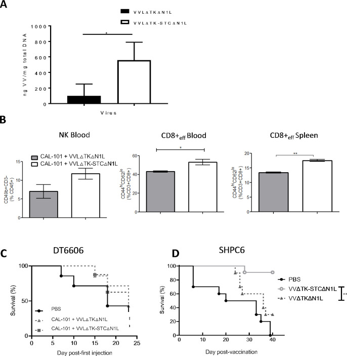
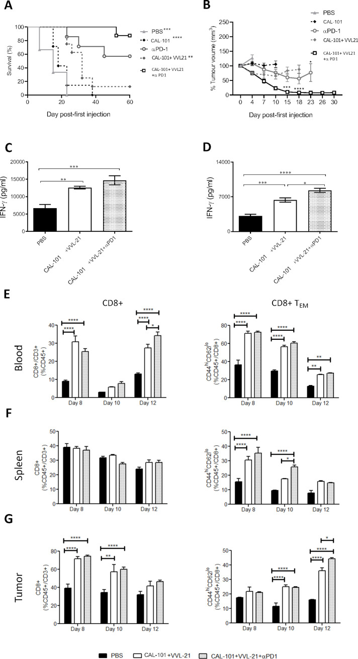
Background: Pancreatic cancer remains one of the most lethal cancers and is refractory to immunotherapeutic interventions. Oncolytic viruses are a promising new treatment option, but current platforms demonstrate limited efficacy, especially for inaccessible and metastatic cancers that require systemically deliverable therapies. We recently described an oncolytic vaccinia virus (VV), VVLΔTKΔN1L, which has potent antitumor activity, and a regime to enhance intravenous delivery of VV by pharmacological inhibition of pharmacological inhibition of PI3 Kinase δ (PI3Kδ) to prevent virus uptake by macrophages. While these platforms improve the clinical prospects of VV, antitumor efficacy must be improved.

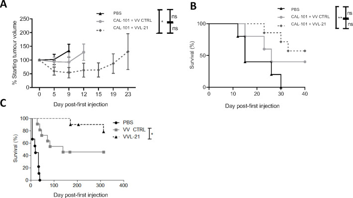
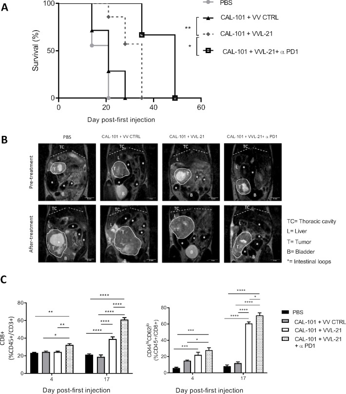
Methods: VVLΔTKΔN1L was modified to improve viral spread within and between tumors via viral B5R protein modification, which enhanced production of the extracellular enveloped virus form of VV. Antitumor immunity evoked by viral treatment was improved by arming the virus with interleukin-21, creating VVL-21. Efficacy, functional activity and synergy with α-programmed cell death protein 1 (α-PD1) were assessed after systemic delivery to murine and Syrian hamster models of pancreatic cancer.

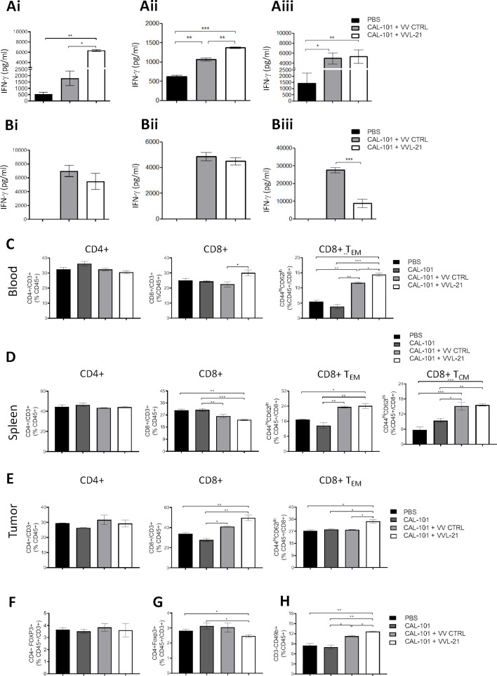
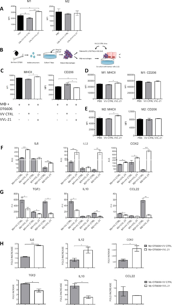
Results: VVL-21 could reach tumors after systemic delivery and demonstrated antitumor efficacy in subcutaneous, orthotopic and disseminated models of pancreatic cancer. The incorporation of modified B5R improved intratumoural accumulation of VV. VVL-21 treatment increased the numbers of effector CD8+ T cells within the tumor, increased circulating natural killer cells and was able to polarize macrophages to an M1 phenotype in vivo and in vitro. Importantly, treatment with VVL-21 sensitized tumors to the immune checkpoint inhibitor α-PD1.

Conclusions: Intravenously administered VVL-21 successfully remodeled the suppressive tumor-microenvironment to promote antitumor immune responses and improve long-term survival in animal models of pancreatic cancer. Importantly, treatment with VVL-21 sensitized tumors to the immune checkpoint inhibitor α-PD1. Combination of PI3Kδ inhibition, VVL-21 and α-PD1 creates an effective platform for treatment of pancreatic cancer.

Keywords: immunomodulation; macrophages; natural killer T-cells; oncolytic viruses; tumor microenvironment.


Conflict of interest statement

Competing interests: None declared.

Figures

Figure 1
Figure 1
VVLΔTK-STCΔN1L is effective at reaching the tumor and inducing antitumor efficacy after CAL-101-potentiated systemic delivery. (A, B) DT6606 tumors were established subcutaneously in immunocompetent C57BL/6 mice. Once palpable (100 mm3), mice were treated with CAL-101 (10 mg/kg) by oral gavage followed 3 hours later by intravenous injection using 1×10⁸ plaque-forming unit (PFU)/injection VVLΔTKΔN1L that does not contain a modified second copy of B5R, or VVLΔTK-STCΔN1L. Treatments were given on days 0, 2 and 4. (A) Five days following the treatment, tumors were excised and viral load analyzed using quantitative PCR shown as ng viral DNA per mg total DNA (n=3/group). (B) Five days following the treatment, blood and spleen were analyzed using flow cytometry for natural killer (NK) or effector CD8+ T cell populations (n=3/group). Mean±SEM is shown and significance analyzed using an unpaired Student’s t-test. (C) DT6606 tumors were established and treated with CAL-101 (10 mg/kg) by oral gavage followed 3 hours later by intravenous injection using 1×10⁸ PFU/injection on days 0, 2 and 4 and 2×10⁸ PFU/injection on days 13, 15 and 17 and survival monitored. Kaplan-Meier survival analysis with log rank (Mantel-Cox) tests were used to assess survival (n=7–8/group). (D) SHPC6 tumors were established intraperitoneally in Syrian Hamsters. Hamsters were treated intraperitoneally with 2×10⁷ PFU/mL VVLΔTKΔN1L or VVLΔTK-STCΔN1L on days 4, 6 and 8 post-tumor implantation. Of note, no CAL-101 was delivered prior to intraperitoneal injection of virus. Kaplan-Meier survival analysis with log rank (Mantel-Cox) tests were used to assess survival (n=10/group). *P<0.05; **p<0.01. PBS, phosphate-buffered saline.
Figure 2
Figure 2
Arming VVLΔTK-STCΔN1L with interleukin (IL)-21 (VVL-21) improves in vivo antitumor efficacy in murine and hamster models of pancreatic cancer. (A, B) DT6606 tumors were established subcutaneously in immunocompetent C57BL/6 mice. Once palpable (100 mm3), mice were treated with CAL-101 (10 mg/kg) by oral gavage followed 3 hours later by intravenous injection using 1×10⁸ plaque-forming unit (PFU)/injection VVLΔTK-STCΔN1L-IL21 (VVL-21) or VV CTRL (no IL-21). Treatments were given on days 0, 2 and 4 (n=5–7/group). (A) Tumor size was monitored twice weekly and the mean±SEM is shown. Significance was assessed using a two-way analysis of variance with Tukey’s multiple comparison post-test and is shown for day 9. (B) Kaplan-Meier survival analysis with log rank (Mantel-Cox) tests were used to assess survival. (C) Disseminated SHPC6 tumors were established intraperitoneally in Syrian Hamsters. Hamsters were treated intraperitoneally with 2×10⁷ PFU/mL VV-CTRL or VVL-21 (in this case, the virus expressed a human version of IL-21) on days 4, 6 and 8 post-tumor implantation. Kaplan-Meier survival analysis with Gehen-Breslow-Wilcoxon tests were used to assess survival (n=9–11/group). *P<0.05; **p<0.01. ns, not significant; PBS, phosphate-buffered saline.
Figure 3
Figure 3
VVL-21 induces robust antitumor adaptive immune responses after systemic delivery. DT6606 tumors were established as previously and treated on days 0, 2 and 4 with 1×10⁸ plaque-forming unit (PFU) VV CTRL or VVL-21. (A–B) After treatment, the response of splenocytes to mitomycin C killed tumor cells (A) or viral protein (B8R epitope) (B) was examined ex vivo using tumor cell restimulation assays on days 7 (i), 10 (ii) or 12 (iii) after the first treatment. Interferon (IFN)-γ production in response to stimulation was determined after incubation for 72 hours using ELISA. Mean production±SEM is shown and a one-way analysis of variance (ANOVA) with Tukey’s multiple comparison post-test used to determine statistical significance (n=3/group). (C–H) 10 days after the first treatment, blood (C), spleen (D) and tumor (E) was collected and analyzed using flow cytometry for the presence of CD4+, CD8+, effector CD8+ (TEM) or central memory CD8+ (TCM) T cells. Mean populations±SEM are shown and a one-way ANOVA with Tukey’s multiple comparison post-test was used to determine statistical significance (n=3/group). (F–G) Intratumoral (F) and splenic (G) regulatory T populations were analyzed using flow cytometry 10 days after the first treatment. Mean populations±SEM are shown and a one-way ANOVA with Tukey’s multiple comparison post-test was used to determine statistical significance (n=3/group). (H) Natural killer (NK) cells responded rapidly to treatment and an elevation was detected in the blood using flow cytometry 4 days after the first treatment. Mean populations±SEM are shown and a one-way ANOVA with Tukey’s multiple comparison post-test was used to determine statistical significance (n=3/group). *P<0.05; **p<0.01; ***p<0.001. PBS, phosphate-buffered saline.
Figure 4
Figure 4
VVL-21 augments M1 macrophage polarization in vivo and in vitro. (A) DT6606 tumors were established as previously and treated on days 0, 2, 4 with 1×10⁸ plaque-forming unit (PFU) VV CTRL or VVL-21. Ten days after the first treatment, tumors were excised and macrophages analyzed using flow cytometry to determine major histocompatibility complex (MHC)II expression. MHCIIhi macrophages were considered M1 polarized and CD206hi considered M2 polarized. Median fluorescence intensity (MFI)±SEM is shown and results analyzed using a one-way analysis of variance (ANOVA) with Bonferroni post-test (n=3/group). (B) Schematic detailing in vitro isolation and culture of bone marrow-derived macrophages. (C) Naïve macrophages were co-cultured with DT6606 tumor cells±VV CTRL or VVL-21. MHCII expression (for M1 phenotype) and CD206 expression (for M2 phenotype) were assessed using flow cytometry. MFI±SEM is shown and results analyzed using a one-way ANOVA with Bonferroni post-test (n=3/group). (D–E) Macrophages were polarized to an M1 () or M2 (E) phenotype and incubated with VV CTRL or VVL-21 at a multiplicity of infection (MOI) (of 1 PFU/cell for 24 hours. Expression of M1 (MHCII) and M2 (CD206) markers in each group was analyzed using flow cytometry. Mean MFI±SEM is shown and results analyzed using a one-way ANOVA with Bonferroni post-test (n=3/group). (F–G) Naïve, M1-polarized or M2-polarized macrophages were incubated with virus as above (MOI 1 PFU for 24 hours) and the expression of M1 markers (F) interleukin (IL)6, IL12 and COX2 or M2 markers (G) transforming growth factor (TGF)β, IL10 and CCL22 analyzed using quantitative PCR (qPCR). Arbitary Units (AU)±SEM is shown and results analyzed using a one-way ANOVA with Bonferroni post-test (n=3/group). (H) Naïve macrophages co-cultured with virus-infected DT6606 cells (using MOI of 1 PFU) were assessed for expression of M1 or M2 markers using qPCR 24 hours after incubation. The fold increase with respect to GAPDH is shown (±SEM) and results analyzed using a one-way ANOVA with Bonferroni post-test (n=3/group). *P<0.05; **p<0.01; ***p<0.001. PBS, phosphate-buffered saline.
Figure 5
Figure 5
α-Programmed cell death protein 1 (α-PD1) can augment the antitumor efficacy of CAL-101 potentiated intravenous-delivered VVL-21 in vivo. (A–B) DT6606 subcutaneous tumors were established as previously and treated using 1×10⁸ plaque-forming unit (PFU) for injections on days 0, 2, 4 and 2×10⁸ on days 13, 15, 17. α-PD1 was administered by intraperitoneal injection on days 2, 5, 7, 15, 19 and 20 (200 µg/injection). (A) Kaplan-Meier survival analysis with log rank (Mantel-Cox) tests were used to assess survival. Significance in relation to CAL-101+VVL-21+α-PD1 group is shown (n=6–8/group). (B) Tumors were measured twice weekly and the mean tumor sixe (±SEM) plotted. A two-way analysis of variance (ANOVA) with Tukey’s multiple comparison post-test was used to measure significance. Significance of CAL-101+VVL-21+α-PD1 compared with CAL-101+VVL-21 is shown at days 15 and 18 and significance of CAL-101+VVL-21+α-PD1 compared with α-PD1 is shown at day 23. (C–G) DT6606 subcutaneous tumors were established as previously and treated using 1×10⁸ PFU for injections on days 0, 2, 4. α-PD1 was administered by intraperitoneal injection on days 2, 5 and 7 (200 µg/injection). (C, D) After treatment, the response of splenocytes to tumor cells was examined ex vivo using tumor cell restimulation assays on days 8 (C) or 10 (D) after the first treatment. Interferon (IFN)-γ production in response to stimulation was determined after 72 hours using ELISA. Mean production±SEM is shown and a one-way ANOVA with Tukey’s multiple comparison post-test used to determine statistical significance (n=3/group). (E–G) Eight, 10 and 12 days after the first treatment, blood (E), spleen (F) and tumor (G) was collected and analyzed using flow cytometry for the presence of CD8+ and effector CD8+ (TEM) cells. Mean populations±SEM are shown and a two-way ANOVA with Tukey’s multiple comparison post-test was used to determine statistical significance (n=3/group). *P<0.05; **p<0.01; ***p<0.001; ****p<0.0001.
Figure 6
Figure 6
CAL-101-potentiated intravenous delivery of VVL-21 with concurrent α-programmed cell death protein 1 (α-PD1) treatment improves survival in a murine orthotopic model of cancer. Immunocompetent C57BL/6 mice had DT6606 tumor cells implanted in the tail of the pancreas. Ten days postimplantation, mice were treated according to the regime indicated in online supplemental figure S7, using 1×10⁸ plaque-forming unit (PFU)/injection on days 0, 2, 4 and 2×10⁸ PFU/injection on days 13, 15, 17. (A) Kaplan-Meier survival analysis with log rank (Mantel-Cox) tests were used to assess survival. Phosphate-buffered saline (PBS) n=9/group: VV CTRL n=7/group; VVL-21 n=7/group; VVL-21+α-PD1 n=3/group. *P=0.0355; **p=0.0084. (B) Representative images of MRI scanning that was conducted weekly. Pretreatment scans occurred 10 days post-tumor implantation. After treatment refers to scans taken in the last week of treatment. (C) Blood was drawn from the tail vein of mice at days 4 and 17 after the first day of treatment and analyzed for CD8CD8+ T cells and effector CD8+ T cells using flow cytometry. A two-way analysis of variance and Bonferroni post-test was used to determine statistical significance at each time-point. *P<0.05; **p<0.01; ***p<0.001; ****p<0.0001.

Similar articles

Cited by

References

    1. Bray F, Ferlay J, Soerjomataram I, et al. . Global cancer statistics 2018: GLOBOCAN estimates of incidence and mortality worldwide for 36 cancers in 185 countries. CA Cancer J Clin 2018;68:394–424. 10.3322/caac.21492 - DOI - PubMed
    1. Sun D, Ma J-X, Wang J, et al. . Clinical observation of immune checkpoint inhibitors in the treatment of advanced pancreatic cancer: a real-world study in Chinese cohort. Ther Clin Risk Manag 2018;14:1691–700. 10.2147/TCRM.S173041 - DOI - PMC - PubMed
    1. Ribas A, Dummer R, Puzanov I, et al. . Oncolytic virotherapy promotes intratumoral T cell infiltration and improves anti-PD-1 immunotherapy. Cell 2017;170:1109–19. e10. 10.1016/j.cell.2017.08.027 - DOI - PMC - PubMed
    1. Mercer J, Helenius A. Vaccinia virus uses macropinocytosis and apoptotic mimicry to enter host cells. Science 2008;320:531–5. 10.1126/science.1155164 - DOI - PubMed
    1. Hiley CT, Yuan M, Lemoine NR, et al. . Lister strain vaccinia virus, a potential therapeutic vector targeting hypoxic tumours. Gene Ther 2010;17:281–7. 10.1038/gt.2009.132 - DOI - PMC - PubMed

Publication types

MeSH terms