. 2021 Feb 2;118(5):e2012793118.
doi: 10.1073/pnas.2012793118.

Tissue-specific dynamic codon redefinition in Drosophila

Affiliations

Tissue-specific dynamic codon redefinition in Drosophila

Andrew M Hudson et al. Proc Natl Acad Sci U S A. .

Abstract

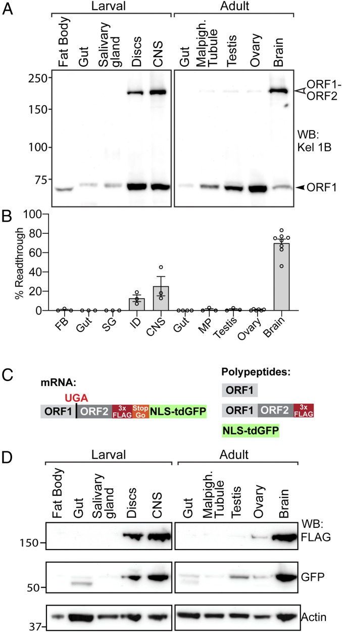
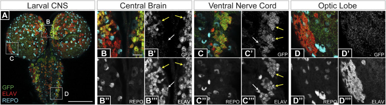
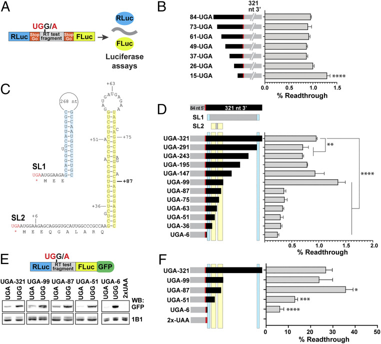
Translational stop codon readthrough occurs in organisms ranging from viruses to mammals and is especially prevalent in decoding Drosophila and viral mRNAs. Recoding of UGA, UAG, or UAA to specify an amino acid allows a proportion of the protein encoded by a single gene to be C-terminally extended. The extended product from Drosophila kelch mRNA is 160 kDa, whereas unextended Kelch protein, a subunit of a Cullin3-RING ubiquitin ligase, is 76 kDa. Previously we reported tissue-specific regulation of readthrough of the first kelch stop codon. Here, we characterize major efficiency differences in a variety of cell types. Immunoblotting revealed low levels of readthrough in malpighian tubules, ovary, and testis but abundant readthrough product in lysates of larval and adult central nervous system (CNS) tissue. Reporters of readthrough demonstrated greater than 30% readthrough in adult brains, and imaging in larval and adult brains showed that readthrough occurred in neurons but not glia. The extent of readthrough stimulatory sequences flanking the readthrough stop codon was assessed in transgenic Drosophila and in human tissue culture cells where inefficient readthrough occurs. A 99-nucleotide sequence with potential to form an mRNA stem-loop 3' of the readthrough stop codon stimulated readthrough efficiency. However, even with just six nucleotides of kelch mRNA sequence 3' of the stop codon, readthrough efficiency only dropped to 6% in adult neurons. Finally, we show that high-efficiency readthrough in the Drosophila CNS is common; for many neuronal proteins, C-terminal extended forms of individual proteins are likely relatively abundant.

Keywords: Drosophila; Kelch; central nervous system (CNS); recoding; stop codon readthrough.


Conflict of interest statement

The authors declare no competing interest.

Figures

Fig. 1.
Fig. 1.
High-level readthrough of kelch mRNA in the Drosophila central nervous system. (A) Western blot of tissues dissected from third-instar larvae and 3- to 5-d-old adults probed with anti-Kelch. The predicted molecular masses for ORF1 and ORF1-ORF2 are 77 kDa and 160 kDa, respectively. (B) Quantification of the percent of ORF1-ORF2 in total Kelch protein present in immunoblots (n ≥ 3, error bars represent SEM). (C) Schematic of reporter system for detecting readthrough of endogenous kelch. Translation through the ORF1 UGA stop codon results in the Kelch ORF1-ORF2 readthrough product tagged at its C terminus with 3× FLAG and a distinct nuclear-localized tandem dimer GFP (NLS::tdGFP) polypeptide released during translation of a viral T2A/StopGo sequence. (D) Western blot of tissues dissected from third-instar larvae and adult animals homozygous for the readthrough reporter insertion in the kelch locus, probed with α-FLAG to detect the readthrough product, α-GFP to detect the released NLS::tdGFP, and α-actin as a loading control.
Fig. 2.
Fig. 2.
Visualization of endogenous kelch readthrough. (A) Analysis of larval CNS revealed the widespread presence of NLS::tdGFP produced following kelch readthrough translation. Three regions of the larval CNS were imaged at a higher magnification: central brain (B), ventral nerve cord (C), and optic lobe (D). NLS::tdGFP accumulated in neurons (ELAV, red) and not glial cells (REPO, blue) in the central brain (BB′′′, yellow arrows) and in the ventral nerve cord (CC′′′, yellow arrows). Some neurons (white arrows) in the central brain (B) and ventral nerve cord (C) did not show evidence of readthrough. (DD′′′) NLS::tdGFP did not accumulate in optic lobe cells. (Scale bar for A, 100 µm; for BD, 10 µm.)
Fig. 3.
Fig. 3.
Analysis of the contribution to readthrough of cis-acting mRNA structural elements near the kelch stop codon. (A) Diagram of pSGDluc reporter construct. StopGo sequences from foot and mouth disease virus (FMDV) flanking the readthrough test fragment resulted in the translation of luciferase polypeptides free of residues encoded by kelch sequence; readthrough efficiency was calculated based on measured luciferase activities (46). (B) Sequences 5′ of the kelch stop codon were analyzed by transfecting the indicated 5′ truncation constructs into HEK-293T cells and measuring readthrough efficiency. (C) Diagram of predicted stem loop structures 3′ of the kelch ORF1 UGA codon. SL1 was previously described (27). For SL2, residues corresponding to the limits of 3′ deletions in D are indicated; location of 87 nt 3′ UTR is marked in bold. (D) Sequence requirements 3′ of the kelch UGA were analyzed as in B using the indicated 3′ deletion constructs. Predicted stem loops SL1 and SL2 are indicated above, with cyan or yellow shading corresponding to the shaded sequences in C. In B and D, n = 3 biological replicates. (E) Diagram of dual-luciferase construct for analysis of readthrough in transgenic Drosophila. GFP coding sequence was fused to the Renilla luciferase gene and readthrough efficiencies in F were calculated from the ratio of GFP produced by UGA constructs compared to matched in-frame UGG controls. Constructs were expressed in adult brains using nSybGal4, and lysates were prepared from heads. Representative Western blots of constructs analyzed are shown Below the diagram. The Top row shows immunoblotting with a GFP antibody and the Bottom row shows immunoblotting with an Adducin antibody (1B1) as a loading control. (F) Quantification of readthrough efficiencies in flies; n ≥ 3. In B, D, and F, mean and SD are plotted. Readthrough efficiencies of truncation constructs were compared to the corresponding in-frame control using ANOVA with multiple comparisons correction. *P < 0.05, **P < 0.01, ***P < 0.001, ****P < 0.0001.
Fig. 4.
Fig. 4.
kelch cDNA reporter reveals cell types supporting kelch readthrough. (AD) α-Tub84BGal4-driven expression in larval tissues of the UGG (in-frame control) reporter construct (A and B) and UGA readthrough reporter (C and D) in the dual-luciferase/GFP construct. Kelch cDNAs correspond to the 84-UGA/G-321 fragment described in Fig. 3.α-Tub84BGal4drove expression throughout the CNS (A) and at high levels in the endocrine prothoracic gland (PG), outlined in yellow and shown at higher magnification in B. (C and D) GFP intensity levels of the UGA readthrough reporter were increased 10-fold to allow the pattern of expression to be compared between the UGA and UGG constructs. (C) GFP produced by readthrough accumulated primarily in the central brain (CB) and the ventral nerve cord (VNC) neuropil. Green dotted line indicates the area occupied by the optic lobes (OL). GFP was not detected in the PG (C) and is shown at higher magnification in D and D′. (EH) Images directly comparing GFP reporter expression in neurons or glia. Expression specifically in neurons using nSybGal4 (E) revealed readily detectable readthrough expression from the UGA readthrough reporter (F). Expression specifically in glial cells using repoGal4 (G) failed to produce detectable readthrough (region occupied by larval CNS outlined in white in H). (I) Western analysis of reporter expression in lysates prepared from heads of adult flies expressing reporter constructs specifically in neurons or glia. (JS) UGG and UGA dual-fluorescent reporters expressed in adult brains reveal readthrough in five neuron subtypes; (J′–S′) GFP displayed with a color look-up table. (J and K) Pan-neural expression with nSybGal4 showed widespread readthrough in adult brains with highest levels in the mushroom bodies (J′ and K′, arrowheads). (L and M) Expression in GABA-producing neurons with Gad1Gal4. (N and O) Expression glutamate-producing neurons with VGlutGal4. (P and Q) Expression in dopamine-producing neurons with DdcGal4. A subset of dopaminergic neurons did not support readthrough (P′ and Q′, arrowheads). (R and S) Expression in serotonin receptor optic lobe neurons with 5-HT1AGal4. Brain structure in PS is evident due to Bruchpilot antibody staining. (Scale bar for whole larval CNS images A, C, and EH, 100 μm.) (Scale bar for prothoracic gland images B and D, 50 μm.) (Scale bar for adult brain images JS, 200 µm.)
Fig. 5.
Fig. 5.
Multiple Drosophila genes show high-level readthrough in neuronal tissue. (AD) Western blots comparing readthrough translation detected in lysates prepared from ovaries and adult brains (AC) or third instar larval (L3) CNS (D). Genes encoding each protein are diagrammed at Top; blue indicates ORF1 coding sequence, red indicates ORF2 coding sequence. Predicted molecular weights for ORF1 and ORF1-ORF2 readthrough products are listed. Ptp10D is a transmembrane receptor tyrosine phosphatase and is known to be glycosylated, which presumably accounts for the shift in apparent molecular weight (70). Blots were probed with antibodies against the ORF1 product of the indicated genes. Readthrough products are indicated with red arrowheads and ORF1 products are indicated with blue arrowheads. Apparent readthrough efficiencies are indicated beneath each lane, calculated from the ratio of readthrough product to the total quantity of protein detected.

References

    1. Ivanov A., et al. , Polyadenylate-binding protein-interacting proteins PAIP1 and PAIP2 affect translation termination. J. Biol. Chem. 294, 8630–8639 (2019). - PMC - PubMed
    1. Brown A., Shao S., Murray J., Hegde R. S., Ramakrishnan V., Structural basis for stop codon recognition in eukaryotes. Nature 524, 493–496 (2015). - PMC - PubMed
    1. Matheisl S., Berninghausen O., Becker T., Beckmann R., Structure of a human translation termination complex. Nucleic Acids Res. 43, 8615–8626 (2015). - PMC - PubMed
    1. Shao S., et al. , Decoding mammalian ribosome-mRNA states by translational GTPase complexes. Cell 167, 1229–1240.e15 (2016). - PMC - PubMed
    1. Brown C. M., Stockwell P. A., Trotman C. N., Tate W. P., Sequence analysis suggests that tetra-nucleotides signal the termination of protein synthesis in eukaryotes. Nucleic Acids Res. 18, 6339–6345 (1990). - PMC - PubMed

Publication types

LinkOut - more resources