. 2021 Jan 26;11(1):2185.
doi: 10.1038/s41598-020-80911-z.

Farnesyl dimethyl chromanol targets colon cancer stem cells and prevents colorectal cancer metastasis

Affiliations

Farnesyl dimethyl chromanol targets colon cancer stem cells and prevents colorectal cancer metastasis

Kazim Husain et al. Sci Rep. .

Retraction in

Abstract

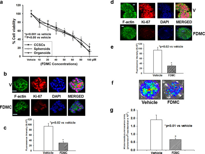
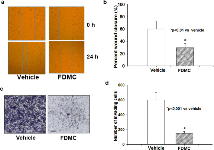
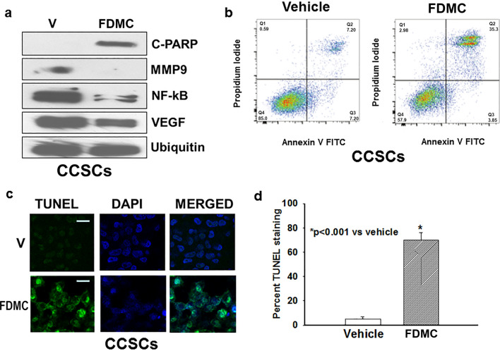
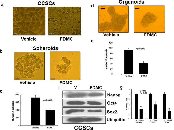
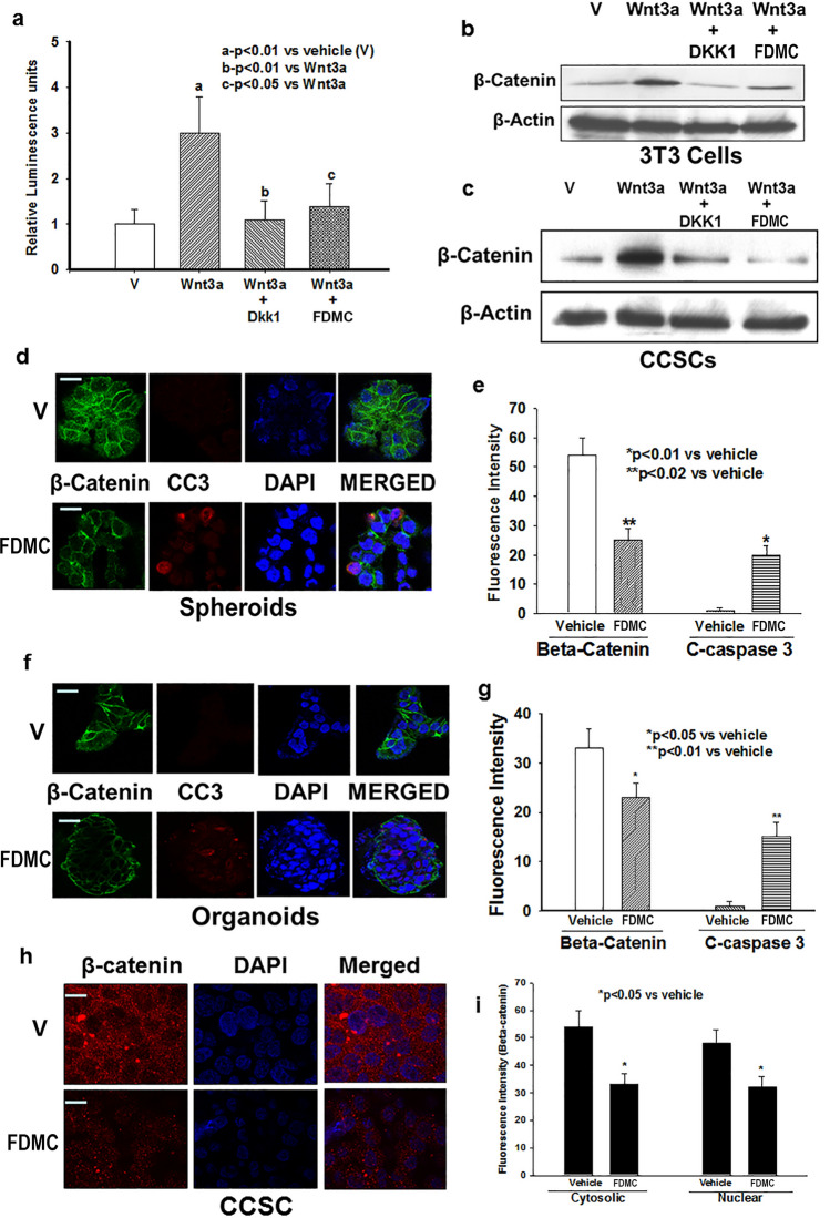
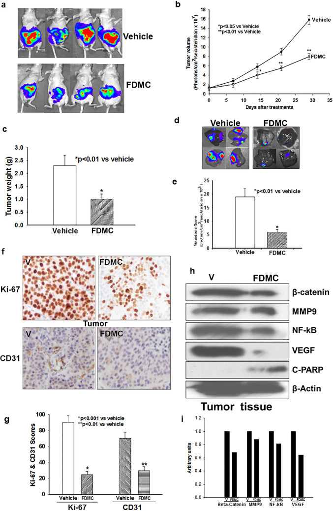
The activation and growth of tumour-initiating cells with stem-like properties in distant organs characterize colorectal cancer (CRC) growth and metastasis. Thus, inhibition of colon cancer stem cell (CCSC) growth holds promise for CRC growth and metastasis prevention. We and others have shown that farnesyl dimethyl chromanol (FDMC) inhibits cancer cell growth and induces apoptosis in vitro and in vivo. We provide the first demonstration that FDMC inhibits CCSC viability, survival, self-renewal (spheroid formation), pluripotent transcription factors (Nanog, Oct4, and Sox2) expression, organoids formation, and Wnt/β-catenin signalling, as evidenced by comparisons with vehicle-treated controls. In addition, FDMC inhibits CCSC migration, invasion, inflammation (NF-kB), angiogenesis (vascular endothelial growth factor, VEGF), and metastasis (MMP9), which are critical tumour metastasis processes. Moreover, FDMC induced apoptosis (TUNEL, Annexin V, cleaved caspase 3, and cleaved PARP) in CCSCs and CCSC-derived spheroids and organoids. Finally, in an orthotopic (cecum-injected CCSCs) xenograft metastasis model, we show that FDMC significantly retards CCSC-derived tumour growth (Ki-67); inhibits inflammation (NF-kB), angiogenesis (VEGF and CD31), and β-catenin signalling; and induces apoptosis (cleaved PARP) in tumour tissues and inhibits liver metastasis. In summary, our results demonstrate that FDMC inhibits the CCSC metastatic phenotype and thereby supports investigating its ability to prevent CRC metastases.


Conflict of interest statement

Dr. Malafa is named as an inventor on US Patent “Delta-Tocotrienol Treatment and Prevention of Pancreatic Cancer” (June 26, 2007; OTML docket number 06A069) but does not have financial interest in the companies that have licensed this patent. Further patents are in development. The other authors declare no competing interests.

Figures

Figure 1
Figure 1
Effects of FDMC on growth of human colon cancer stem cells (CD24+, CD44+, LGR5+), spheroids, and organoids, as evidenced by comparisons with vehicle-treated controls (V = vehicle). (a) FDMC significantly inhibited cell viability of human CCSCs, spheroids, and organoids in a concentration-dependent manner (CellTiter Glo assay). (b,c) Confocal microscopy data show that FDMC (50 µM) significantly inhibited proliferation (Ki-67 red immunofluorescence staining) in spheroids (*p < 0.02). (d,e) Confocal microscopy data show that FDMC (50 µM) significantly inhibited proliferation (Ki-67 red immunofluorescence staining) in organoids (*p < 0.02). (f,g) Colonogenic soft agar assay with luciferase-expressing CCSCs shows that FDMC (50 µM) significantly inhibited anchorage-independent growth (luminescence units) (*p < 0.01). Results are mean ± standard error of the means (bars; n = 3).
Figure 2
Figure 2
Effect of FDMC on migration and invasion of human CCSCs, as evidenced by comparisons with vehicle-treated controls (V = vehicle). (a) FDMC inhibited cell migration after 24 h in CCSCs (wound-healing assay or scratch test). (b) FDMC (50 µM) significantly inhibited cell migration CCSCs (*p < 0.01). (c) FDMC inhibited cell invasion in CCSCs (Matrigel invasion chamber test). (d) FDMC (50 µM) significantly inhibited cell invasion in CCSCs (*p < 0.001). Results are mean ± SE (bars; n = 3).
Figure 3
Figure 3
Effect of FDMC on inflammation, angiogenesis, and metastasis markers and induction of apoptosis in CCSCs, as evidenced by comparisons with vehicle-treated controls (V = vehicle). (a) Western blot analyses show that FDMC (50 µM) inhibited inflammation (NF-kB expression), angiogenesis (VEGF expression), and metastasis (MMP9 expression) and induced apoptosis (C-PARP) in CCSCs. Data are from 3 independent experiments. (b) Flow cytometry analysis of apoptosis (Annexin V/PI) after FDMC and vehicle treatment in CCSCs: FDMC (50 µM) treatment of human CCSCs for 24 h induced 35% apoptosis, whereas 7% apoptosis was seen in the vehicle-treated control. (c) Results of immunofluorescence staining (green) TUNEL assay of apoptosis in CCSCs. (d) FDMC (50 µM) significantly induced apoptosis in CCSCs (*p < 0.001). Results are mean ± SE (bars; n = 3).
Figure 4
Figure 4
Effect of FDMC on morphology, self-renewal capacity, organoid formation, and expression of pluripotency transcription factors in CCSCs, as evidenced by comparisons with vehicle-treated controls (V = vehicle). (a) Microscopic image shows that CCSCs became spherical after 24 h of FDMC (50 µM) treatment, in contrast to vehicle-treated cells. (b) Microscopic image shows that FDMC (50 µM) inhibited self-renewal (spheroid formation) of human CCSCs (CD24+CD44+LGR5+) grown in ultralow non-adherent plate containing stem cell-specific medium in 3D culture. (c) FDMC (50 µM) significantly inhibited the number of spheroids compared to vehicle (*p < 0.0005). (d) Microscopic image shows that FDMC (50 µM) inhibited CCSC-derived organoids formation when grown in Matrigel and stem cell-specific organoid medium in 3D culture. (e) FDMC (50 µM) significantly inhibited the number of organoids compared to vehicle (*p < 0.0002). (f,g) Western blot data show that FDMC (50 µM) significantly inhibited CCSC expression of the stem cell pluripotency transcription factors Nanog, Oct4, and Sox2 compared to vehicle (*p < 0.05 and **p < 0.02). Data are from 3 independent experiments.
Figure 5
Figure 5
Effects of FDMC on Wnt/β-catenin activity in CCSCs, spheroids and organoids, and induction of apoptosis, as evidenced by comparisons with vehicle-treated controls (V = vehicle). (a) Wnt receptor activity (luciferase units) significantly induced by agonist (Wnt3a) (ap < 0.01) and decreased by antagonist (DKK1) (bp < 0.01) in 3T3 cells. FDMC (50 µM) treatment for 24 h significantly suppressed agonist-induced receptor activity (cp < 0.01) in 3T3 cells. (b) Western blot data show that Wnt receptor agonist Wnt3a treatment induced β-catenin expression, whereas Wnt receptor antagonist DKK1 treatment abolished the Wnt3a-induced induction of β-catenin in 3T3 cells. FDMC inhibited Wnt3a-induced β-catenin expression in 3T3 cells. (c) Western blot data show that Wnt receptor agonist Wnt3a treatment induced β-catenin expression, whereas Wnt receptor antagonist DKK1 treatment abolished the Wnt3a-induced induction of β-catenin in CCSCs. FDMC inhibited Wnt3a-induced β-catenin expression in CCSCs. (d,e) Confocal microscopy data show that FDMC (50 µM) treatment to CCSC-derived spheroids significantly depleted β-catenin expression (green immunofluorescence staining) and induced apoptosis (cleaved caspase 3; red immunofluorescence staining; *p < 0.01 and **p < 0.02). (f,g) Confocal microscopy data show that FDMC (50 µM) treatment to CCSC-derived organoids significantly depleted β-catenin expression (green immunofluorescence staining) and induced apoptosis (cleaved caspase 3; red immunofluorescence staining; *p < 0.05 and **p < 0.01). Results are mean ± SEM (bars; n = 3).
Figure 6
Figure 6
Effect of FDMC on colorectal tumour growth, metastasis, inflammation, and angiogenesis markers, β-catenin expression and induction of apoptosis in mice, as evidenced by comparisons with vehicle-treated controls (V = vehicle). (a) Luciferase bioluminescence image shows that treatment with FDMC (200 mg/kg, orally, twice a week) for 4 weeks decreased tumour growth in mice. (b) FDMC significantly decreased tumour volume 2 weeks after treatment (*p < 0.05) and more significantly (**p < 0.01) after 3 to 4 weeks of treatment. (c) FDMC significantly decreased tumour weight 4 weeks after treatment (*p < 0.01). (d) Luciferase bioluminescence image shows that FDMC decreased the numbers of liver metastasis nodules in mice. (e) FDMC significantly decreased liver metastases (luminescence units) mice (*p < 0.01). (f,g) Histology data of colorectal tumours show that FDMC significantly inhibited tumour cell proliferation (Ki-67) and tumour angiogenesis (CD31). (h) Western blot data depict the depletion of markers for inflammatory, angiogenesis, and metastasis (NF-kB, VEGF and MMP9) and induced apoptosis (C-PARP) in the tumour tissue of mice treated with FDMC compared with vehicle. Results are mean ± SEM (bars; n = 3–5).

References

    1. Siegel, R. L. et al. Colorectal cancer statistics, 2017. CA Cancer J. Clin.67, 177–193. 10.3322/caac.21395 (2017). - PubMed
    1. Siegel, R. L., Miller, K. D. & Jemal, A. Cancer statistics, 2018. CA Cancer J. Clin.68, 7–30. 10.3322/caac.21442 (2018). - PubMed
    1. Siegel, R. L., Miller, K. D. & Jemal, A. Cancer statistics, 2019. CA A Cancer J. Clin.69, 7–34. 10.3322/caac.21551 (2019). - PubMed
    1. DeSantis, C. E. et al. Cancer treatment and survivorship statistics, 2014. CA A Cancer J. Clin.64, 252–271. 10.3322/caac.21235 (2014). - PubMed
    1. Torre, L. A., Siegel, R. L., Ward, E. M. & Jemal, A. Global cancer incidence and mortality rates and trends-an update. Cancer Epidemiol. Biomark. Prev.25, 16–27. 10.1158/1055-9965.EPI-15-0578 (2016). - PubMed

Publication types

MeSH terms