

Motor cortical excitability predicts cognitive phenotypes in amyotrophic lateral sclerosis
- PMID: 33500476
- PMCID: PMC7838179
- DOI: 10.1038/s41598-021-81612-x
Motor cortical excitability predicts cognitive phenotypes in amyotrophic lateral sclerosis
Abstract
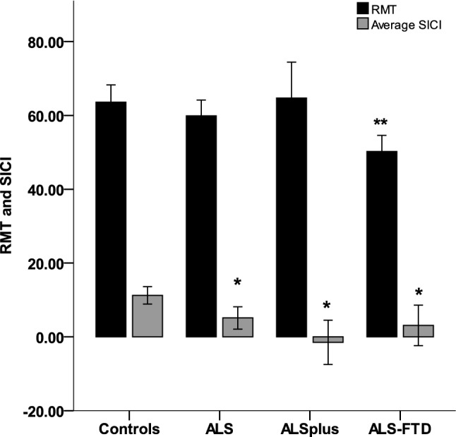
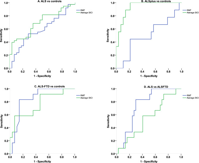
Amyotrophic lateral sclerosis (ALS) and frontotemporal dementia (FTD) are well-recognised as an extended disease spectrum. This study hypothesised that cortical hyperexcitability, an early pathophysiological abnormality in ALS, would distinguish cognitive phenotypes, as a surrogate marker of pathological disease burden. 61 patients with ALS, matched for disease duration (pure motor ALS, n = 39; ALS with coexistent FTD, ALS-FTD, n = 12; ALS with cognitive/behavioural abnormalities not meeting FTD criteria, ALS-Cog, n = 10) and 30 age-matched healthy controls. Cognitive function on the Addenbrooke's cognitive examination (ACE) scale, behavioural function on the motor neuron disease behavior scale (MiND-B) and cortical excitability using transcranial magnetic stimulation (TMS) were documented. Cortical resting motor threshold (RMT), lower threshold indicating hyperexcitability, was lower in ALS-FTD (50.2 ± 6.9) compared to controls (64.3 ± 12.6, p < 0.005), while ALS-Cog (63.3 ± 12.7) and ALS (60.8 ± 13.9, not significant) were similar to controls. Short interval intracortical inhibition (SICI) was reduced across all ALS groups compared to controls, indicating hyperexcitability. On receiver operating characteristic curve analysis, RMT differentiated ALS-FTD from ALS (area under the curve AUC = 0.745, p = 0.011). The present study has identified a distinct pattern of cortical excitability across cognitive phenotypes in ALS. As such, assessment of cortical physiology may provide more precise clinical prognostication in ALS.
Conflict of interest statement
The authors declare no competing interests.
Figures
References
Publication types
MeSH terms
LinkOut - more resources
Full Text Sources
Other Literature Sources
Medical
Research Materials
Miscellaneous
