Successful balloon-assisted coil embolization for a diagnostically difficult case of spontaneous vertebrovertebral arteriovenous fistula
- PMID: 33500812
- PMCID: PMC7827306
- DOI: 10.25259/SNI_803_2020
Successful balloon-assisted coil embolization for a diagnostically difficult case of spontaneous vertebrovertebral arteriovenous fistula
Abstract
Background: We describe a rare case of idiopathic lower cervical vertebro-vertebral arteriovenous fistula (VVAVF) with compression of the nerve roots and spinal cord, successfully treated with detachable coils utilizing the transarterial balloon-assisted technique without complication of coil mass.
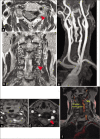
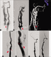
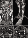
Case description: A 68-year-old woman was admitted for numbness of the left upper limb and pain in the left neck. Cervical magnetic resonance imaging (MRI) revealed compression of nerve roots and spinal cord by a large vascular lesion. The left vertebral angiography demonstrated a VVAVF draining into the vertebral venous plexus at C5 level. Under general anesthesia, the fistula site was accessed with a microcatheter through the right femoral artery, and successful embolization performed by compactly placing several detachable coils using balloon-assisted technique. The patient made a full recovery, with long-term MRI-documented left vertebral artery patency and no fistular leakage, and without postoperative complications.
Conclusion: Target occlusion utilizing the balloon-assisted technique in this case of VVAVF with compression of nerve roots and spinal cord, was effective in improving neurological symptoms, and achieved long-term occlusion when preservation of the VA was desired.
Keywords: Balloon-assisted technique; Cervical portion of vertebral artery; Endovascular embolization; Radiculopathy; Vertebro-vertebral arteriovenous fistula.
Copyright: © 2020 Surgical Neurology International.
Conflict of interest statement
There are no conflicts of interest.
Figures


Similar articles
-
Case of Iatrogenic Vertebro-Vertebral Arteriovenous Fistula Treated by Combination of Double-Catheter and Balloon Anchoring Techniques.World Neurosurg. 2019 Aug;128:98-101. doi: 10.1016/j.wneu.2019.04.236. Epub 2019 May 7. World Neurosurg. 2019. PMID: 31075492
-
Traumatic vertebro-vertebral arteriovenous fistula manifesting as radiculopathy. Case report.Neurol Med Chir (Tokyo). 2008 Apr;48(4):167-70. doi: 10.2176/nmc.48.167. Neurol Med Chir (Tokyo). 2008. PMID: 18434695
-
Endovascular treatment of high-flow cervical direct vertebro-vertebral arteriovenous fistula with detachable coils and Onyx liquid embolic agent.Acta Neurochir (Wien). 2011 Feb;153(2):347-52. doi: 10.1007/s00701-010-0850-z. Epub 2010 Nov 9. Acta Neurochir (Wien). 2011. PMID: 21058042
-
Pediatric cervical kyphosis in the MRI era (1984-2008) with long-term follow up: literature review.Childs Nerv Syst. 2022 Feb;38(2):361-377. doi: 10.1007/s00381-021-05409-z. Epub 2021 Nov 22. Childs Nerv Syst. 2022. PMID: 34806157 Review.
-
Endovascular management of V3 segment vertebro-vertebral fistula: case management and literature review.Br J Neurosurg. 2024 Oct;38(5):1188-1192. doi: 10.1080/02688697.2022.2071416. Epub 2022 May 3. Br J Neurosurg. 2024. PMID: 35502703 Review.
Cited by
-
Vertebro-Vertebral Arteriovenous Fistulae: A Case Series of Endovascular Management at a Single Center.Diagnostics (Basel). 2024 Feb 13;14(4):414. doi: 10.3390/diagnostics14040414. Diagnostics (Basel). 2024. PMID: 38396452 Free PMC article.
-
Vertebral-Venous fistulas: Single center experience and practical treatment approach.Interv Neuroradiol. 2025 Aug;31(4):464-474. doi: 10.1177/15910199231170079. Epub 2023 Apr 18. Interv Neuroradiol. 2025. PMID: 37073124 Free PMC article.
References
-
- Aljobeh A, Sorenson TJ, Bortolotti C, Cloft H, Lanzino G. Vertebral arteriovenous fistula: A review article. World Neurosurg. 2019;122:1388–97. - PubMed
-
- Beaujeux RL, Reizine DC, Casasco A, Aymard A, Rüfenacht D, Khayata MH, et al. Endovascular treatment of vertebral arteriovenous fistula. Radiology. 1992;183:361–7. - PubMed
-
- Briganti F, Tortora F, Elefante A, Volpe A, Bruno MC, Panagiotopoulos K. An unusual case of vertebral arteriovenous fistula treated with electrodetachable coil embolization. Minim Invasive Neurosurg. 2004;47:386–8. - PubMed
Publication types
LinkOut - more resources
Full Text Sources
Miscellaneous
