

Minimally invasive intervention in external cervical resorption: a case report with six-year follow-up
- PMID: 33500844
- PMCID: PMC7811936
Minimally invasive intervention in external cervical resorption: a case report with six-year follow-up
Abstract
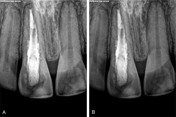
Root resorption consists of the loss of mineralized tissue (enamel, dentin, and cementum) of the inner or outer surface of the tooth due to the action of clastic cells. The correct diagnosis, the location, degree of tissue destruction, and the type of treatment are obstacles to the resolution of these lesions. The external cervical resorption is initiated in the amelocemental region progressively resorbing cementum, dentin, and enamel, constituting multiples ducts of resorption in an apical direction. This study reports a clinical case of treatment of a tooth affected by external cervical resorption with six-year clinical and radiographic follow-up. A 28-year-old male patient attended the clinic reporting a stain in the element 11. On clinical examination, there was a pinkish stain in the cervical lingual region, small cavitation in the enamel cervical region, and gingival bleeding with no insertion loss. Radiographically was observed a change at the root in the right central incisor, which was diagnosed as external root resorption. The negative response to the pulp sensitivity test confirmed the condition of pulp necrosis, indicating the need for endodontic treatment. To the treatment, it was opted for a minimally invasive approach, with endodontic access, instrumentation, and monthly exchanges of calcium hydroxide, for three months. After this period, the root canal has been filled with gutta-percha and sealer 26, in the apical third. The cervical and medium third were filled with MTA (mineral trioxide aggregate) leaving a central space for later fiberglass posts placing. The fiberglass post has been cemented with resinous cement and the tooth restored with resin composite. After six years of a radiographic control semiannual and annual, noticed normality in the periradicular tissues and disruption of the resorption process, was observed. The clinical management minimally invasive adopted reported in this case presents a viable treatment for external root resorption of the cervical third, especially in anterior teeth.
Keywords: Dental trauma; MTA; external cervical resorption; external root resorption; minimally invasive intervention; treatment of reabsorption.
IJBT Copyright © 2020.
Conflict of interest statement
None.
Figures


References
-
- Darcey J, Qualtrough A. Root resorption: simplifying diagnosis and improving outcomes. Prim Dent J. 2016;5:36–45. - PubMed
-
- Sasaki T, Watanabe C, Shimizu T, Debari K, Segawa K. Possible role of cementoblasts in the resorbant organ of human deciduous teeth during root resorption. J Periodontal Res. 1990;25:143–151. - PubMed
-
- Ferreira MM, Carrilho EV, Leitão J. Mecanismo e classificação das reabsorções radiculares. Rev. Portuguesa de Estomatol. Med Dent Cir Maxilofac. 2006
-
- Lyroudia KM, Dourou VI, Pantelidou OC, Labrianidis T, Pitas IK. Internal root resorption studied by radiography, stereomicroscope, scanning electron microscope and computerized 3D reconstructive method. Dent Traumatol. 2002;18:148–152. - PubMed
Publication types
LinkOut - more resources
Full Text Sources